Tee Mitral Valve Anatomy
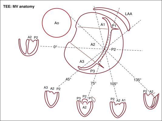
The comprehensive tee examination of the mitral valve consists of a series of eight cross sectional views. Transesophageal echocardiography tee is performed intraoperatively in all patients undergoing valve surgery and is critical to assess and localize valvular dysfunction.
 Finally Mitral Valve Orientation Explained
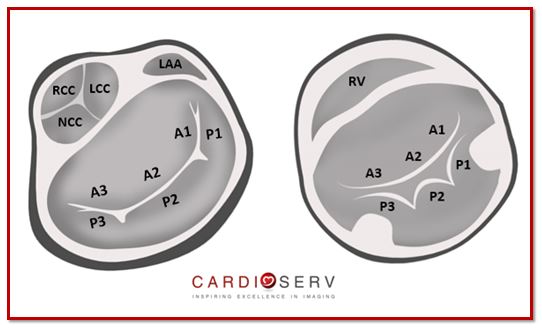
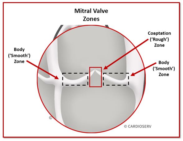
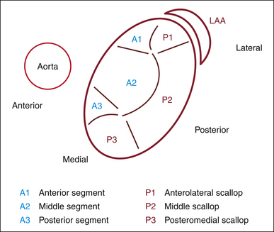
If we zoom in on the mitral leaflets from the atrial surface we can identify two zones that are used for describing location of pathology seen.
Tee mitral valve anatomy. Anatomy of mitral valve mitral valve apparatus mitral valve annulus. Normal mitral valve anatomy leaflets. This document will review the comprehensive 2d examination of the mv.
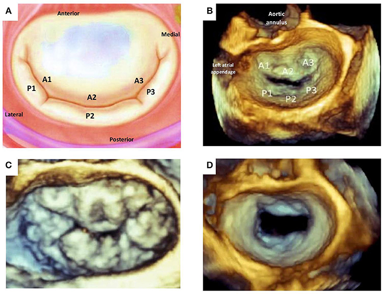
Assessment of mitral valve anatomy by real time 3 dimensional 3d transesophageal echocardiography tee has proven to be superior compared to 2 dimensional tee 121. Figure 20 1 mitral valve anatomy looking toward the left ventricle from posterior to anterior. Surgeons skill and experience 2.
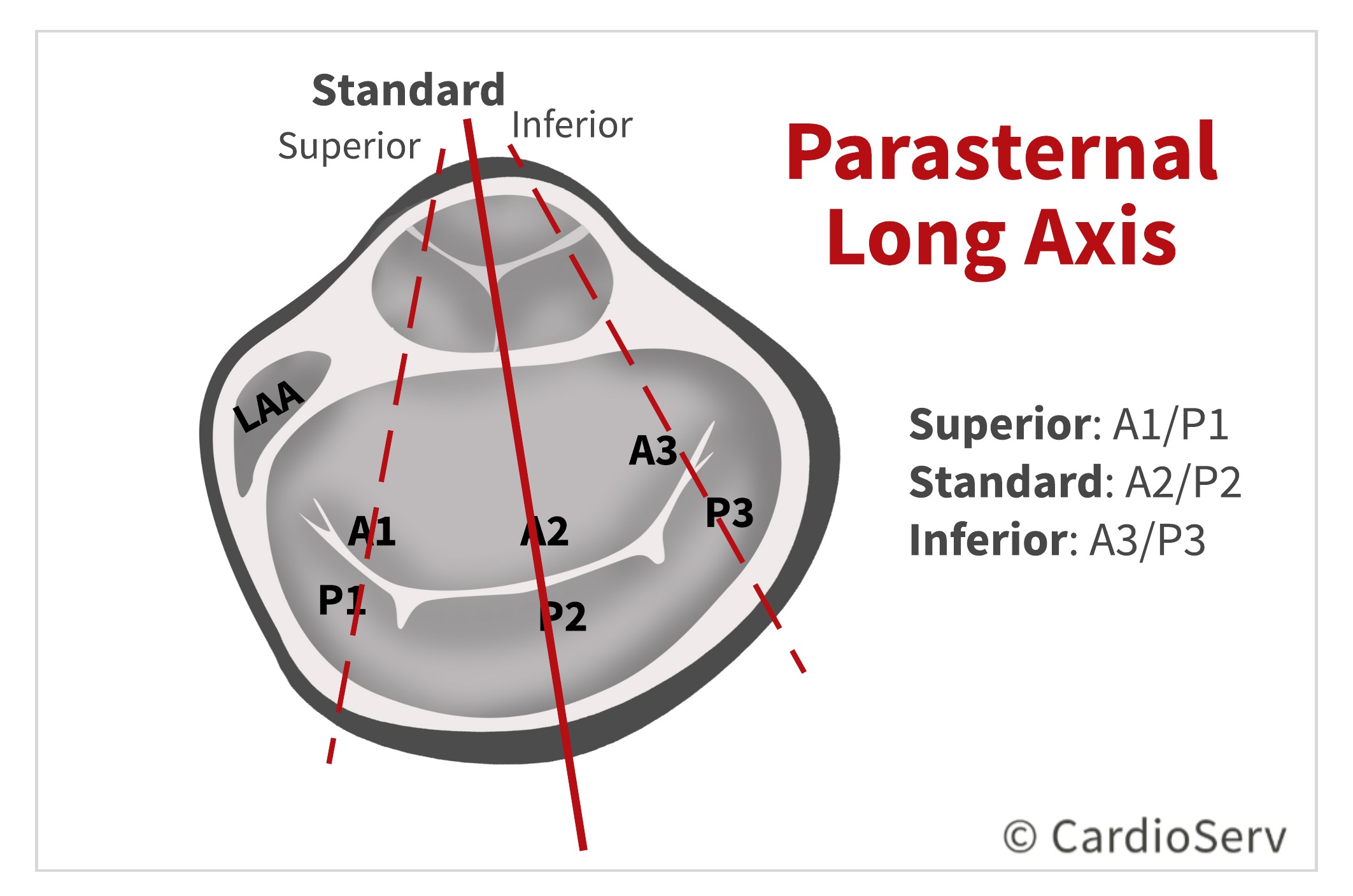
The mv comprises two leaflets annular attachment at the atrioventricular junction tendinous chords and the papillary muscles pms. Mitral leaflets with commissures. Learn the best echo views for visualizing the mitral valve scallops.
The standard modalities of real time 3d tee have recently been described 32. Altogether called as mitral. Learn the mitral valve apparatus anatomy as visualized by echocardiography transthoracic and tee.
The two leaflets of the mv are noticeably different in structure and are referred to as the anterior and posterior leaflets by clinicians. Surface area on leaflet body. Enhance your knowledge on the various structures and the relation to the function of the mitral valve.
The mitral apparatus has very specific details that make up the large picture of the mitral valve. The mitral valve consists of the mitral annulus anterior and posterior leaflets chordae tendineae and the papillary muscles. Accurate identification the anatomic lesions of the mitral valve echocardiography is pivotal in defining the functional anatomy of the mitral valve surgeon and echocardiographer speaking a common language mutual respect and honesty knowing when to send the.
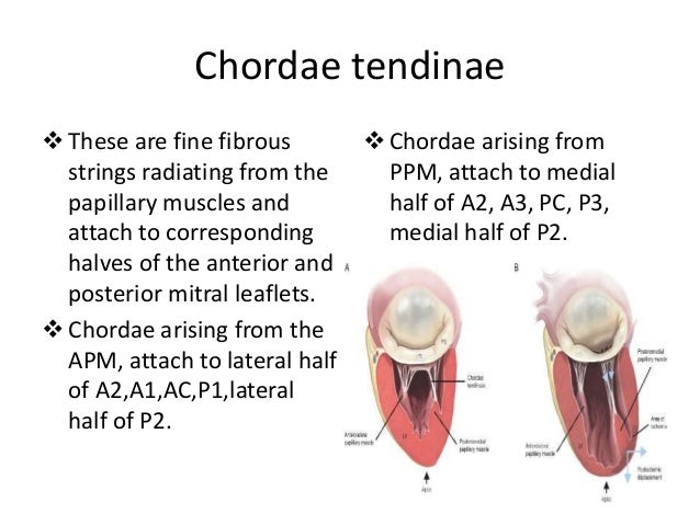
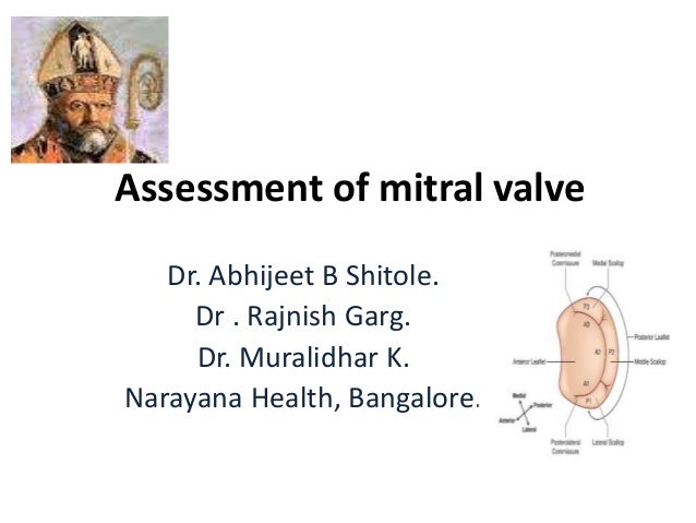
Feasibility of mitral repair 1. Via chordae tendineae small tendons which ensure that the leaflets do not prolapse the valve leaflets are attached to two major papillary muscles anterolateral en posteromedial in the left ventricle. Assessment of mitral valve by tee 1.
The mitral valve consists of two valve leaflets the anterior leaflet amvl and the posterior leaflet pmvl which together have a surface of 4 6 cm 2. Assessment of mitral valve dr. Describe the detailed anatomy of the mitral valve mv using two dimensional 2d transesophageal echocardiography tee based on the american society of echocardiographysociety of cardiovascular anesthesiology guidelines.
 Cardiac Interventions Today Echocardiographic Imaging In


 Mitral Valve Repair Chapter 16 Core Topics In
 Pdf Value Of Transesophageal Echocardiography Tee

 Mitral Valve Anterior Leaflet Prolapse
 Finally Mitral Valve Orientation Explained
 Mitral Valve Anatomy Name 5 Components
Role Of Transesophageal Echocardiography In Mitral Valve

 Assessment Of Mitral Valve Anatomy According To The Wilkins


 Assessment Of Mitral Valve Anatomy According To The Wilkins
 Schematic Left And 3d Tee Image Middle Demonstrating The
 Echocardiographic Guidance For Transcatheter Mitral Valve
 Anatomy Of Tricuspid Valve
 Tee Lv Anatomy

 Assessment Of Mitral Valve By Tee
 Myxomatous Mitral Valve Disease Comparison Of Different
The Geometric Model Of The Human Mitral Valve
 Role Of Transesophageal Echocardiography In Mitral Valve
 Multimodality Imaging In The Context Of Transcatheter Mitral
 Echocardiography Of The Mitral Valve Sciencedirect
 Assessment Of Mitral Valve By Tee
 Ischemic Mitral Regurgitation From Echo Assessment To
Mitral Valve Morphology Assessment Three Dimensional
 Mitral Valve Anatomy Schematic Representation Of The Mitral
 Frontiers Percutaneous Mitral Valve Interventions Repair
 Box Plots With The Cumulative Data Of The Four Mitral Valve
 Perioperative Echocardiography Education Apil
Belum ada Komentar untuk "Tee Mitral Valve Anatomy"
Posting Komentar