Diaphragm Anatomy
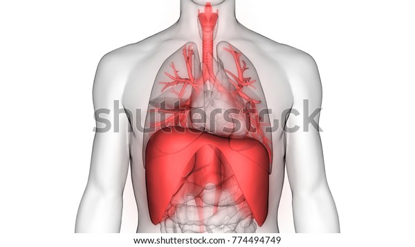
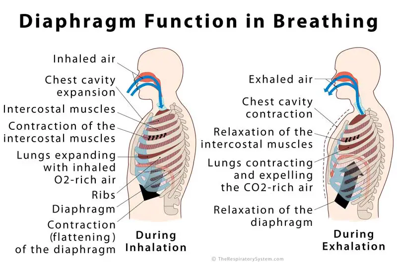
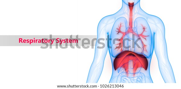
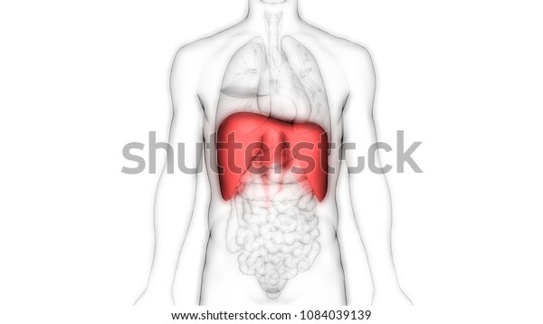
Contraction of the diaphragm increases the internal height of the thoracic cavity thus lowering its internal pressure and causing inspiration of air. It acts as the floor of the thoracic cavity and the roof of the abdominal cavity.
![]() Diaphragm Muscle Anatomy Innervation And Function Kenhub
Diaphragm Muscle Anatomy Innervation And Function Kenhub
Gross anatomy the muscular fibers of the diaphragm originate around the circumference of the inferio.

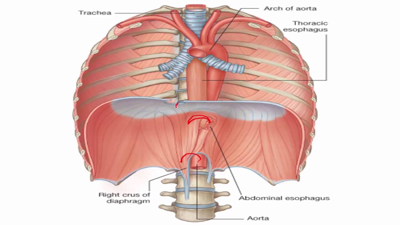
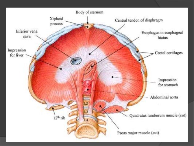
Diaphragm anatomy. Also known as the thoracic diaphragm it serves as an important anatomical landmark that separates the thorax or chest from the abdomen. The thoracic spinal levels at which the three major structures pass through the diaphragm can be remembered by the number of letters contained in each structure. Diaphragm anatomy and function the diaphragm is a thin skeletal muscle that sits at the base of the chest and separates the abdomen from the chest.
The diaphragm is the dome shaped muscle that separates the thoracic cavity from the abdominal cavity enclosing the inferior thoracic aperture. The diaphragm is one of the main muscles of respiration. It contracts and flattens when you inhale.
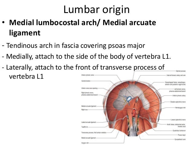
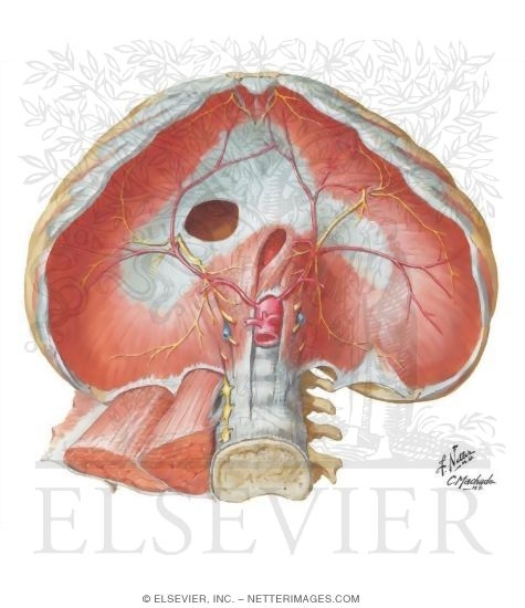
The attachments of diaphragm can be divided into peripheral and central attachments. It has three peripheral attachments. Lumbar vertebrae and arcuate ligaments.
Oesophagus 10 letters passes through the diaphragm at t10. Motor innervation of the diaphragm comes from the phrenic. The diaphragm is located at the inferior most aspect of the ribcage filling the inferior thoracic aperture.
Diaphragm anatomy parts of the main structure the peripheral muscle. The right crus arises from the bodies of first three lumbar vertebrae and their intervertebral discs. Diaphragm dome shaped muscular and membranous structure that separates the thoracic and abdominal cavities in mammals.
It is the principal muscle of respiration. There are three parts of the peripheral muscle sternal costal and lumbar depending on the location of the peripheral attachment. The diaphragm is a musculotendinous structure with a peripheral attachment.
Vena cava 8 letters passes through the diaphragm at t8. The diaphragm is the dome shaped sheet of muscle and tendon that serves as the main muscle of respiration and plays a vital role in the breathing process. The diaphragm is a musculotendinous sheet.
Lateral to the crura on both sides the diaphragm arises from the medial and lateral arcuate ligaments.
 Human Respiratory System Lungs Diaphragm Anatomy Stock Image
 Vector Illustration Diaphragm Anatomical Vector
 Anatomy Of The Diaphragm
 Diaphragm Definition Location Anatomy Function Diagram
Yoga Anatomy To Tap The Real Power Of Your Breath With Belly
 Anatomy 1 C2 L15 Diaphragm
 Anatomy Of The Normal Diaphragm Semantic Scholar
 Illustration Human Diaphragm Anatomy Stock Photo
 Structures Passing Through The Diaphragm As Seen From The
 Image Result For Diaphragm Anatomy Thoracic Duct Xiphoid
 Comparative Anatomy Of Diaphragms In Echidna Tachyglossus
 Human Respiratory System Diaphragm Anatomy Stock Photo
 Diaphragm Human Anatomy
 Diaphragm Muscle Anatomy Origin Insertion Action And
 Diaphragm Sciencedirect
 Diaphragm In Respiratory System
![]() Diaphragm Muscle Anatomy Innervation And Function Kenhub
Diaphragm Muscle Anatomy Innervation And Function Kenhub
![]() Diaphragm Muscle Anatomy Innervation And Function Kenhub
Diaphragm Muscle Anatomy Innervation And Function Kenhub
 The Diaphragm Yogabody Anatomy Kinesiology And Asana
 Diaphragm And Posterior Abdominal Wall Human Anatomy 1
Chest Breath Vs Belly Breath What S The Deal In
 Diaphragm Anatomy Stock Illustrations 1 021 Diaphragm
 Diaphragm Muscle
 Diaphragm Anatomy Quiz By Thebrend88
 Diaphragm Abdominal Surface
 Human Respiratory System Lungs Diaphragm Anatomy Stock
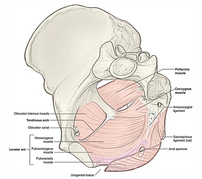 Easy Notes On Pelvic Diaphragm Learn In Just 4 Minutes
 Human Body Organs Diaphragm Anatomy 3d Stock Illustration
 The Diaphragm Anatomy Embryology


Belum ada Komentar untuk "Diaphragm Anatomy"
Posting Komentar