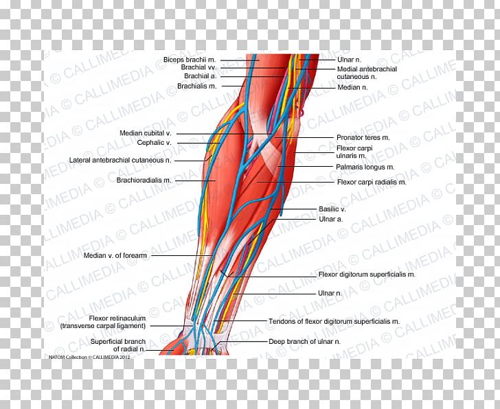
Anatomy Of Arm Veins
Veins of the upper limb hand. This web of veins extends across the back of the hand.
 Venous Cannulation Sites In The Arm Illustration Stock
12 jan arm anatomy part 1 muscles arteries and veins.

Anatomy of arm veins. Labels include cephalic vein brachial arteryvein basilic vein musculoskeletal nerve ulnar collateral artery radial collateral artery ulnar nervearteryvein interosseous arteryvein median nerve and radial nervearteryvein. As their name implies these veins are close to the skins surface. From the palmar aspect of the hand.
Making its way through the basilic vein is the median basilica vein located in the lower part of the elbow which works as a communicator in the arm. This is the space between the upper. This large vein travels through the upper arm before branching near.
They are located within the subcutaneous tissue of the upper limb. Veins of the upper limb. Their anatomy is discussed in the following sections see figure 23 1.
As you reach the proximal arm the axillary vein will divide into the basilic and brachial veins. The forearm is drained by numerous deep veins which form double venae comitantes. Some of the veins in the arm include.
The major superficial veins of the upper limb are the cephalic and basilic veins. The superficial veins of the upper limb are the veins selected for most elective venipuncture. Like in the forearm the arm is drained by the brachial veins.
In human anatomy the arm is the upper limb of the body that starts from the glenohumeral joints to the elbow joint. Continuing upwards through the forward part of the elbow the cephalic vein makes its way through the valley created by the biceps brachii and the brachioradialis on either side of it. There are two prominent superficial veins of the upper limb.
Anatomy of the nerves arteries and veins of the arm upper extremity. The basilic vein originates from the dorsal venous network of the hand and ascends the medial aspect of the upper limb. Continue from the axillary vein checking in transverse that the basilic and brachial veins of the upper arm are compressible.
At the border of the teres major the vein moves deep into the arm. Blood to the digits is drained through an anastomosis of palmar and dorsal digital veins. Usually single but may be duplicated.
In common usage the arm extends to the hand it can be divided in the upper arm the brachium the forearm and the hand. Upper arm veins brachial basilic the basilic vein is the larger and is more superficial.
 Thumb Elbow Vein Forearm Anatomy Png 600x600px Watercolor
 Diagram Pictures Neurovasculature Of The Arm And The
 Clinical Anatomy Of The Cephalic Vein For Safe Performance
 Veins Of The Arm Anatomy Study Buddy
 Torso And Arm Veins Dc Anatomy With Mrs Fisher At
 Cutaneous Nerves And Superficial Veins Of Shoulder And Arm
 Venipuncture Part 1 Anatomy Of The Arm And Vein Location
 Names Of Veins In The Arm Anatomy And Physiology Medical
 Anterior Compartment Of The Forearm Nerve Muscle Vein Png
 Arm Veins For Lower Extremity Arterial Reconstruction
 World S Best Anatomy Of Arm Veins Stock Illustrations
 Cephalic Vein Anatomy And Clinical Points Kenhub
 Medical Illustration Of Human Arm Muscles Veins And Nerves Canvas Print
 Arm Veins For Venipuncture Veins Dorsal Aspect Of The
 The Veins Of The Upper Extremity And Thorax Human Anatomy
 Illustrations Of The Blood Vessels Cleveland Clinic
 Lab 6 2a 7 Deep Veins Of Right Arm Veins Diagram Quizlet
 Cardiovascular System Of The Arm And Hand
 Veins Arm Stock Photos Veins Arm Stock Images Alamy
 The Cardiovascular System Of The Upper Limbs Anatomy Of
 Circulatory Routes Boundless Anatomy And Physiology
 Phlebotomy Related Vascular Anatomy
 Cephalic Vein Circulatory System Anatomy Human Body Arm
 Veins Of The Upper Limb Anatomy Kenhub
 Anatomy Atlases Illustrated Encyclopedia Of Human Anatomic

Belum ada Komentar untuk "Anatomy Of Arm Veins"
Posting Komentar