Thigh Mri Anatomy
Eye diagram muscles anatomy 16 photos of the eye diagram muscles anatomy diagram back muscles diagram leg muscles diagram of eye anatomy diagram of muscles in arm diagram of muscles in lower back diagram of muscles in the knee diagram of muscles in the neck human muscles diagram back muscles diagram leg muscles. Use the mouse scroll wheel to move the images up and down alternatively use the tiny arrows on both side of the image to move the images on both side of the image to move the images.
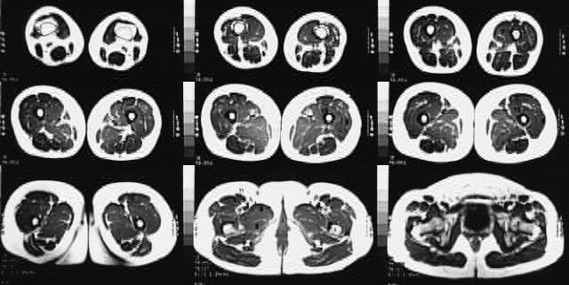
 Mri Thigh Calf Anatomy Normal Anatomy Dr Ahmed Eisawy
Online mri ct sectional anatomy kenneth k.

Thigh mri anatomy. Ho bachelor of medicine bachelor of surgery university of hong kong fellow hong kong college of radiologists fellow hong kong academy of medicine radiology real time interface human sectional anatomy. This mri hip joint axial cross sectional anatomy tool is absolutely free to use. Anatomy of the thigh.
The medical information on this site is provided as an information resource only and is not to be used or relied on for any diagnostic or treatment purposes. Welcome to online mri ct sectional anatomy. Injuries such as anterior cruciate ligament meniscus and rotator cuff tears are all easily diagnosed when there is a firm understanding and knowledge of human anatomy.
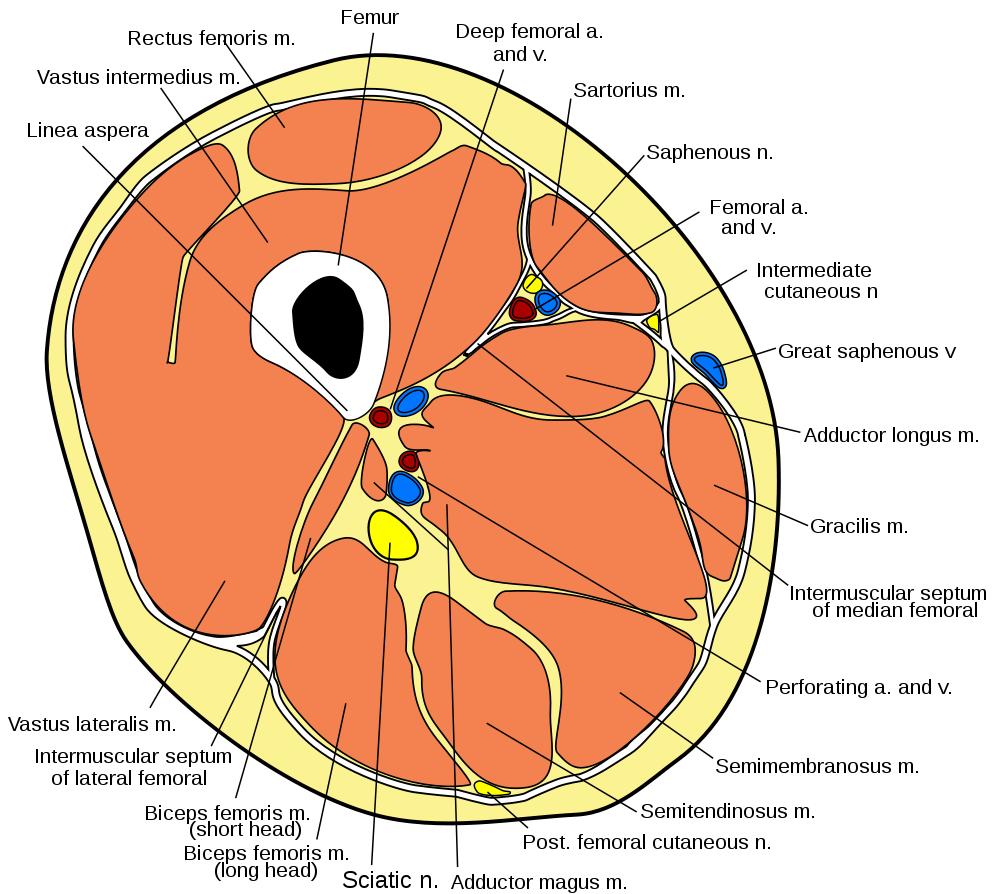
Upper two thirds of the medial margin and proximal margin of the patella medial condyle of the tibia and investing deep fascia of the leg with the tendons of vastus intermedius lateralis and rectus and through the patellar ligament onto the front of the tibial tuberosity. Online mri ct sectional anatomy omcsa k anatomy is probably one of the most user friendly. Unable to process the form.
2 vastus medialis intermedius muscles. Stanford bone tumor bayesian network issssr msk lectures for residents ocad msk cases from around the world stanford msk mri atlas has served almost 800000 pages to users in over 100 countries. Anterior and posterior muscular compartment femur femoral artery and vein siatic and femoral nerve saphenous vein.
1 vastus lateralis muscle. Magnetic resonance imaging is particularly well suited for the medical evaluation of the musculoskeletal msk system including the knee shoulder ankle wrist and elbow. A magnetic resonance imaging mri was performed on a healthy subject.
Check for errors and try again. Related posts of thigh muscle anatomy mri eye diagram muscles anatomy. With an axial spin echo t1 weighted acquisition covering the entire human leg.
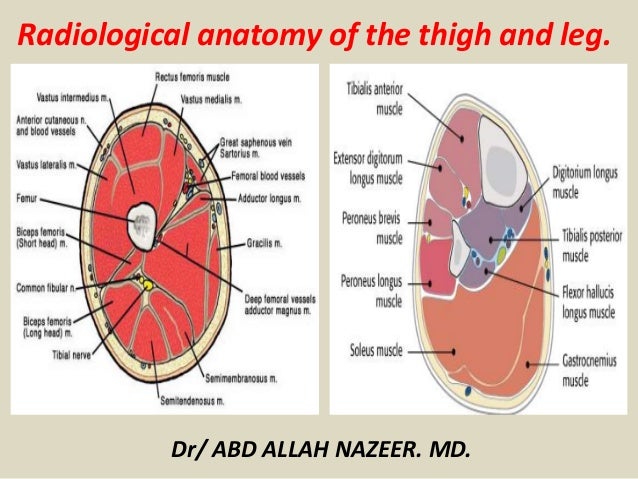 Presentation1 Pptx Radiological Anatomy Of The Thigh And Leg
 Mri Of The Thigh Detailed Anatomy
 Mri Anatomy Of Hip Joint Free Mri Axial Hip Anatomy
 Muscle Mri
 Mri Hip Anatomy
 Muscles Of The Posterior Thigh Hamstrings Damage
 Figure 4 From Normal Mr Imaging Anatomy Of The Thigh And Leg
 Lower Extremity Mri Of Anatomical Atlas
 The Hip Anatomy On 3t Mr And 3d Pictures
 Stanford Msk Mri Atlas 1 0
 Radiology Images
 X Rays Ct Scans And Mris Orthoinfo Aaos
 Muscles Of The Anterior Thigh Quadriceps Teachmeanatomy
 Thigh Anatomy
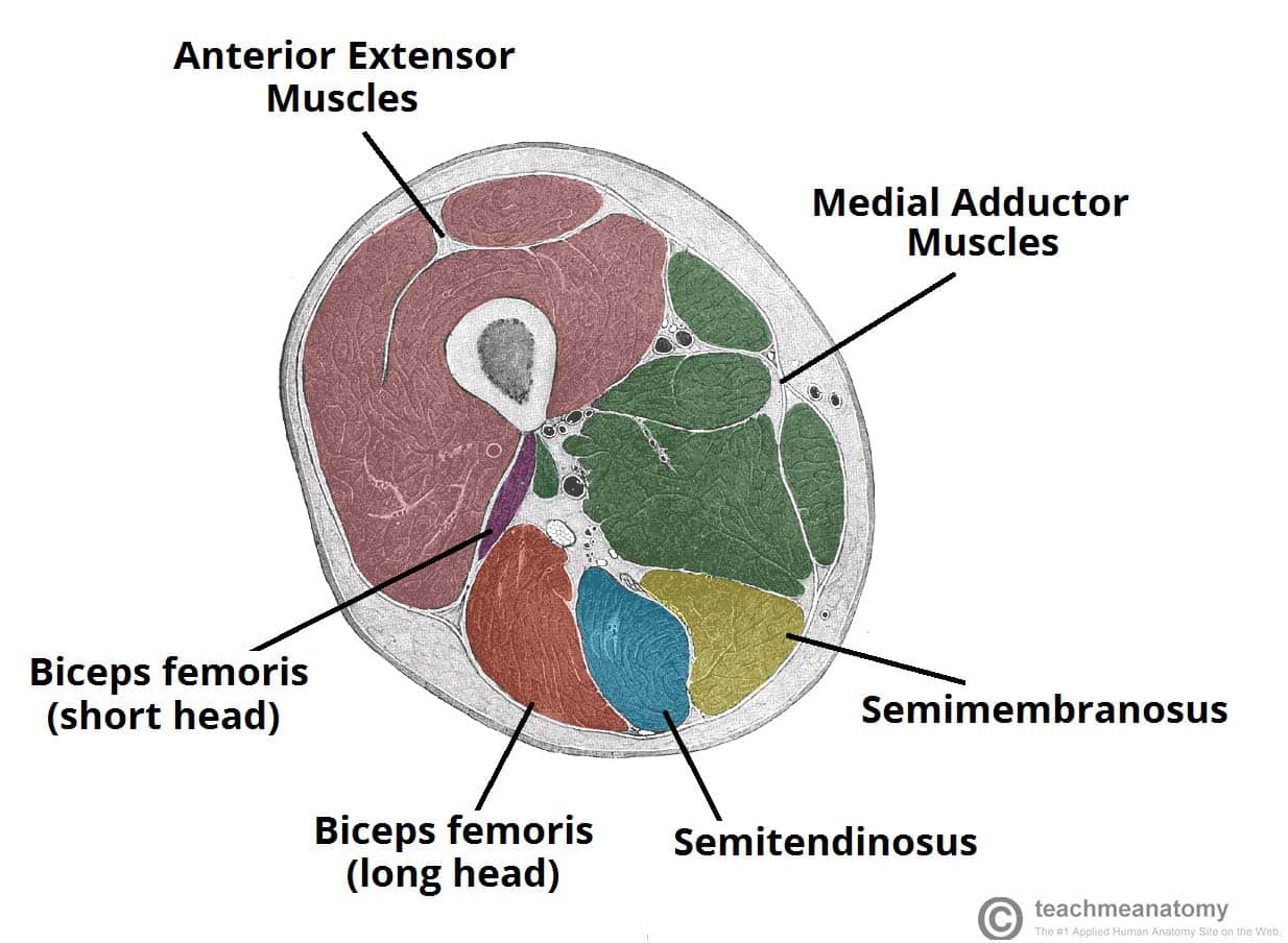
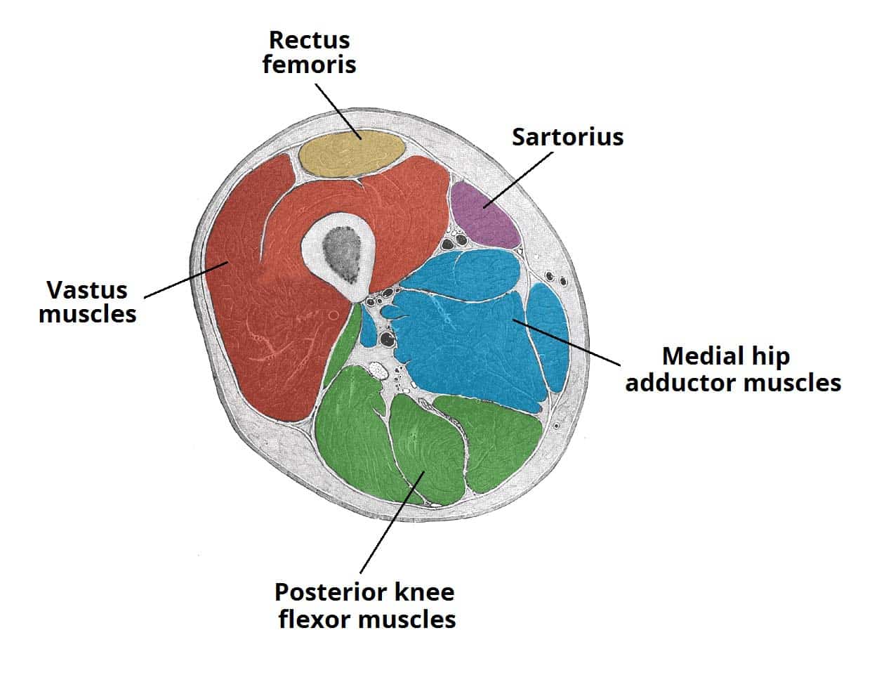
 Thigh Muscles Cross Sectional Anatomy Radiology Case
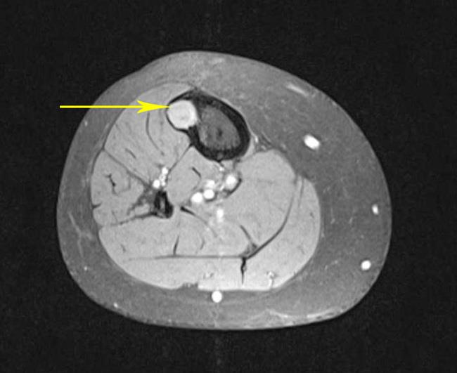
 Example Of Mri Slice Mid Thigh With The Knee Extensors And
 Lower Extremity Mri Of Anatomical Atlas
 The Hip Anatomy On 3t Mr And 3d Pictures
 Vastus Lateralis Anatomy Orthobullets
 The Radiology Assistant Muscle Mr Traumatic Changes
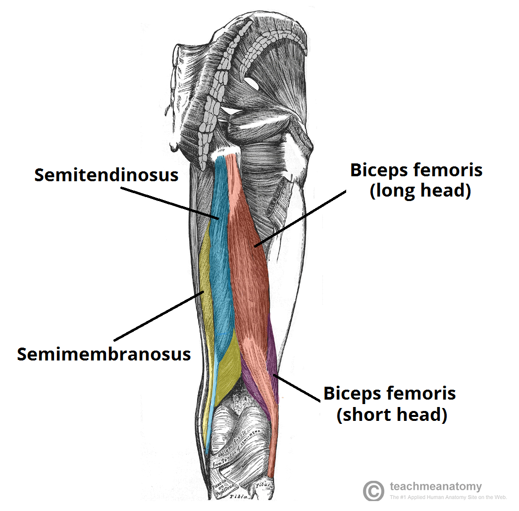 Muscles Of The Posterior Thigh Hamstrings Damage
 Cross Sectional Anatomy Of The Knee Based On Mri Articular
 Mri Of The Hip Detailed Anatomy
 Hamstring Tears Radsource

Belum ada Komentar untuk "Thigh Mri Anatomy"
Posting Komentar