Maxillary Sinus Anatomy
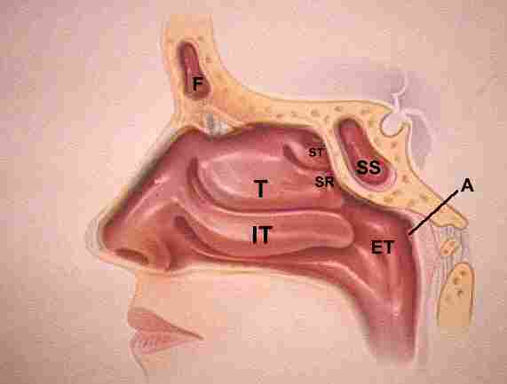
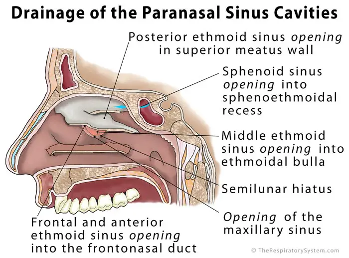
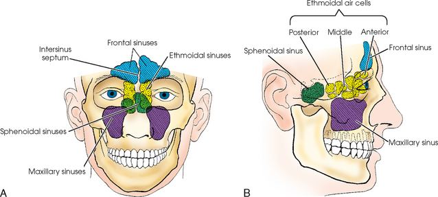
The anterior ostiomeatal unit omu is comprised of the frontal sinus ostium frontal sinus drainage pathway fsdp maxillary sinus ostium infundibulum and middle meatus. Introduction paranasal sinuses air containing bony spaces present.
 Beautifully Designed Maxillary Sinus Photographs Fine Art
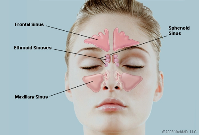
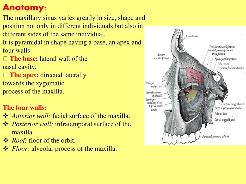
The maxillary sinuses are the largest of the paranasal sinuses located one on each side as well as totally fill the bodies of the maxillae.

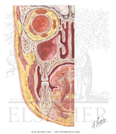
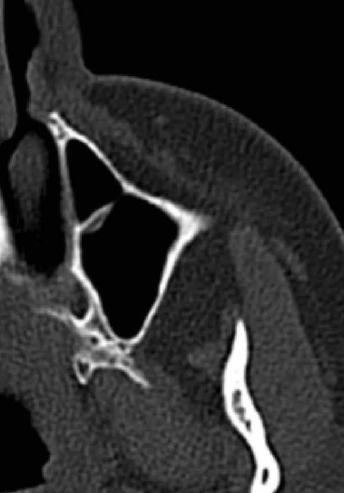
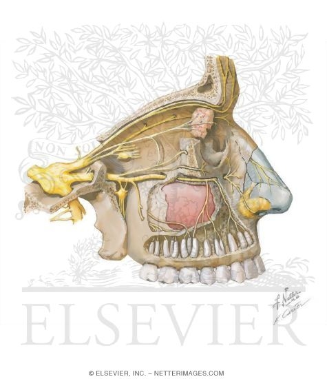
Maxillary sinus anatomy. The symptoms of sinusitis are headache usually near the involved sinus and foul smelling nasal or pharyngeal discharge possibly with some systemic signs of infection such as fever and weakness. The maxillary sinus or antrum of highmore lies within the body of the maxillary bone and is the largest and first to develop of the paranasal sinuses figure 22 9. Venous drainage anteriorly is via the sphenopalatine vein and posteriorly via.
The maxillary sinus is the largest of the paranasal sinuses. The skin over the involved sinus can be tender hot and even reddened due to the inflammatory process in the area. The maxillary sinuses are shaped like a pyramid and each contain three cavities which point sideways inwards and downwards.
Surgical anatomy of maxillary sinus note on oaf dr. These important structures connect the frontal anterior ethmoid and maxillary sinuses. Anatomy of maxillary sinus4.
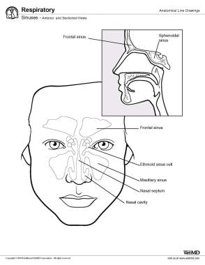
The two maxillary sinuses are located below the cheeks above the teeth and on the sides of the nose. The maxillary sinuses were first illustrated and described by leonardo da vinci in 1489 and later documented by the english anatomist nathaniel highmore in 1651. Each is pyramid shaped with the laterally pointed top and the base deep towards the lateral wall of the neighboring nasal cavity.
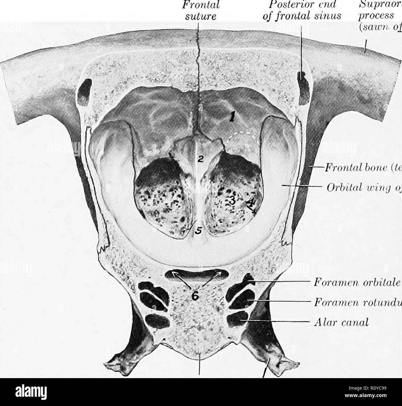
Embryology of maxillary sinus3. Paranasal air sinuses the maxillary sinuses are not only the largest of the air sinuses but also the first to appear being present in the fourth month of intrauterine life. Surgical anatomy of maxillary sinus note on 2 1.
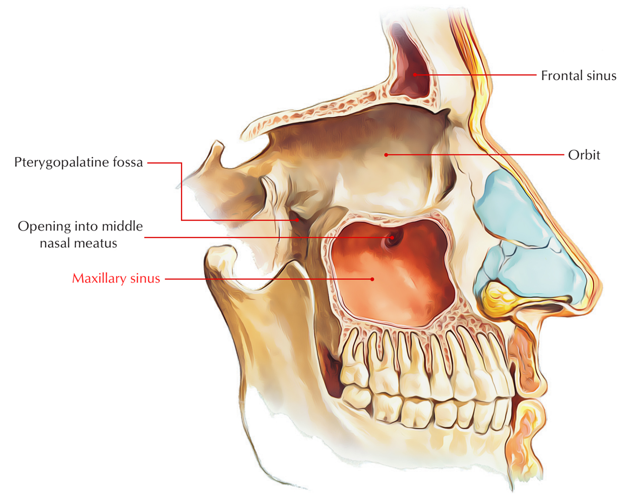
Small arteries from the facial maxillary infraorbital. Described as a pyramid the maxillary sinuses have a base on the lateral border. Each is a pyramidal space its roof formed by the floor of the eye socket and its floor by.
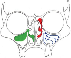
The maxillary sinus ostium drains into the infundibulum which joins the hiatus semilunaris and drains into the middle meatus.
 What Are The Sinuses Pictures Of Nasal Cavities
 Sinus Infection Sinusitis Symptoms Signs Treatment
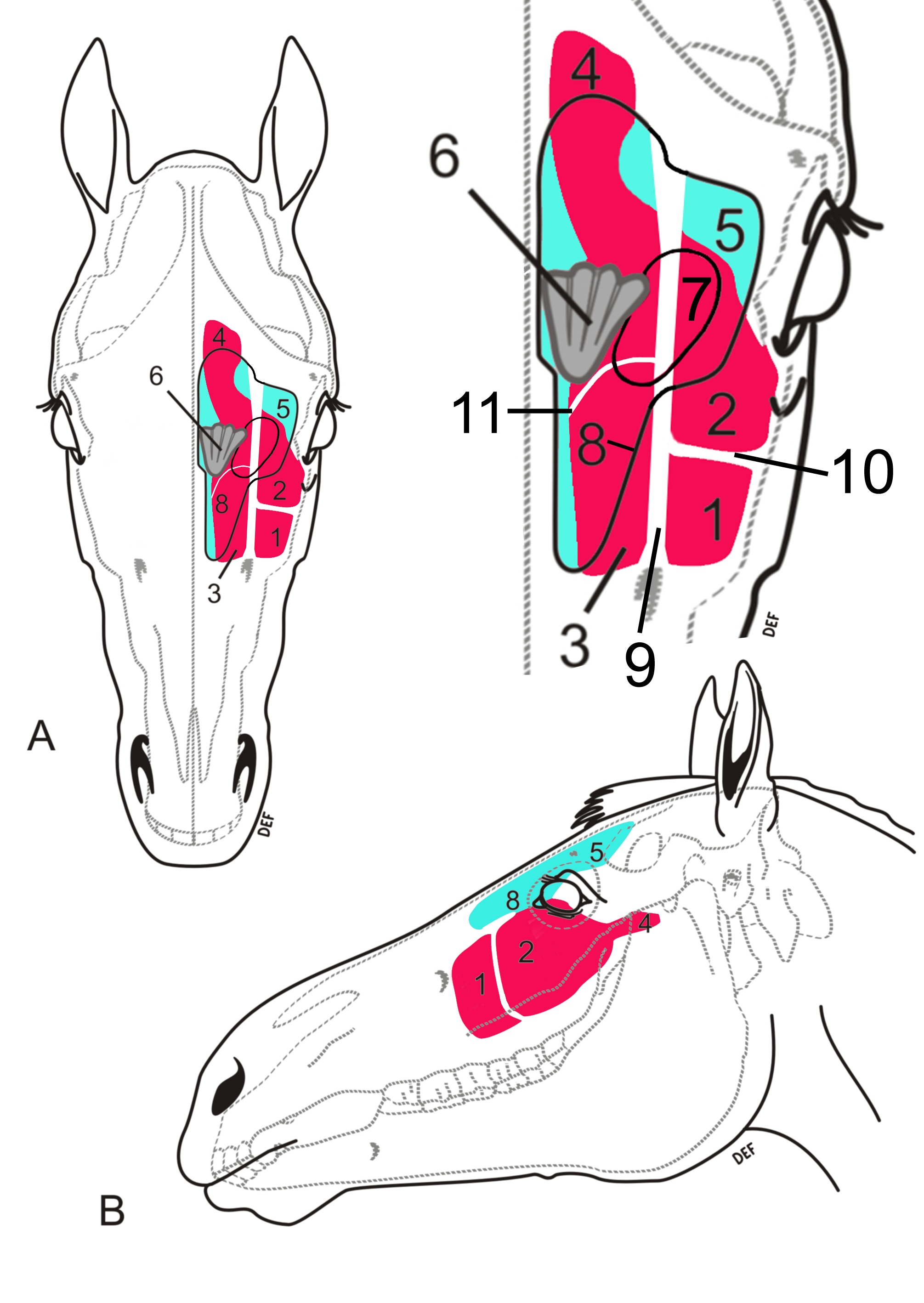 Equine Sinus Conditions Large Animal Hospital College Of
 Medial Maxillectomy
 Maxillary Sinus Disease Diagnosis And Treatment British
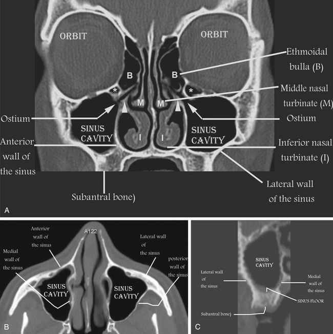
 Sinus Ct Scan Sinusitis W S Tichenor M D
 Maxillary Sinus Disease Ppt Download
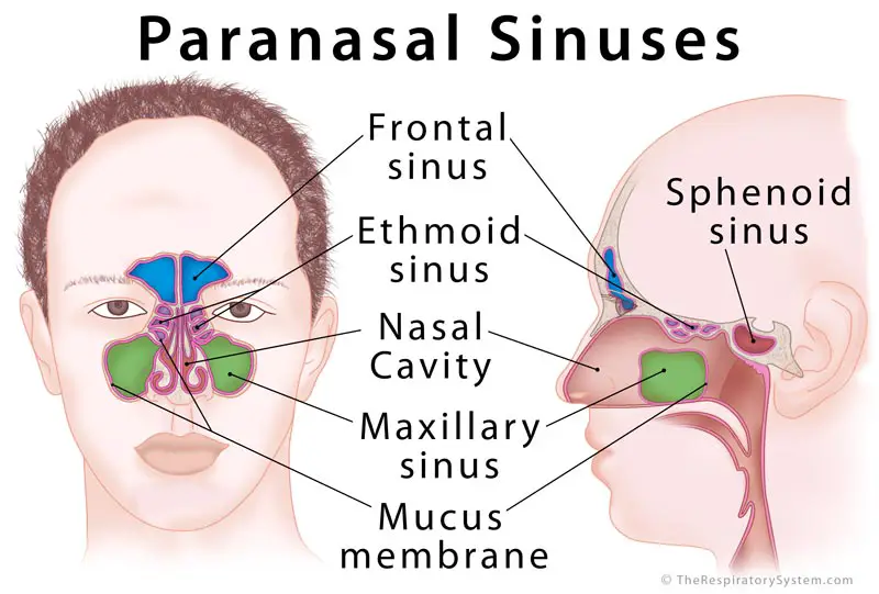 Paranasal Sinus Definition Location Anatomy Function
 Paranasal Sinus Definition Location Anatomy Function
 Paranasal Sinus Anatomy What The Surgeon Needs To Know
 Maxillary Sinus Earth S Lab
 Applied Anatomy Of M Sinus Authorstream
 Maxillary Hiatus Wikipedia
 Protrusion Of The Infraorbital Nerve Into The Maxillary
 Ethmoid Sinus An Overview Sciencedirect Topics
 Maxillary Sinus Stock Photos Maxillary Sinus Stock Images
 Sinus Grafting For Dental Implants Pocket Dentistry
 Definition Of Maxillary Sinus Nci Dictionary Of Cancer
 Maxillary Sinus
 File Maxillary Sinus Posterior View Png Wikimedia Commons
 Paranasal Sinus Anatomy Overview Gross Anatomy
 Maxillary Sinus Maxillary Sinus Anatomy 3d Anatomy
 The Paranasal Sinuses Structure Function Teachmeanatomy
 Paranasal Sinus Anatomy What The Surgeon Needs To Know
 Maxillary Sinus Nerve Supply
 Paranasal Sinuses Radiology Key
 Maxillary Sinus Anatomy Function Function Body Maps


Belum ada Komentar untuk "Maxillary Sinus Anatomy"
Posting Komentar