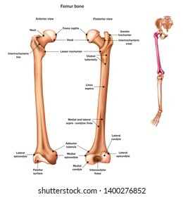
Femur Bone Anatomy
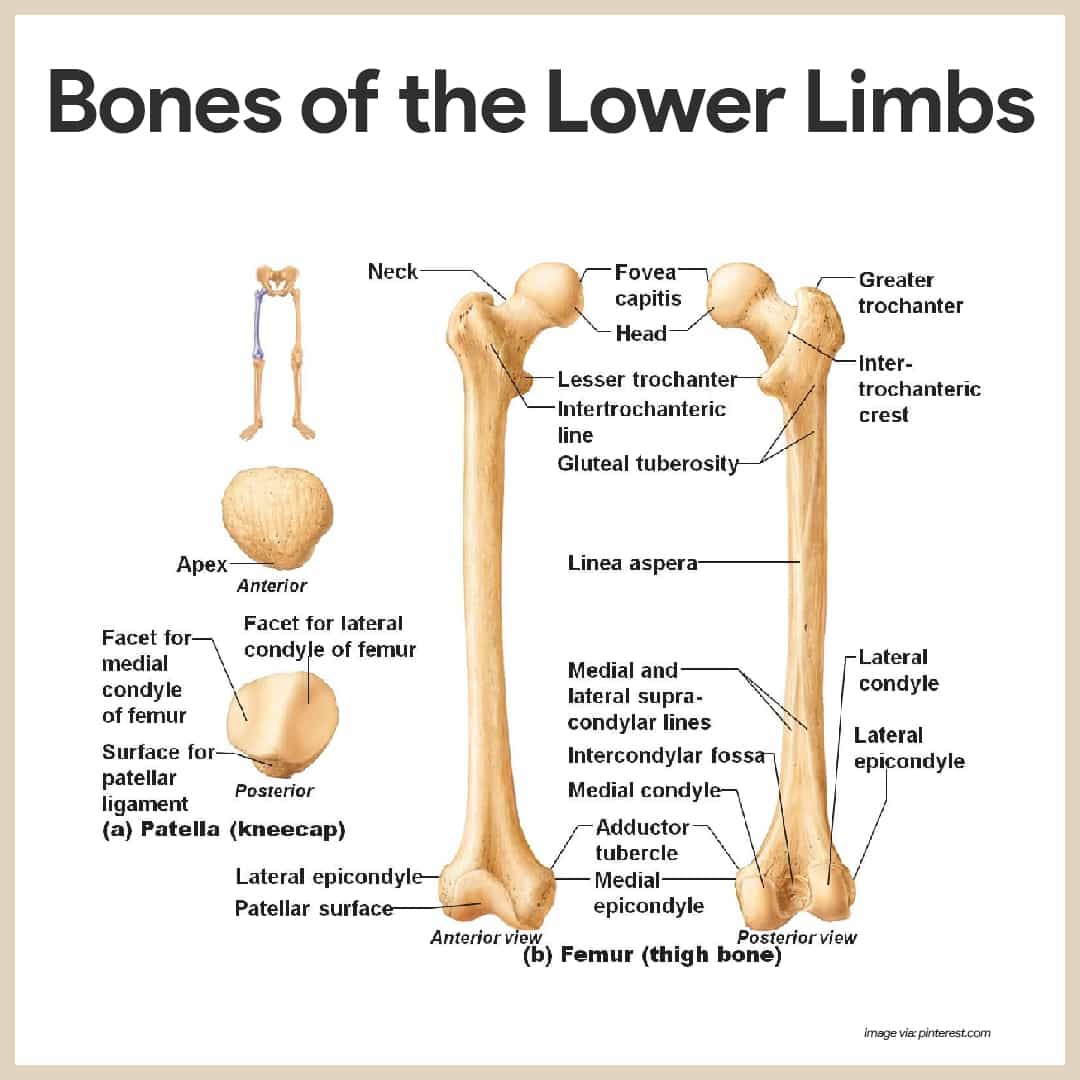
The femur or thigh bone is the longest heaviest and strongest bone in the entire human body. Condyles lateral medial condyle lateral condyle is flat laterally.
 Knee Wikipedia
The femur ˈ f iː m ər pl.

Femur bone anatomy. All of the bodys weight is supported by the femurs during many activities such as running jumping walking and standing. It can account for about a quarter of someones height. The femur is the largest bone in the human body.
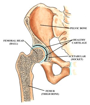
It is commonly known as the thigh bone femur is latin for thigh and reaches from the hip to the knee. Its also one of the strongest. The head of the femur articulates with the acetabulum in the pelvic bone forming the hip joint while the distal part of the femur articulates with the tibia and kneecap forming the knee joint.
Lower end of the femur includes the following parts. The cylindrical shaft is convex forwards. The upper leg is often called the thigh.
The femur is also called the thigh bone and is the longest and strongest bone of the body. Upper leg anatomy and function. Also called the thigh bone this is the longest bone in the body.
Neck connects the head of the femur with the shaft. A human male adult femur is about 19 inches long and weighs a little more than 10 ounces. The two condyles are partially covered by a large articular.
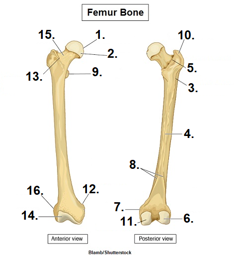
Important features of this bone include the head medial and lateral condyles patellar surface medial and lateral epicondyles and greater and lesser trochanters. Lesser trochanter smaller than the greater trochanter. It is both the longest and the strongest bone in the human body extending from the hip to the knee.
The femur bone is the strongest and longest bone in the body occupying the space of the lower limb between the hip and knee joints. Proximal head articulates with the acetabulum of the pelvis to form the hip joint. The upper and bears a rounded head whereas the lower end is widely expanded to from two large condyles.
Separates the lower and posterior parts. The head forms a ball and socket joint with the hip at the acetabulum being held in place by a ligament ligamentum teres femoris within the socket and by strong surrounding ligaments. Femur anatomy is so unique that it makes the bone suitable for supporting the numerous muscular and ligamentous attachments within this region in addition to maximally extending the limb during ambulation.
Femur also called thighbone upper bone of the leg or hind leg. Its the area that runs from the hip to the knee in each leg. The femur is the only bone located within the human thigh.
By most measures the femur is the strongest bone in the body. The head is directed medially. Greater trochanter the most lateral palpable projection of bone that originates from.
In humans the neck of the femur connects the shaft and head at a 125 angle. It is composed of an upper end a lower end and a shaft. Femurs or femora ˈ f ɛ m ər ə or thigh bone is the proximal bone of the hindlimb in tetrapod vertebrates and of the human thigh.
Intercondylar fossa or intercondylar notch.
 Anatomy Of The Human Proximal Femur Upper Third Of The
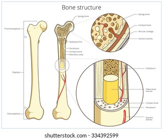
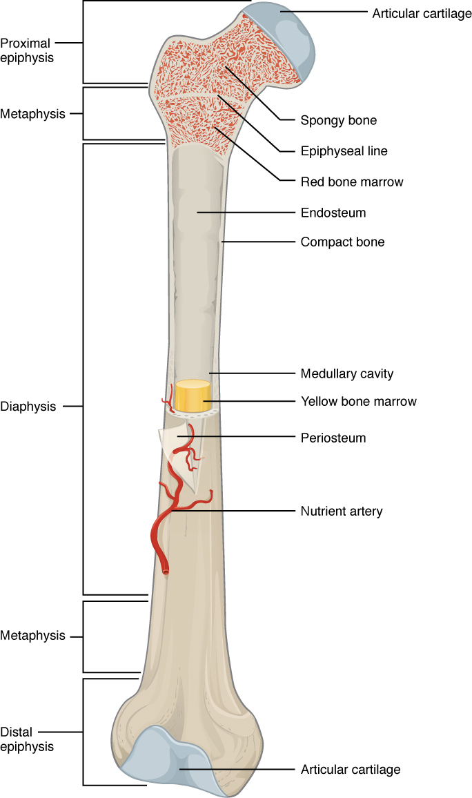 6 3 Bone Structure Anatomy And Physiology
 Femur Bone Images Stock Photos Vectors Shutterstock
 Hip Anatomy Yoga Understanding The Hips For Yoga Jason
 Femur Bone Structure Stock Vector Illustration Of Health
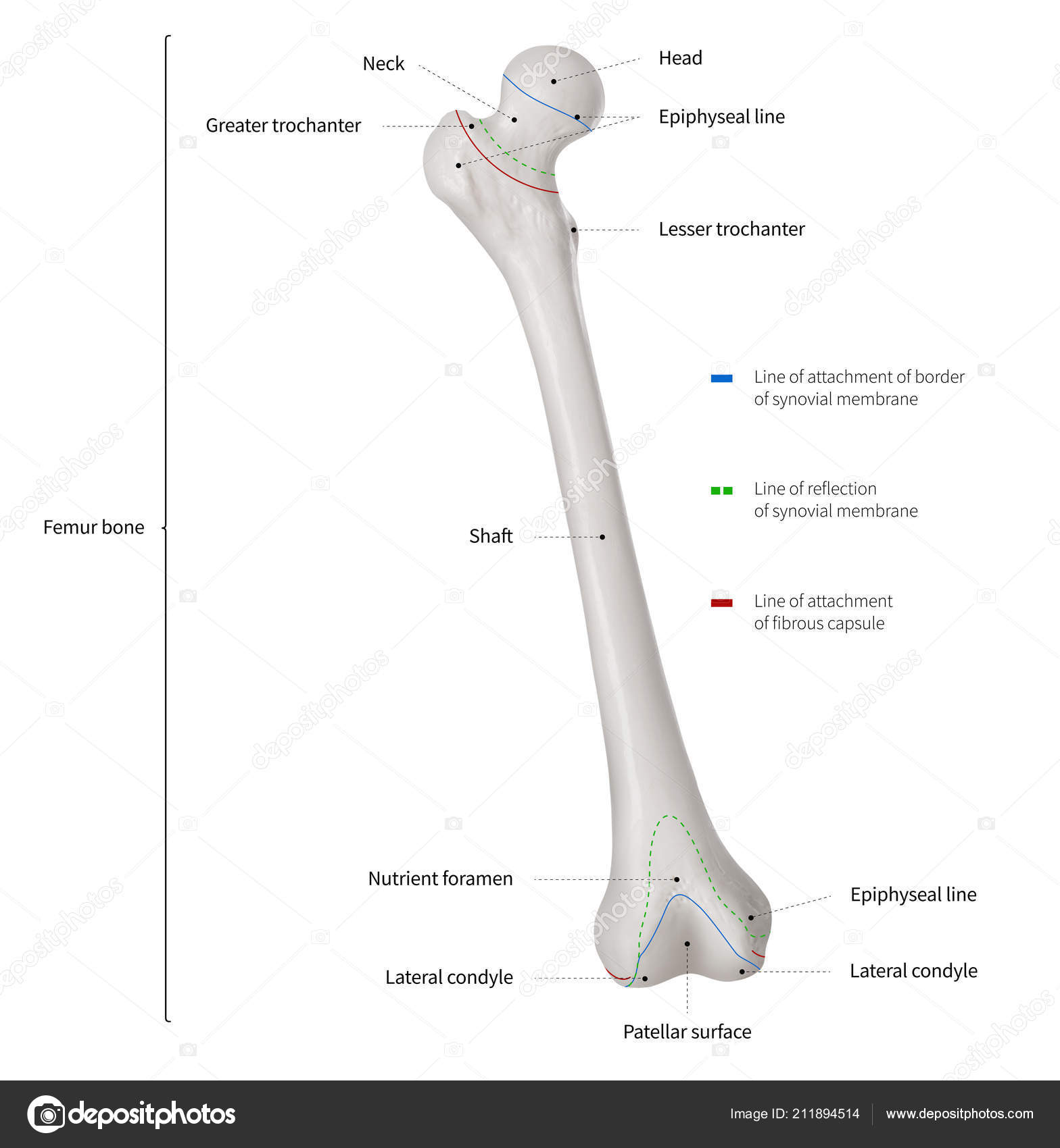 Anterior View Of Femur Infographic Diagram Human Femur
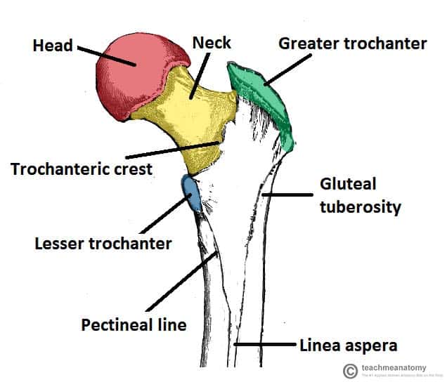 The Femur Proximal Distal Shaft Teachmeanatomy
 Femur Bone Structure Canvas Print
 Vector Illustration Bone Anatomy The Femur Eps Clipart
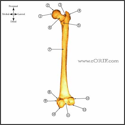 Femur Anatomy Eorif
 Femur Bone Labeled Differences Between Femur Humerus
 Femur An Overview Sciencedirect Topics
 Femur Anatomy Quiz
 Femoral Head Watercolor Print Femur Bone Poster Head Of Femur Print Orthopedic Art Skeletal System Anatomy Art Print Medical Art Wall Decor
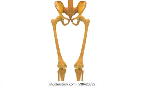
 Bones Of The Leg And Foot Interactive Anatomy Guide
 Broken Leg Treatment Symptoms Recovery Time Pictures
 Royalty Free Femur Stock Images Photos Vectors Shutterstock
 Bones Of The Lower Limb Anatomy And Physiology I
 Why People Have To Squat Differently The Movement Fix
 Femur Labeling Quiz
 Overview Of The Anatomy Of The Femur Which Is A Long Bone
 World S Best Leg Bone Stock Illustrations Getty Images
![]() Hip And Thigh Bones Joints Muscles Kenhub
Hip And Thigh Bones Joints Muscles Kenhub
 Skeletal System Anatomy And Physiology Nurseslabs
 The Femur Human Anatomy
Amazon Com Moslion Bone Mouse Pad Human Femur Bone
Infographic Diagram Of Human Femur Bone Or Leg Bone Anatomy
 Femur Bone Diagram Google Search In 2019 Bones Skull
 Free Art Print Of Bone Anatomy The Femur
 The Lower Limb Boundless Anatomy And Physiology
 Femoral Bone Anatomy Medical Image And Geometrical Modeling
 Bones Of The Lower Limbs Course Hero
 Royalty Free Femur Stock Images Photos Vectors Shutterstock
 Hip Impingement A Patient S Guide To Mobility Arthroscopy


Belum ada Komentar untuk "Femur Bone Anatomy"
Posting Komentar