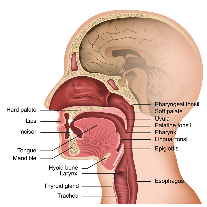
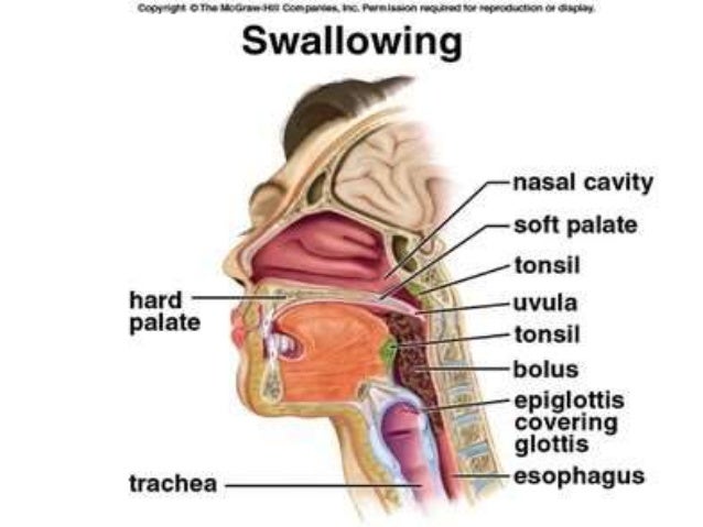
Diagram Of Swallowing Anatomy
The swallowing center then outputs signals to initial and control the next two phases of swallowing the pharyngeal phase and the esophageal phase. The throat anatomy diagram divides the pharynx into three parts the nasopharynx the oropharynx and the laryngopharynx.
 Muscles Of Chewing Swallowing I Purposegames
Swallowing consists of three phases.

Diagram of swallowing anatomy. Eating and swallowing are compex behaviors including both volitional and reflexive activities involving more than 30 nerves and muscles. The nasopharynx is the uppermost part of the pharynx and extends down from the base of the skull to the nasal passages hence the name. The anatomy of the oral cavity pharynx larynx and innervations of the muscles are shown in figure 1 and table 1.
Swallowing is the process by which food moves from the mouth to the stomach. During the oral phase the upper esophageal sphincter is closed and food will not be able to pass in the esophagus until it is open. This process is under neural control of several areas of cerebral cortex including the motor cortex.
The tongue has both oral and pharyngeal surfaces. Deglutition process of swallowing oropharyngeal dysphagia oral pharyngeal espohageal anatomy esopharyngeal dysphagia. The swallow reflex is initiated and is under involuntary neuromuscular control.
This movement is controlled by the nervous system and involves both voluntary and involuntary muscle contractions. Unlike the oral phase the pharyngeal phase is an involuntary process. Swallowing refers to the entire act of deglutition from placement of food in the mouth through the oral and pharyngeal stages of the swallow until the material enters the esophagus through the cricopharyngeal juncture.
Next is the pharyngeal phase of swallowing. Learn vocabulary terms and more with flashcards games and other study tools. Pharyngeal phase starts with stimulation of tactile receptors in the oropharynx by the food bolus.
 Dysphagia Difficulty Swallowing New England Ent Facial
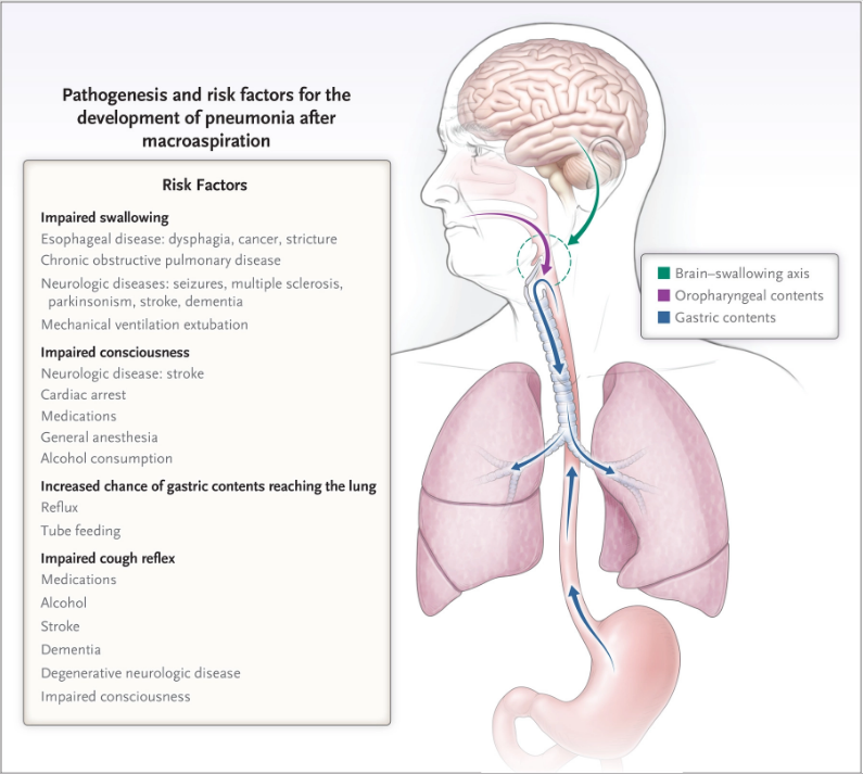 Nejm On Twitter Aspiration Is Often The Result Of Impaired
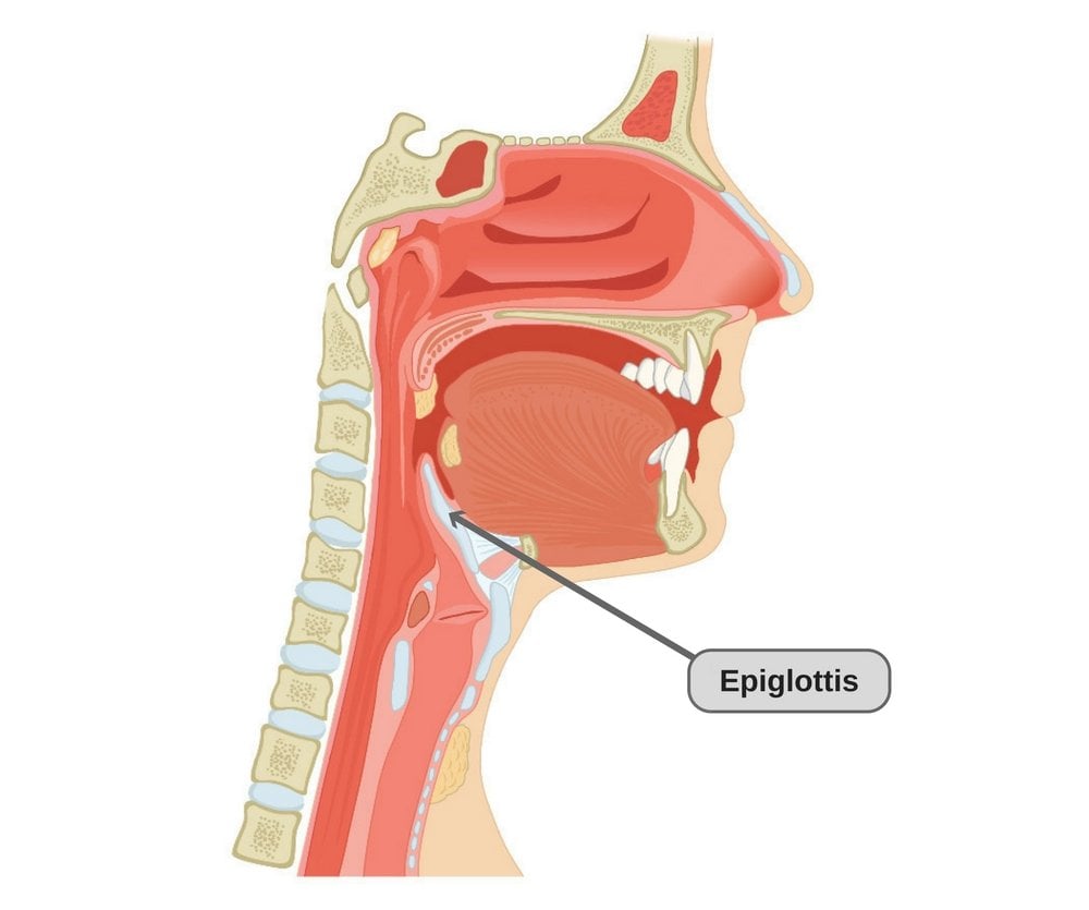
 Epiglottitis Harvard Health
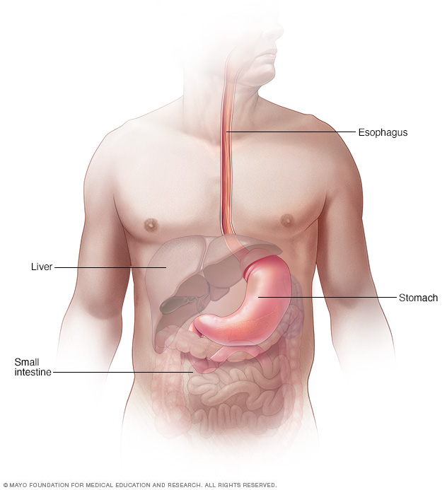
 What Are Esophagus And Trachea Why Are They Located Close
 Basic Anatomy Of The Human Swallowing Apparatus Download
 Feeding And Swallowing Processes And Disorders Pearls And
 Swallowing Anatomy Lateral View Pt 1 Diagram Quizlet
 Inpatient Dysphagia A Case Study Elite Learning

 Anatomy And Physiology Of Feeding And Swallowing Normal And
 Anatomy Of Swallowing Anatomy Organs Throat Anatomy Neck
 Anatomy Of Throat Cancer Headandneckcancerguide Org
![]() Stages Of Swallowing Deglutition Kenhub
Stages Of Swallowing Deglutition Kenhub
 Anatomy Of The Throat And Esophagus
 Dysphagia Disease Reference Guide Drugs Com
 Swallowing Anatomy Diagram Quizlet
 Diagram Of The Swallowing Mechanism
![]() Stages Of Swallowing Deglutition Kenhub
Stages Of Swallowing Deglutition Kenhub
 Anatomy And Physiology Of Feeding And Swallowing Normal And
 Anatomy Swallowing Images Stock Photos Vectors Shutterstock

 Radiation Dysphagia
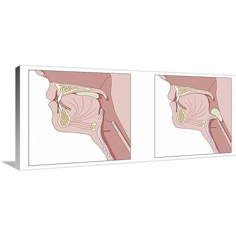 Amazon Com Greatbigcanvas Gallery Wrapped Canvas Entitled
 What Are Esophagus And Trachea Why Are They Located Close
 Essential Body Parts For Speech And Swallowing Poster
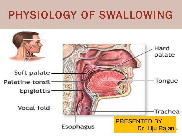 Physiology Of Swallowing
 Epiglottis Anatomy Britannica
Tablet W Swallowing Diagrams Awesome Speech Pathology
Belum ada Komentar untuk "Diagram Of Swallowing Anatomy"
Posting Komentar