Ct Anatomy Of Brain
Brainstem and cerebellum without evidence of focal lesions. The brain consists of grey and white matter structures which are differentiated on ct by differences in density.
:max_bytes(150000):strip_icc()/coloured-ct-scan-of-a-healthy-brain--side-view--680799273-5973bb6d6f53ba00107c1993.jpg) A Guide To The Anatomy Of The Brain
A Guide To The Anatomy Of The Brain
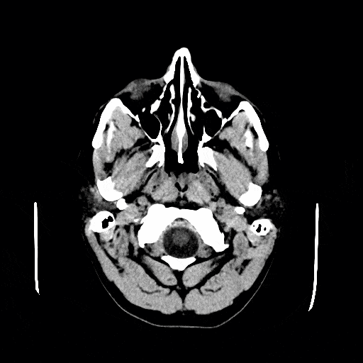
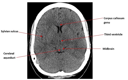
Basal subarachnoid cisterns normal configuration.

Ct anatomy of brain. Ct images of the brain are conventionally viewed from below as if looking up into the top of the head. White matter has a high content of myelinated axons. Grey matter contains relatively few axons and a higher number of cell bodies.
Brain bones of cranium sinuses of the face. Adequate gray matter white matter differentiation. Brain and face ct.
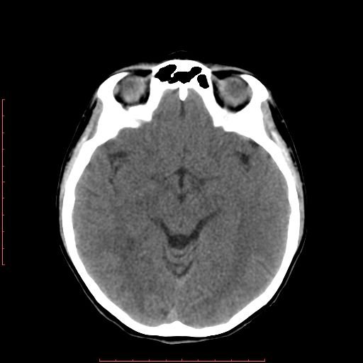
Welcome to online mri ct sectional anatomy. Anatomy of the head on a cranial ct scan. Focal abnormalities are not observed in the brain parenchyma.
Brain bones of cranium sinuses of the face. As myelin is a fatty substance it is of relatively low density compared to the cellular grey matter. Cerebral lobes basal ganglia lentiform nuclei thalamus vascular territories internal capsule calcifications fossa posterior.
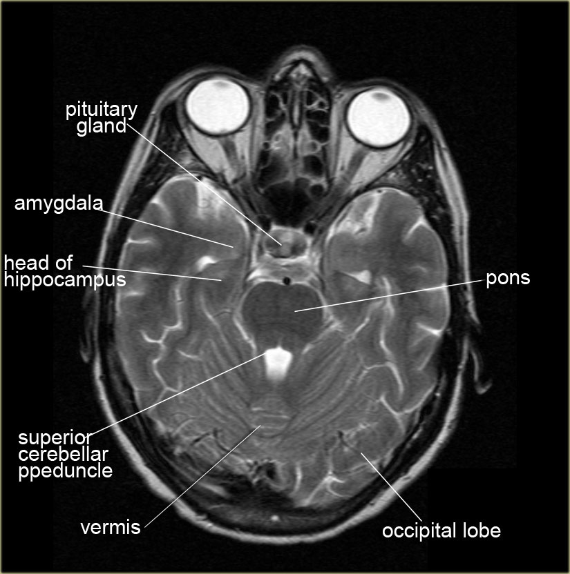
This means that the right side of the brain is on the left side of the viewer. Amygdala on ct and mr images the amygdala is a large region of gray matter contiguous with the uncus of the medial temporal lobe and the most anterior portion of the hippocampus the pes hippocampi. Ct cross sectional anatomy of brain chest abdomen paranasal sinusus neck temporal bone heart slideshare uses cookies to improve functionality and performance and to provide you with relevant advertising.
Anatomy of the head on a cranial ct scan. Online mri ct sectional anatomy omcsa k anatomy is probably one of the most user friendly and convenient online interface for human anatomy atlas. They lie on the ventricular surface of the hippocampus and become the fimbria of the fornix medially.
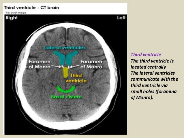
Real time interface human sectional anatomy. Third and fourth ventricles in midline. Lateral ventricles of normal volume.
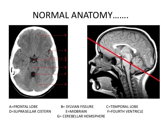
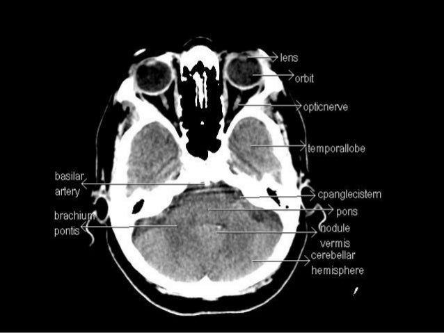
Anatomy ct axial brain form no 18. 6 frontal bone 27 occipital bone 32 optic nerve 37 basilar artery 40 hemisphere of cerebellum 43 frontal sinus 45 sigmoid sinus 46 internal carotid artery 47 sphenoid bone 49 medulla oblongata 50 external auditory meatus 51 spinal central canal. The anterior part of the head is at the top of the image.
Ct anatomy of the brain.
 Head Ct
 Ai Detects Tiny Brain Hemorrhages On Ct Scans Outperforms
 Ct Brain Hemorrhage Startradiology
 Presentation1 Pptx Radiological Anatomy Of The Brain
 Ct Scans Interpretation Principles Basics Teachmeanatomy

 Head Ct Scan Procedure Radtechonduty
 Radiology Basics Head Anatomy
 Basics Of Ct Head
 Normal Brain Ct Mri Imaging In Arabic Prof Dr Mamdouh Mahfouz
 Brain And Face Ct Interactive Anatomy Atlas
 Figure 69 5 From How To Read A Head Ct Scan Semantic Scholar
 Brain Ct Neurologyneeds Com
 Ct Anatomy
 The Radiology Assistant Brain Anatomy


 Normal Brain Anatomy Ct And Mri Youtube
 Ct Brain Hemorrhage Startradiology
 The Ct Anatomy Tutor
 Brain Ischemic Stroke Ct Scan Reconstruction Stock Image
 Basic Anatomy Of Ct Brain Hku E Learning Platform In
 The Radiology Assistant Brain Anatomy
 Basic Ct Anatomy Of The Brain
 Brain Ct Anatomy And Basic Interpretation Part Ii Ppt
 The Ct Anatomy Tutor

 Brain And Face Ct Interactive Anatomy Atlas
Brain Lobes Annotated Mri Radiology Case Radiopaedia Org
Belum ada Komentar untuk "Ct Anatomy Of Brain"
Posting Komentar