Ultrasound Neck Anatomy
The infrahyoid region of the neck includes the visceral anterior cervical posterior cervical carotid retropharyngeal and perivertebral spaces. Find out more from alaska family sonograms.

In radiology the head and neck refers to all the anatomical structures in this region excluding the central nervous system that is the brain and spinal cord and their associated vascular structures and encasing membranes ie the meninges.

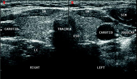
Ultrasound neck anatomy. While the vast majority of patients are supine on the exam table with a pillow supporting the shoulders to allow gentle neck extension keep in mind that some patients have beautiful anatomy d that allows ultrasound exam even in a sitting position. The visceral space contains the thyroid parathyroid glands larynx hypopharynx the cervical trachea and esophagus the recurrent laryngeal nerve. This can be confirmed from ultrasound guided fnac allowing appropriate clinical management and treatment.
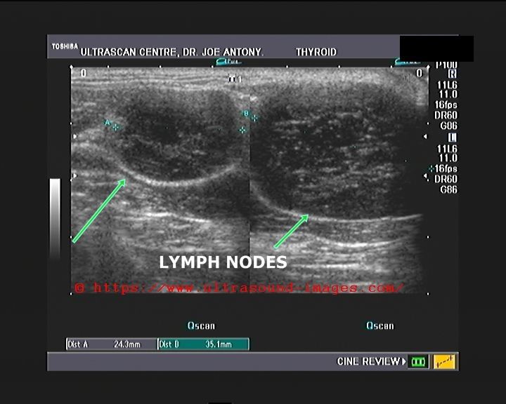
Optimal positioning and exposure of the neck for ultrasound of the thyroid and parathyroid glands a b and lateral neck for lymph node examination and mapping c. An enlarged cervical lymph node is the most commonly encountered neck lump. Head and neck anatomy is important when considering pathology affecting the same area.
A common neck ultrasound is ultrasound of the thyroid which uses sound waves to produce pictures of the thyroid gland within the neck. These include the masseter muscle the zygomatic arch and the outer cortex of the ramus of the mandible and the suprazygomatic portion of the temporalis muscle. It does not use ionizing radiation.
A neck ultrasound is performed to diagnose potential problems of the thyroid lymph nodes and carotid arteries. In the infrahyoid neck it is surrounded by the anterior cervical space anteriorly by the visceral and retropharyngeal spaces medially and by the perivertebral and posterior cervical spaces posteriorly. Anterior neck anatomy false vocal cords true vocal cords paraglottic fat.
Because most lesions in the neck are site specific once a lesion has been located specific ultrasound features can be used to establish the diagnosis. Only some parts of the masticator space can be explored sonographically. The carotid space in the suprahyoid region of the neck contains the internal carotid artery the internal jugular vein cranial nerves ix to xii and the sympathetic plexus.
 The Radiology Assistant Neck Masses In Children
 Diagnostic Ultrasound Head And Neck 9781937242169

 Chapter 25 Overview Of The Neck The Big Picture Gross
 The Radiology Assistant Neck Masses In Children
.jpg) Ent Ultrasound Applications
Ent Ultrasound Applications
 Ultrasound Anatomy Of The Neck The Infrahyoid Region

 Ultrasound Anatomy Of The Neck A Top Panoramic
 How To Identify Structures Of The Neck Using Ultrasound
 Dr Matt Bull On Twitter Case Neck Ultrasound Anatomy
 Neck Masses Pediatrics Clerkship The University Of Chicago
 A Gallery Of High Resolution Ultrasound Color Doppler 3d
![]() Normal Thyroid Gland Transverse Ultrasound Through The Neck
Normal Thyroid Gland Transverse Ultrasound Through The Neck
 Figure 7 From Head And Neck Anatomy And Ultrasound
 The Radiology Assistant Infrahyoid Neck
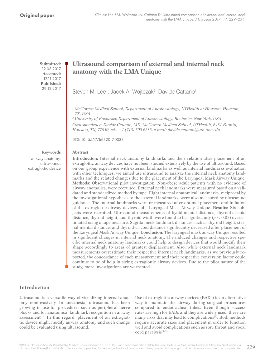
 Normal Neck Anatomy And Method Of Performing Ultrasound
Parotid Gland An Overview Sciencedirect Topics
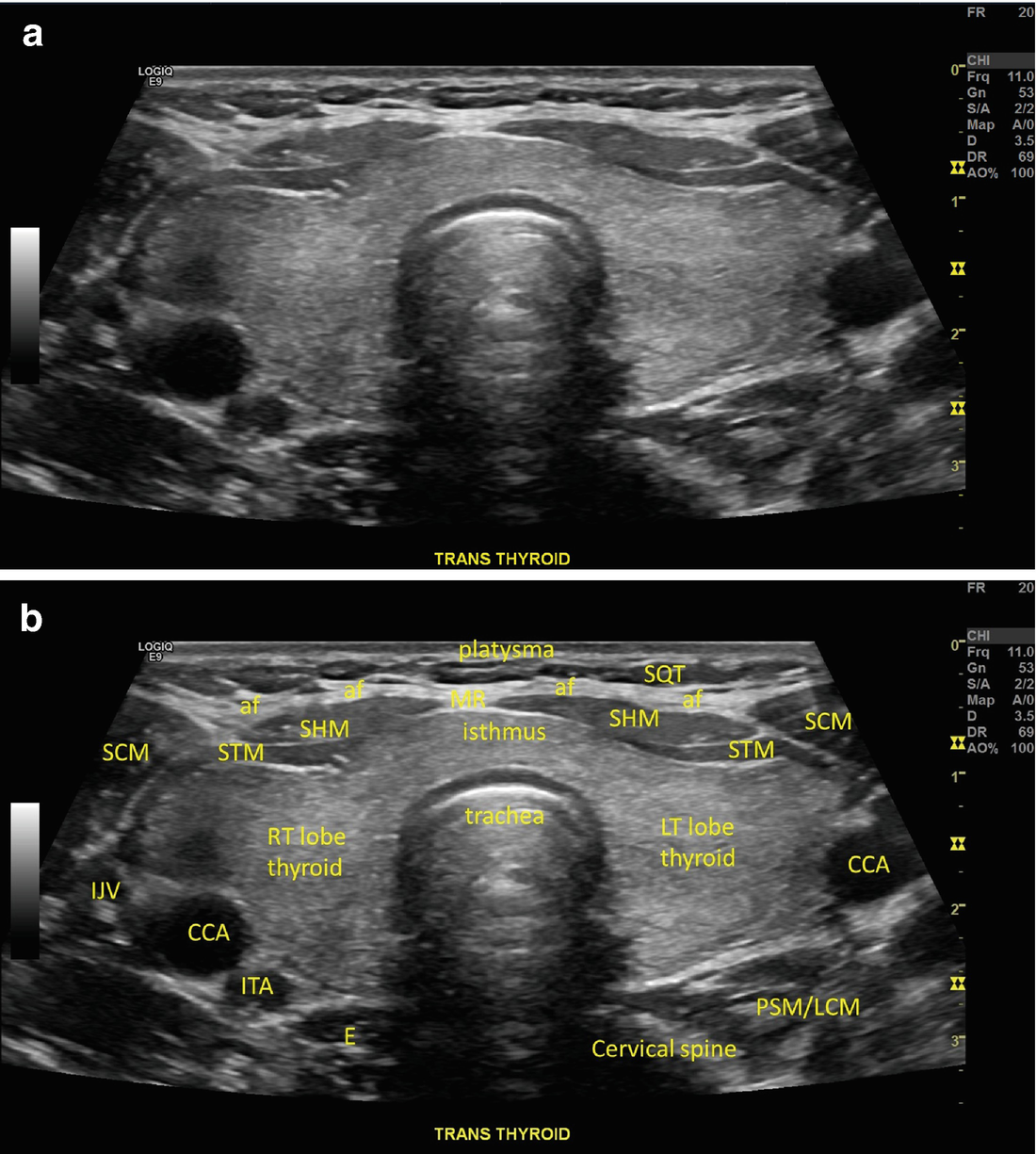 Normal Neck Anatomy And Method Of Performing Ultrasound
 Normal Neck Anatomy And Method Of Performing Ultrasound
 Pdf Ultrasound Comparison Of External And Internal Neck
 Ultrasound Of The Shoulder
 Using Ultrasound In Central Line Placement Uk Emig Quickhit
 Neck Ultrasonography And Ultrasound Assisted Procedures Esaote
 Brachial Plexus
 A Gallery Of High Resolution Ultrasound Color Doppler 3d
 Ultrasound Guided Cervical Plexus Block Nysora
 Normal Carotids Ultrasound How To
 Thyroid Normal Ultrasoundpaedia
 Normal Neck Anatomy And Method Of Performing Ultrasound
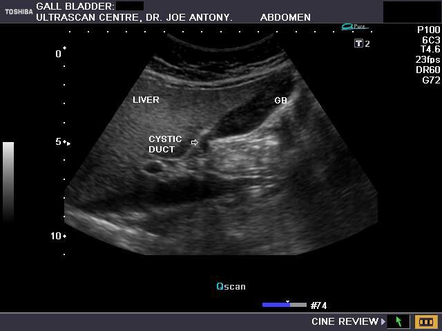
 Biliary Ultrasound Core Em
 The Radiology Assistant Infrahyoid Neck

Belum ada Komentar untuk "Ultrasound Neck Anatomy"
Posting Komentar