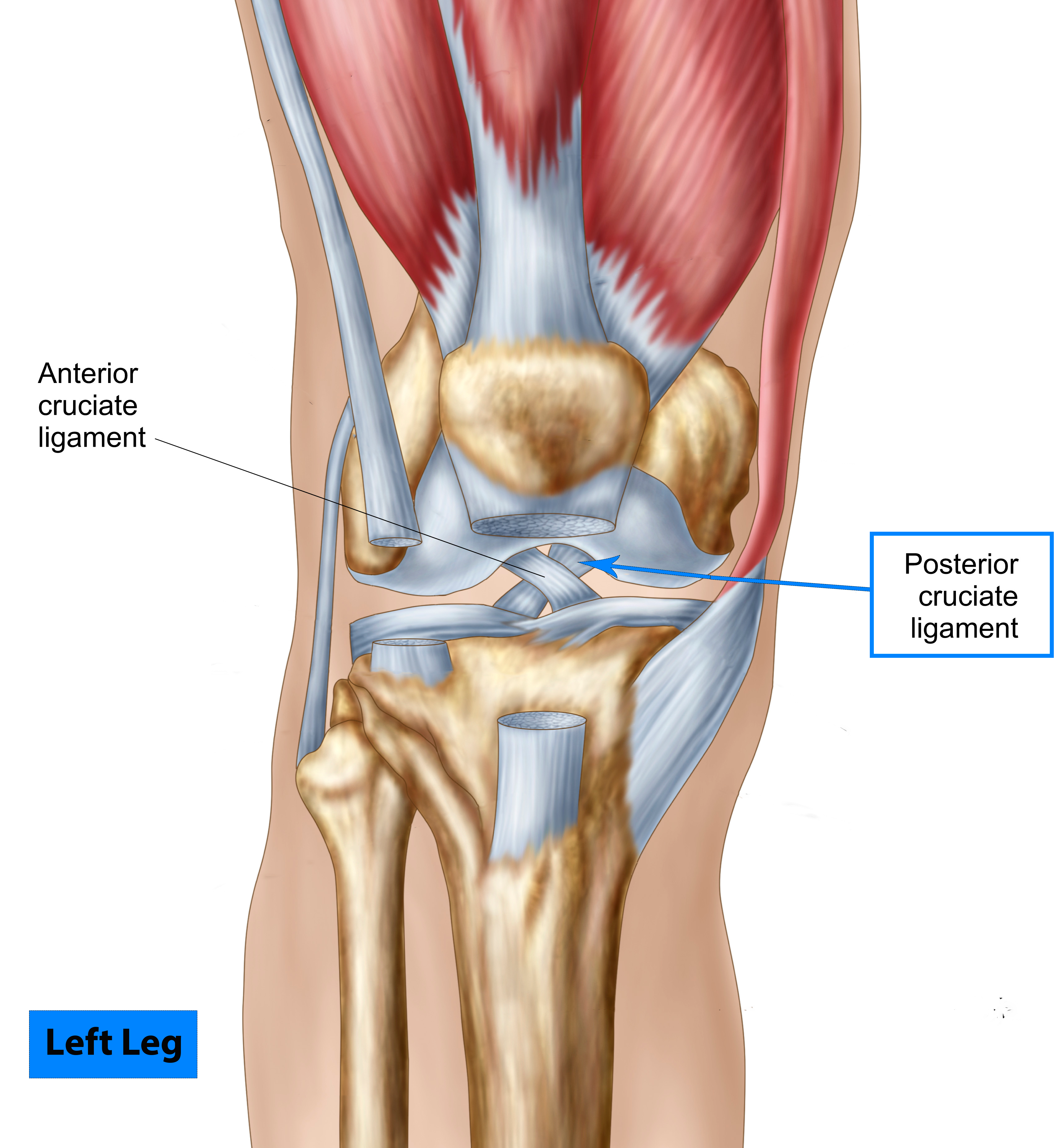
Pcl Anatomy
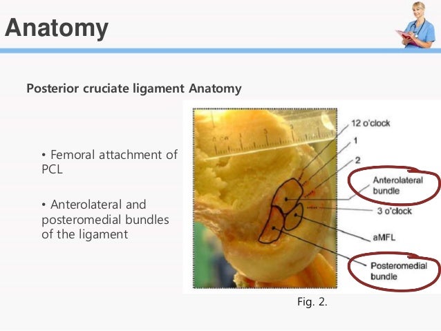
The clock face method to identify the femoral posterior cruciate ligament pcl attachment has poor accuracy and reproducibility. The posterior cruciate ligament or pcl is one of the four major ligaments of the knee.
Posterior Cruciate Ligament Injuries Orthoinfo Aaos
It connects the posterior intercondylar area of the tibia to the medial condyle of the femur.

Pcl anatomy. Measurements of clinically relevant anatomic structures would provide more useful surgical guidance. It also functions to prevent hyper extension and limits internal rotation adduction and abduction at the knee joint. The primary function of the pcl is to resist posterior displacement of the tibia in relation to the femur.
The pcl is one of the two cruciate ligaments of the knee. And prevents the tibia from excessive posterior displacement in relation to the femur. Pcl tibial attachment anterolateral bundle attaches proximal to edge of shelf posteromedial bundle attaches over edge of shelf most posterior fibres dissipate periosteally andrew amis biomechanics section of imperial college london the most posterior fibres of the pcl wrap over the edge of the shelf and dissipate into the.
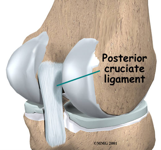
The posterior cruciate ligament pcl is one of four ligaments that hold the knee in place and provide added stability. The posterior cruciate ligament pcl is one of the two cruciate ligaments that stabilize the knee joint. Its secondary function is to prevent hyperextension and limit internal and varusvalgus rotation.
This configuration allows the pcl to resist forces pushing the tibia posteriorly relative to the femur. Ligaments are fibrous tissue that usually connect bones. It acts as the major stabilizing ligament of the knee.
Gross anatomy the pcl attaches to the posterior intercondylar area and passes anterosuperiorly to insert into the lateral surface of the. More specifically the pcl helps to ensure proper alignment of the femur and tibia also called the thighbone and shinbone. 5 the pcl is an extrasynovial structure that lies behind the intra articular portion of the knee.
 A Anterior And B Posterior Views Of The Native Posterior
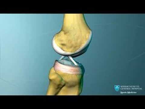 Knee Ligament Anatomy Animation

 Posterior Cruciate Ligament An Overview Sciencedirect Topics
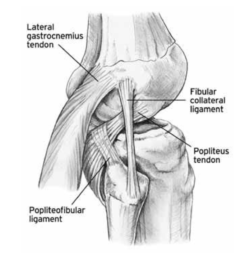 The Knee Resource Posterolateral Corner Injury
Graft Choices In Acl Surgery
 Pcl Tear Brisbane Knee And Shoulder Clinic Dr
 Pcl Anatomy Showing The Relationship Of The Posteromedial
 Pcl Injuries What Happens And What Treatment Options Are
 What Is An Example Of A Ligament And What Is Its Purpose In
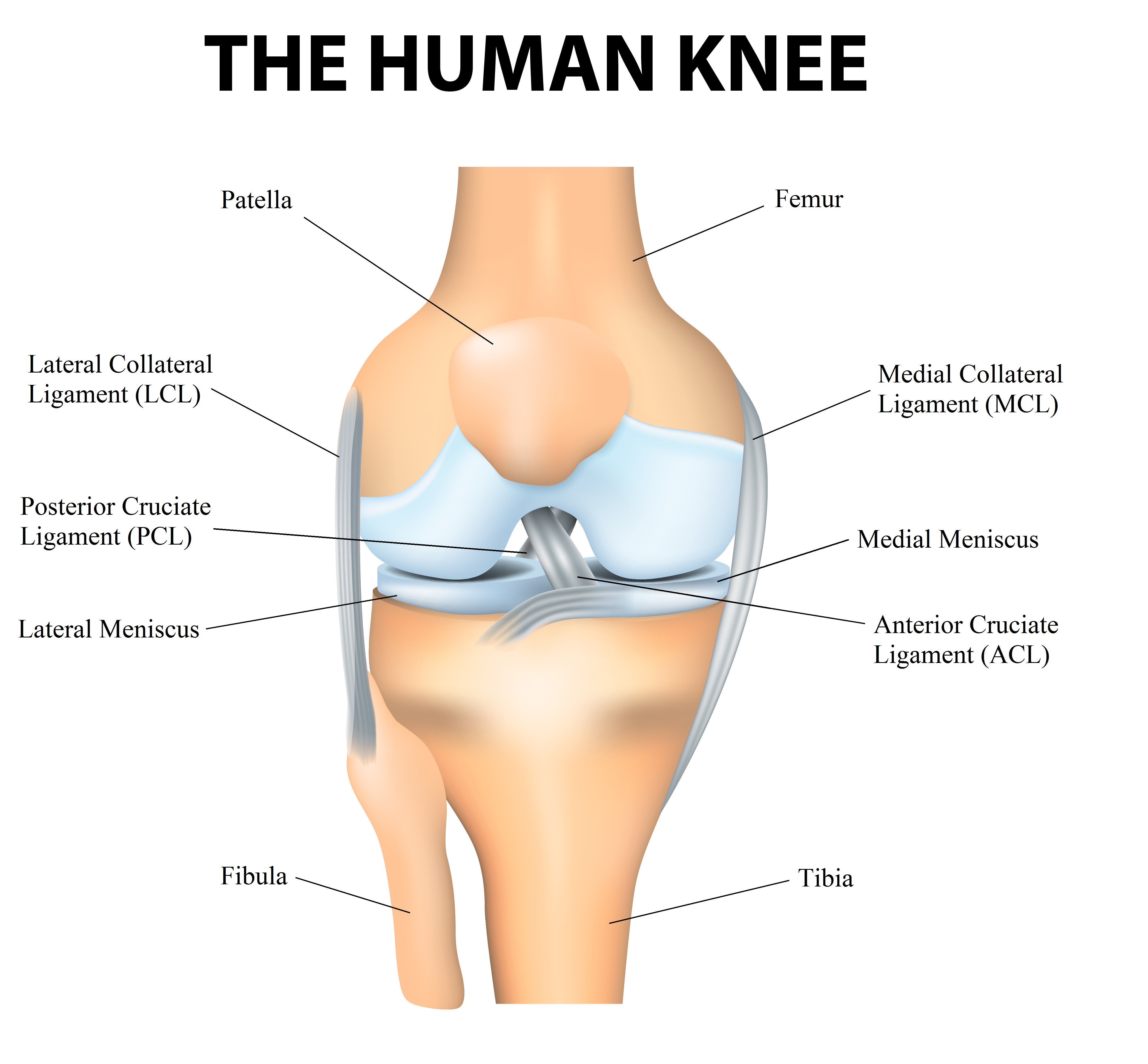
Pcl Acl Lcl Mcl Meniscus Anatomy
Graft Choices In Acl Surgery
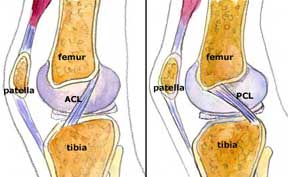
 Diagram Of The Posterior View Of The Knee Demonstrating The
 Posterior Cruciate Ligament Pcl Injuries Thermoskin
 Posterior Cruciate Ligament Pcl Injury Orthopedics
 Posterior Cruciate Ligament Reconstruction By Emad M Qasem
 Knee Joint Anatomy Diagram Focusing On The Knee Ligaments
 Pcl Tear The Complete Injury Guide Vive Health

 Physical Therapy In Indialantic For Knee Posterior Cruciate

 Ligament Injuries Miami Fl Acl Mcl Pcl Tear Miami Fl

 Pcl Reconstruction The Noyes Knee Institute
Belum ada Komentar untuk "Pcl Anatomy"
Posting Komentar