Diverticulum Anatomy
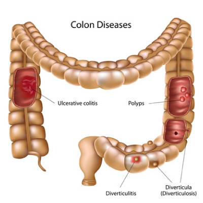
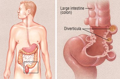
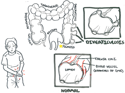
Understanding the anatomy of the large and small intestine can help. Diverticula can occur throughout the colon but are most common near the end of the left side of the colon the sigmoid colon.
Generally detected after middle age.
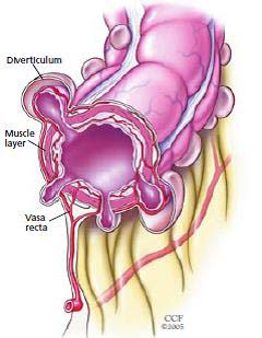
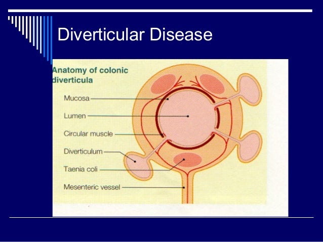
Diverticulum anatomy. Diverticulosis is the name give to this manifestation and most patients with these diverticula will not have any symptoms. As a person ages pressure within the large intestine colon causes pockets of tissue sacs that push out from the colon walls. The diverticula pictured here in yellow can be seen via x ray or a colonoscopy.
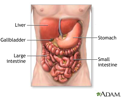
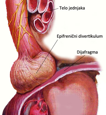
A diverticulum may be present in the stomach the small intestine or most commonly the colon. The pouch is best identified during swallowing and is best seen on the lateral view on which the. An overview of diverticular disease large intestine anatomy.
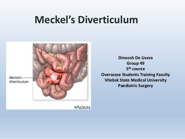
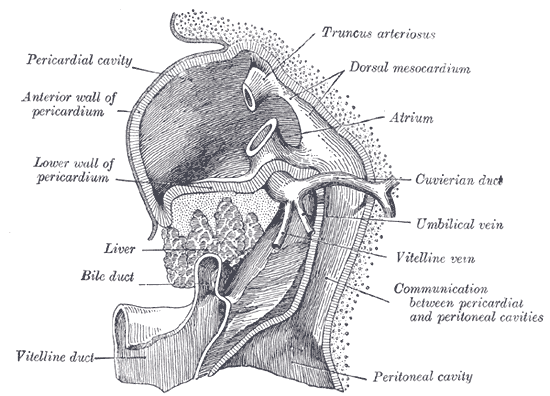
See also diverticulitis diverticulosis meckels diverticulum. No difference in incidence between the sexes. The omphalomesenteric vitelline duct typically arises from a point about 60 cm proximal.
Its not well understood why diverticula develop though there are some theories. Meckel diverticulum surgery anatomy. Ideally a barium swallow examination is performed which may show.
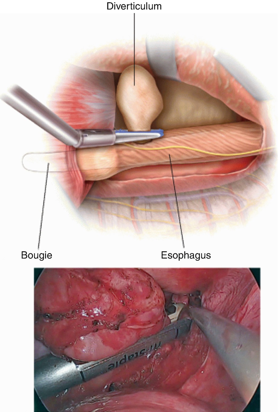
A diverticulum arising from the midline of the posterior wall of the distal pharynx near the pharyngoesophageal junction. The colon or large intestine is a muscular tube that begins at the end of the small intestine and runs to the rectum. A diverticulum is an outpouching of the mucosa of the lining of the bowel.
Anatomy and physiology of diverticular disease. More than 75 of cases occur concomitantly with esophageal motility disorders. The plural is diverticula.
A pouchlike herniation through the muscular wall of a tubular organ. When diverticulitis leads to a painful abscess an ultrasound and ct scan of the abdomen and pelvis can show. The colon absorbs water from liquid stool that is delivered to it from the small intestine.
Diverticula usually dont cause any symptoms. Diverticulum of kommerell. Is an out pouchinganeurysm of the aorta where an aberrant right subclavian artery is located.
The symptoms of diverticular disease in westernised countries usually relate to the sigmoid colon. It is unusual nomenclature in that focal dilatations of a blood vessel are properly referred to as aneurysms. The right wall of the distal esophagus is the most common site.
Medical definition of diverticulum. The pathophysiology varies depending on the etiology of symptoms. The existence of a meckel diverticulum or one of its variants is due to.
It is typically detected by radiography after the ingestion of a radiopaque substance.
 Diverticulum
 Figure 1 From Surgical Treatment Of Zenker S Diverticulum
 Diverticulitis Digestive Disorders Merck Manuals
 Medicine Human Anatomy Diverticulum Drawing Stock Photo
 5 Diverticulitis Symptoms Diverticulum Pain Diet
 Diverticulosis Diverticulitis Jackson Siegelbaum
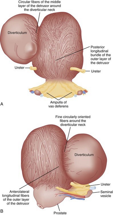 Bladder And Female Urethral Diverticula Abdominal Key
 How To Treat Diverticulitis Diverticulitis Anatomy
Merkel S Diverticulum
 Diverticulosis And Diverticulitis Guide Causes Symptoms
 Epiphrenic And Mid Esophageal Diverticula Springerlink
 Diverticular Disease Familydoctor Org
 Bladder Diverticulum Symptoms Diagnosis Treatment
 Meckel Diverticulum Lima Memorial Health System
 How Diverticulitis Affects The Sigmoid Colon Everyday Health
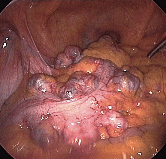
 Laparoscopic Diverticulectomy Of Perforated Duodenal Diverticulum
 Diverticular Disease Clinical Features Management
 Diverticular Disease Of Colon
/GettyImages-506837637-59a7016b685fbe001067278a.jpg) Diverticular Disease Diverticulosis And Diverticulitis
Diverticular Disease Diverticulosis And Diverticulitis
 Diverticular Disease And Common Anorectal Disorders
 Diverticula Metro Health Hospital Metro Health
 Hepatic Diverticulum Wikipedia
Belum ada Komentar untuk "Diverticulum Anatomy"
Posting Komentar