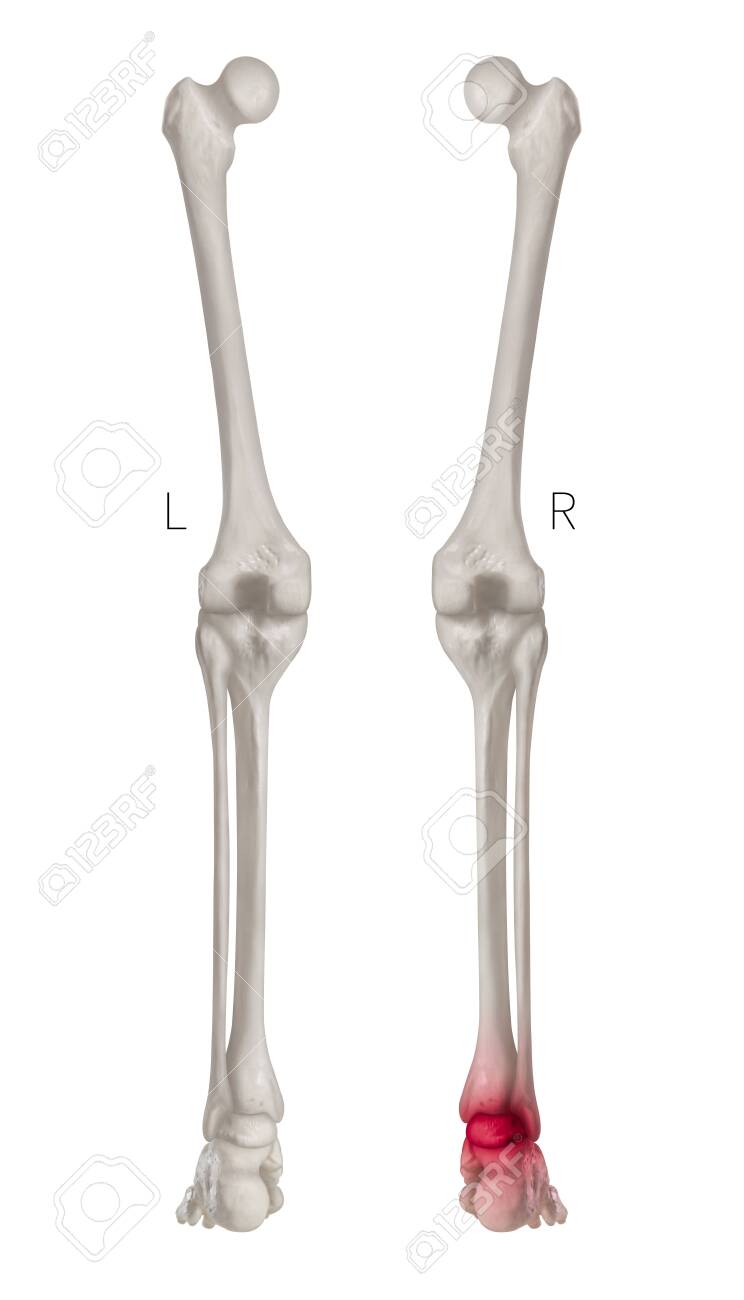
Right Ankle Anatomy
Or dorsiflexion and downward toes down. Consequently the ankle joint mainly only allows for upward toes up.
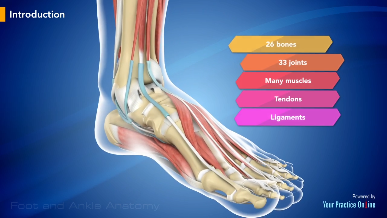
 Ankle Foot Anatomy
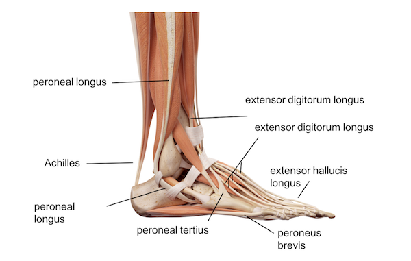
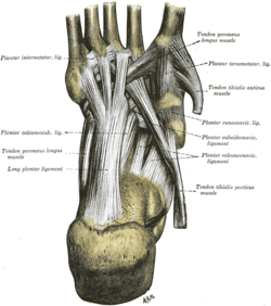
There are also multiple muscles in the ankle that can be strained as follows.

Right ankle anatomy. Click on a link to get sagittal view t1 axial view t2fatsat coronal view t2fatsat sagittal view t2fatsat. The peroneal muscles peroneus longus and peroneus brevis on the outside edge. The ankle joint is a hinged synovial joint with primarily up and down movement plantarflexion and dorsiflexion.
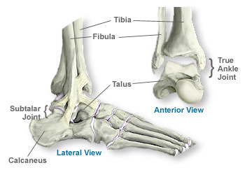
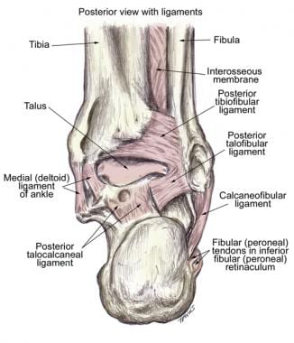
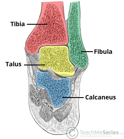
In this article we shall look at the anatomy of the ankle joint. The ankle joint or talocrural joint is a synovial joint formed by the bones of the leg and the foot the tibia fibula and talus. The ankle joint is both a synovial joint and a hinge joint.
The articulating surfaces ligaments movements and any clinical correlations. The unique design of the ankle makes it a very stable joint. Normal ankle function is needed to walk with a smooth and nearly effortless gait.
The ankle is actually made up of several important structures. This webpage presents the anatomical structures found on ankle mri. When thinking about foot and ankle anatomy we usually divide the foot bones into three categories.
Or plantarflexion movements of the foot in relation to the tibia. However when the range of motion of the ankle and subtalar joints talocalcaneal and talocalcaneonavicular is taken together the complex functions as a universal joint see the image below. The posterior tibialis muscle which supports.
The ankle joint allows up and down movement of the foot. Hinge joints typically allow for only one direction of motion much like a door hinge. The hindfoot comprises of the ankle joint found at the bottom of the leg and is where the end of the tibia and fibula meet the ankle bone known as the talus.
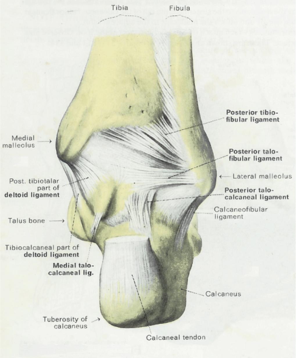
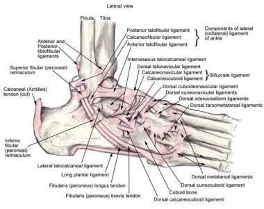
Ankle ligament injury is the most frequent cause of acute ankle pain. Chronic ankle pain often finds its cause in laxity of one of the ankle ligaments. Learn about ankle sprains.
This joint has to be stable in order to withstand 15 times your body weight when you walk and up to eight times your body weight when you run. The calf muscles gastrocnemius and soleus which are connected to the calcaneus via. The hindfoot midfoot and forefoot the hindfoot.
Mri of the ankle. The subtalar joint sits below the ankle joint and allows side to side motion of the foot. Understanding the anatomy of the ankle ligaments is important for correct diagnosis and treatment.
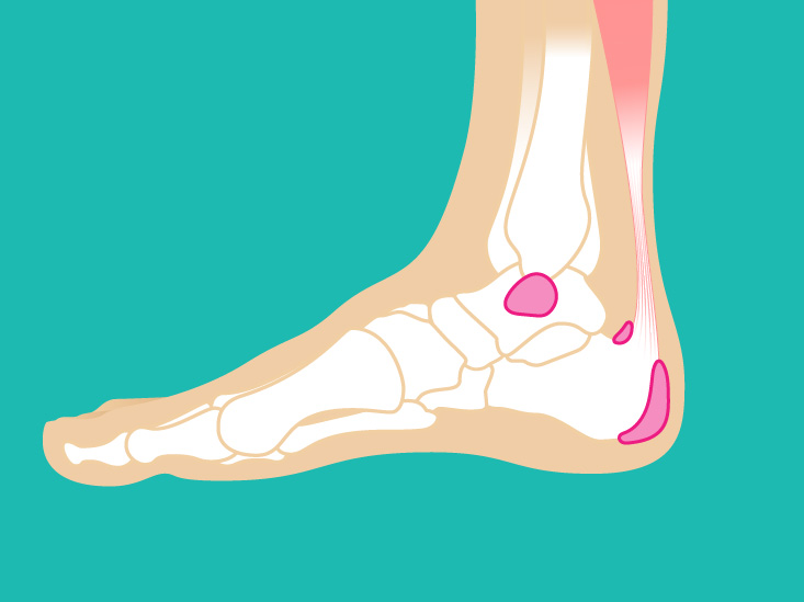
 Bursitis Ankle Bursa Care And Prevention
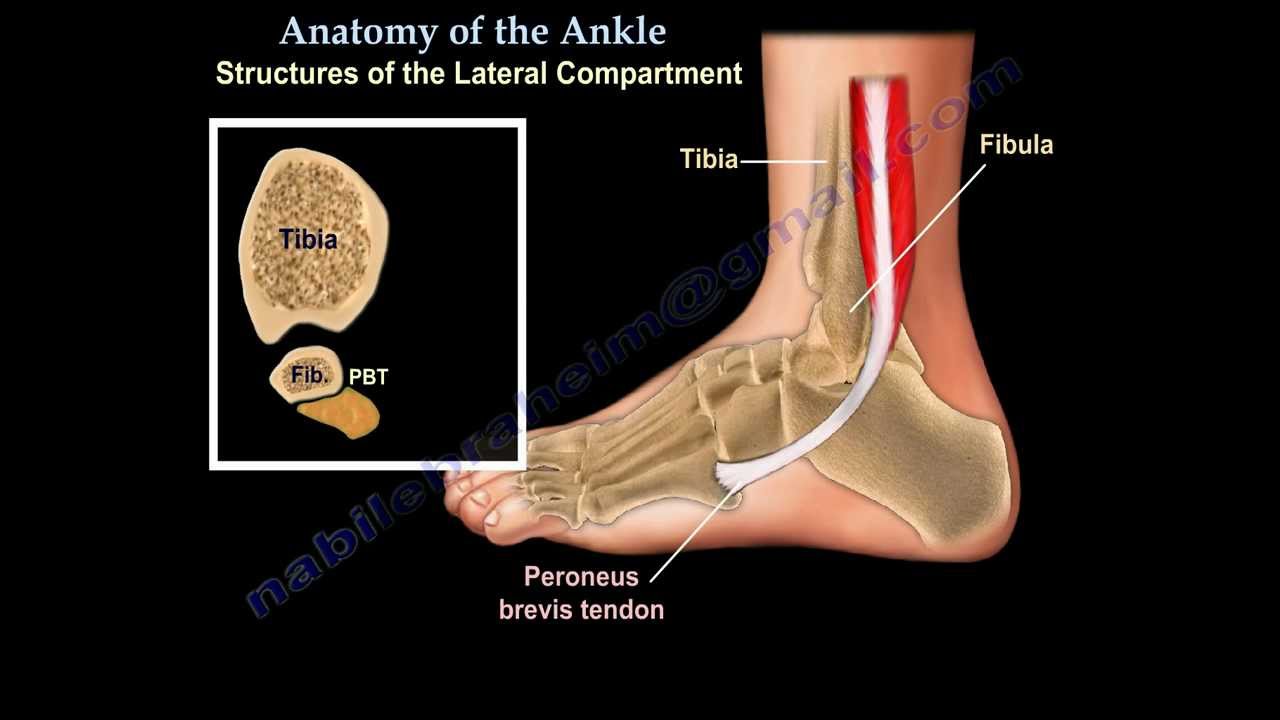 Anatomy Of The Foot Ankle Everything You Need To Know Dr Nabil Ebraheim
 Human Muscle System Foot Anatomy Ankle Anatomy Anatomy
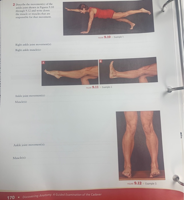 Solved 2 Describe The Movement S Of The Ankle Joint Show
 Ankle Joint Anatomy Overview Lateral Ligament Anatomy And
 Why Ankle Pain Treatments Chronic Ankle Pain Ankle Joint
 Left Ankle Edge Contracture Anatomy Contracture And
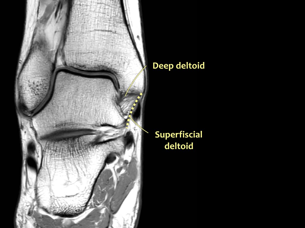
 The Radiology Assistant Ankle Mri Examination
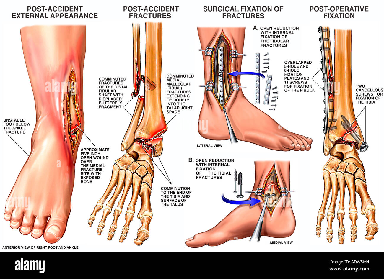
 This Trial Exhibit Depicts A Bimalleolar Right Ankle
 Legal Art Works Injuries To Right Ankle
 Foot And Ankle Anatomy
 Anatomy Of The Ankle Southern California Orthopedic Institute

 Stock Illustration
 Plantar Calcaneonavicular Ligament Wikipedia
 Muscles And Tendons Of The Foot And Ankle Myfootshop Com
 The Radiology Assistant Ankle Mri Examination

 Ankle Joint Anatomy Overview Lateral Ligament Anatomy And
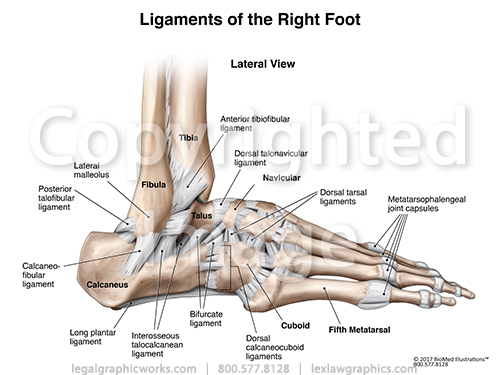 Lateral Right Ankle Ligaments
 Ankle Pain Achilles Tendinitis Home Remedies Treatment
 Lateral Malleolus Fracture Stock Photos Lateral Malleolus
 The Ankle Joint Articulations Movements Teachmeanatomy
 Ankle Joint Anatomy Overview Lateral Ligament Anatomy And
 Ankle Mri Anatomy
 Foot And Ankle Musculoskeletal Key
 Bursitis Ankle Bursa Care And Prevention
 Ankle Foot Atlas Of Anatomy
 Ankle Foot Atlas Of Anatomy
Belum ada Komentar untuk "Right Ankle Anatomy"
Posting Komentar