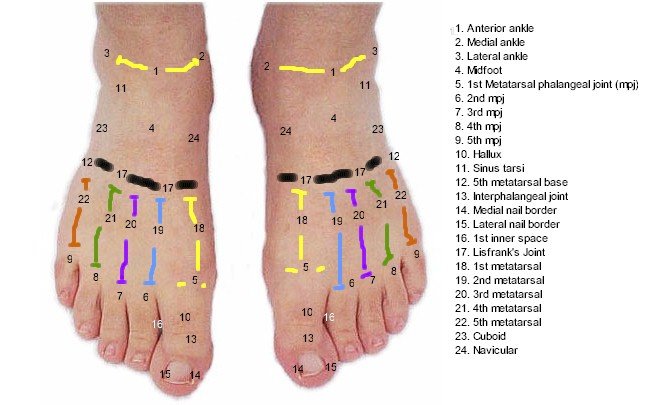
Foot Anatomy Dorsal
Dorsal fascia of the foot. In veterinary anatomy pertaining to the back or upper surface of an animal.
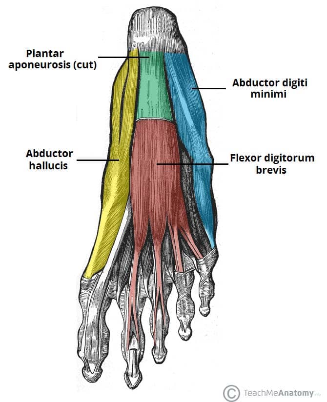
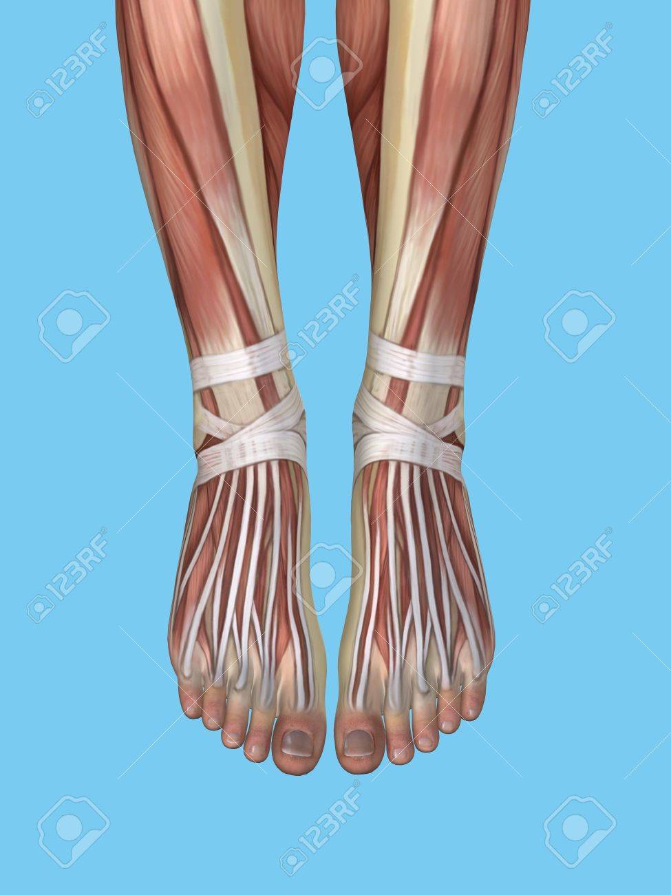
 Muscles Of The Foot
It is the terminal portion of a limb which bears weight and allows locomotion.

Foot anatomy dorsal. Digiti mini little toe plantar region sole. That is nearer the back surface of the body. Its ok there are only a couple of them.
The foot is an anatomical structure found in many vertebrates. Little toe is digit v hallux big toe. More on sole than dorsal.
The dorsal fascia of the foot is the continuation of the deep fascia of the leg crural fascia. In that sense the top of the foot faces front and the sole of the foot faces backwards when the foot is extended. Old term meaning thoracic in a limited sense.
Muscles of the foot that originate on the foot and insert anteriorly on the foot. We looked at the plantar muscles of the foot last week so wed better take a look at the muscles on the other side too. For a random.
Heel part of the rounded superior articular surface of the talus that artic a transverse elevation on the plantar surface of the calcaneus arises from the antero portion of the calcaneus. Often used to indicate the position of one structure relative to another. Volar can also be used to refer to the underside of the palm or sole which are themselves also sometimes used to describe location as palmar and plantar.
Dorsal region dorsum. The structure of the foot is similar to that. In many animals with feet the foot is a separate organ at the terminal part of the leg made up of one or more segments or bones generally including claws or nails.
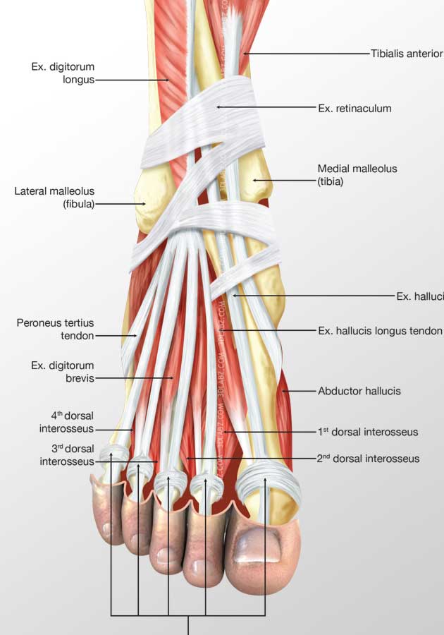
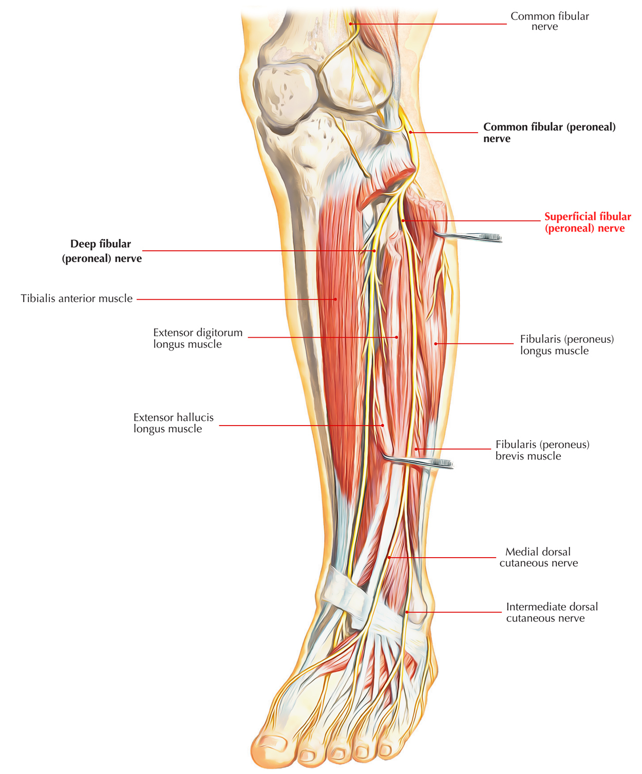
Apart from the muscles of the dorsum of the foot it incloses the tendons of the anterior muscles of the leg the dorsalis pedis vessels continuation of the anterior tibial artery and branches of the deep peroneal nerve. The palmar fascia is palmar to the tendons of muscles which flex the fingers and the dorsal venous arch is so named because it is on the dorsal side of the foot. The foots complex structure contains more than 100 tendons ligaments and muscles that move nearly three dozen joints while bones provide structure.
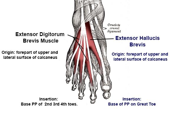
Dorsal aspect whilst many of the extrinsic muscles attach to the dorsum of the foot there are only two intrinsic muscles located in this compartment the extensor digitorum brevis and the extensor hallucis brevis. Big toe is digit i. Dorsal means back spinal or posterior side and ventral means front abdominal or anterior side.
For example dorsal vertebrae.
 Anatomy Of The Dorsal Aspect Of The Foot Myfootshop Com
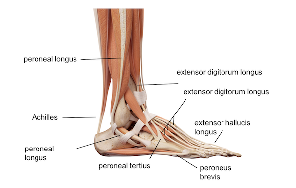
 Ankle Foot Anatomy
 Anatomy Of The Foot And Ankle Orthopaedia
Functions Of The Dorsal Muscles Of The Foot Preview 3d Human Anatomy Kenhub
 Dorsalis Pedis Artery An Overview Sciencedirect Topics
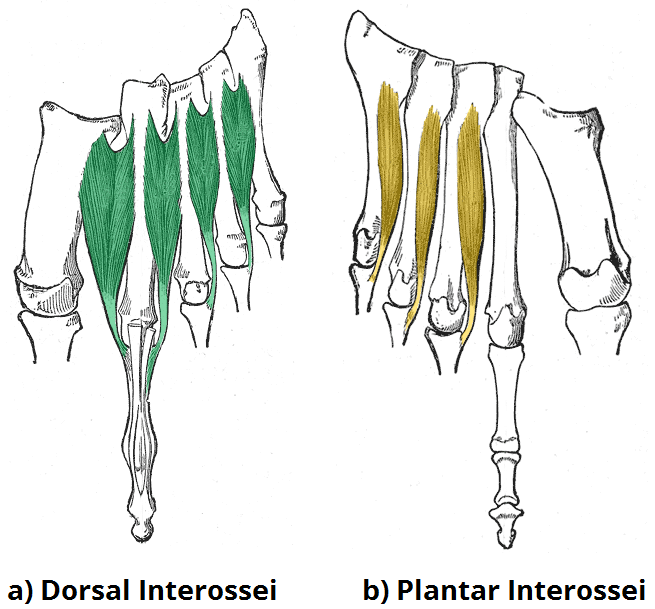
 Dorsal Interossei Lpn Anatomy Orthobullets
 Duke Anatomy Lab 2 Pre Lab Exercise
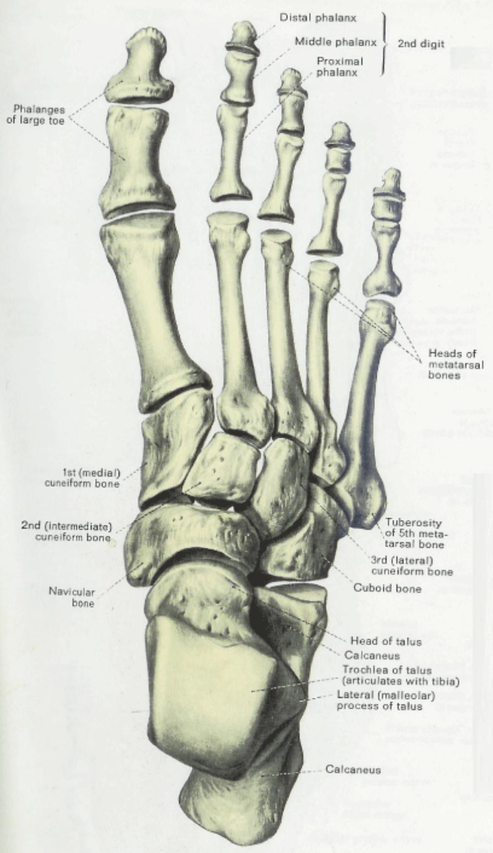
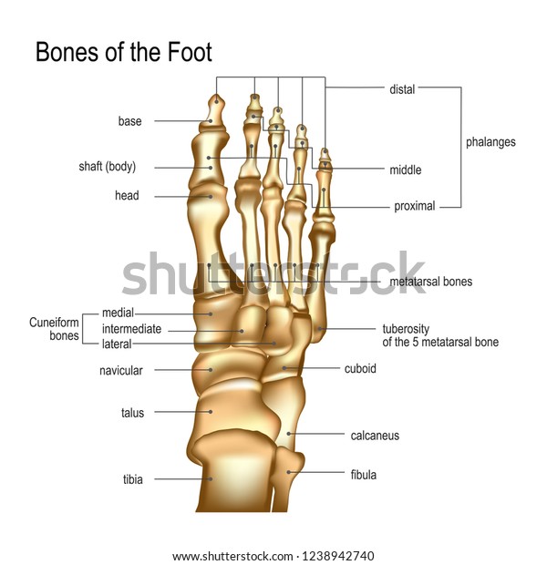
 Anatomical Drawings Foot Anatomy Dorsal View Bones And
 Bones The Of Foot Stock Vector Illustration Of Healthcare
 Tarsometatarsal Joint An Overview Sciencedirect Topics
 Muscles Of The Foot Dorsal Plantar Teachmeanatomy
![]() Second Toe Flap
Second Toe Flap
 Ligaments Of The Foot
 1 10 Foot Anatomy Anatomy Flashcards Memorang
 Foot X Rays
 Foot Dorsal Muscles 3d Illustration Price
 Ankle Foot Anatomy
 Muscles Arteries And Nerves Of Front Of Ankle And Dorsum
 Foot Muscles Attachment Nerve Supply Action Anatomy Info
 Anatomy Of The Foot And Ankle Orthopaedia
 Nerves Of Foot Earth S Lab
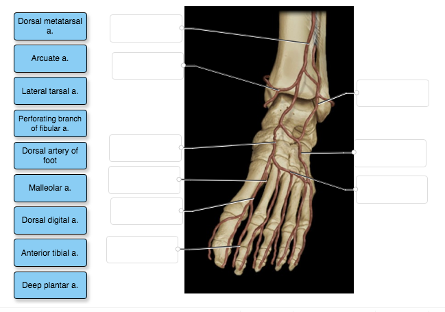 Solved Dorsal Metatarsal A Arcuate A Lateral Tarsal A P
 Dorsal Anatomy Of The Human Foot
 Anatomy Physiology Illustration
 Muscles Of The Foot Dorsal Plantar Teachmeanatomy
![]() Dorsal Interossei Of Foot Attachments Supply Action Kenhub
Dorsal Interossei Of Foot Attachments Supply Action Kenhub
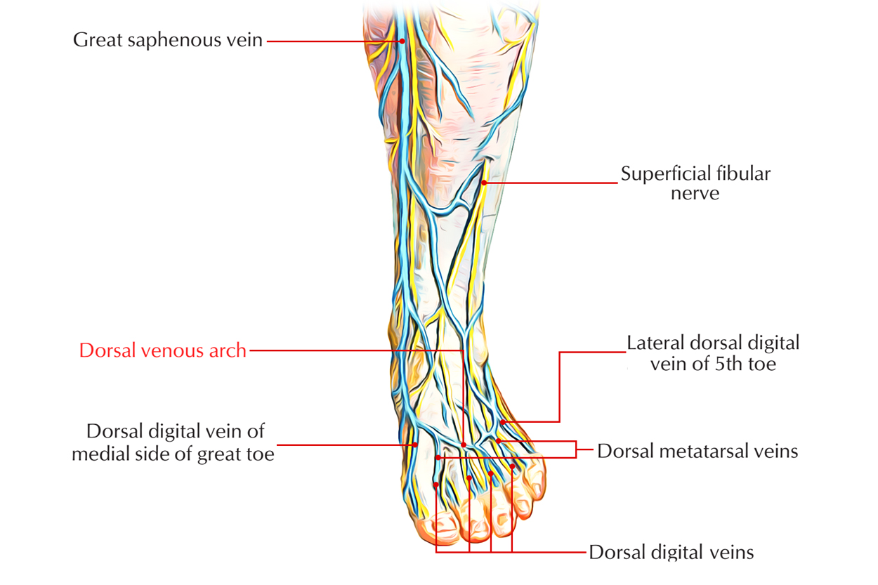 Easy Notes On Dorsal Venous Arch Learn In Just 3 Minutes
 Anatomy Of The Foot And Ankle Orthopaedia
 Anatomy Of Foot Featuring Extensor Digitorum Longus Tendons
 Dorsal View Of The Bone Anatomy Of Rabbit Feet Left Front
 Realistic Skeleton Human Leg Titles Bones Stock Vector
 Ankle Foot Anatomy


Belum ada Komentar untuk "Foot Anatomy Dorsal"
Posting Komentar