The Anatomy Of The Heart
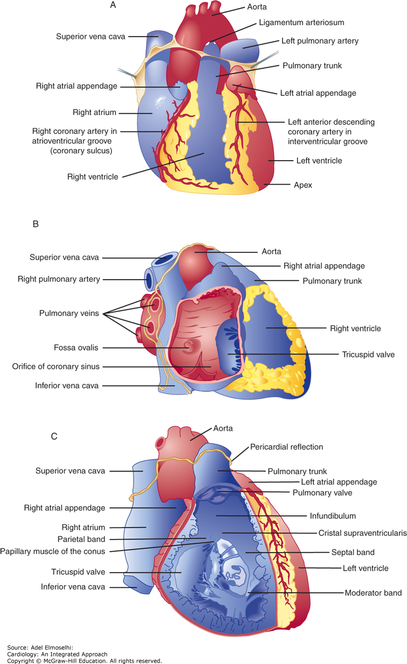
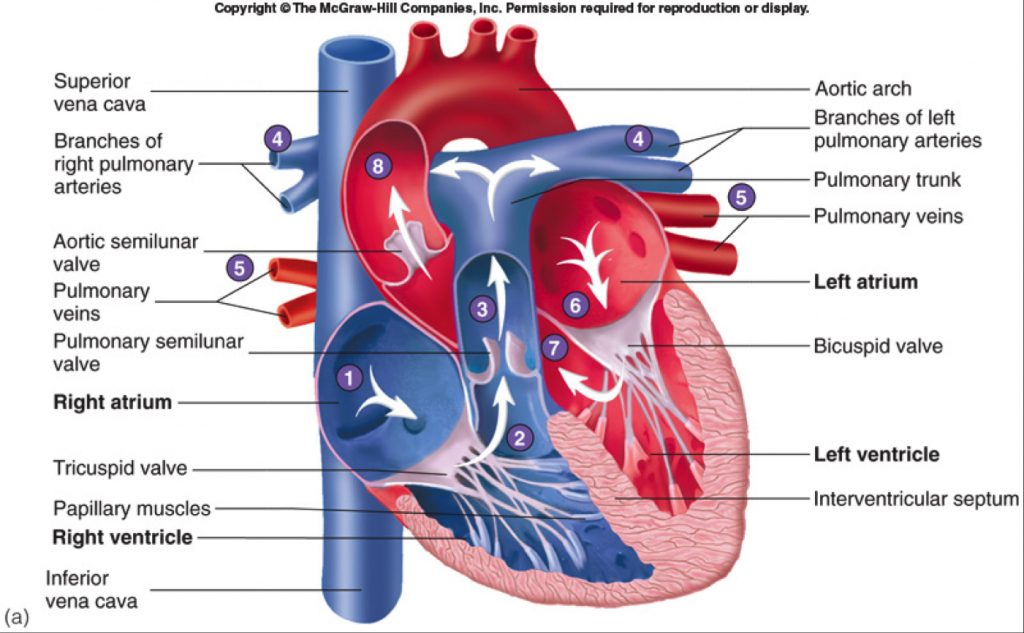
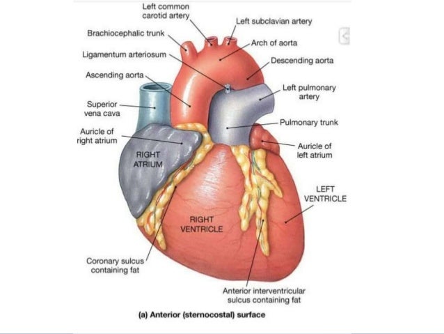
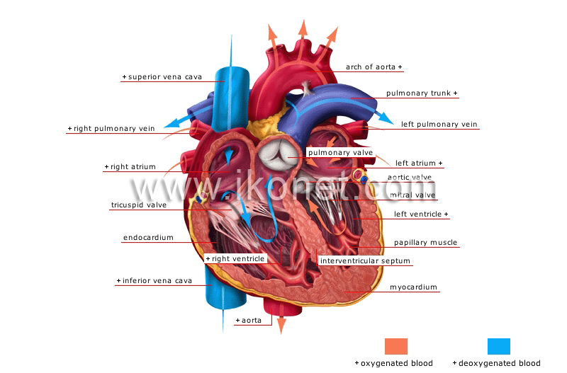
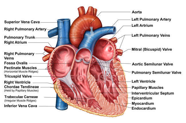
The walls and lining of the pericardial cavity are a special membrane known as the pericardium. The structures initially seen from this perspective include the superior vena cava right atrium right ventricle pulmonary artery and aorta.
 The Heart Anatomy Physiology And Function
The base of the heart is located at the level of the third costal cartilage as seen in figure 1.

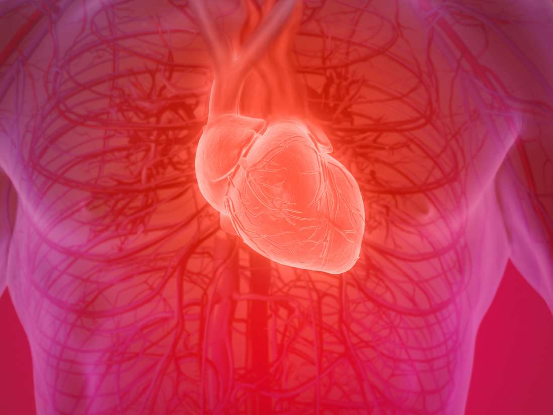
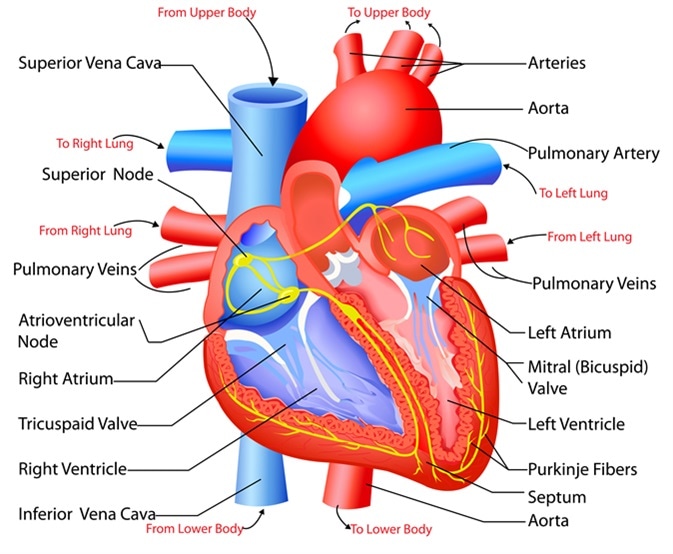
The anatomy of the heart. This amazing muscle produces electrical impulses that cause the heart to contract. Anatomy of the heart pericardium. In fact each day the average heart beats 100000 times pumping about 2000 gallons 7571 liters of blood.
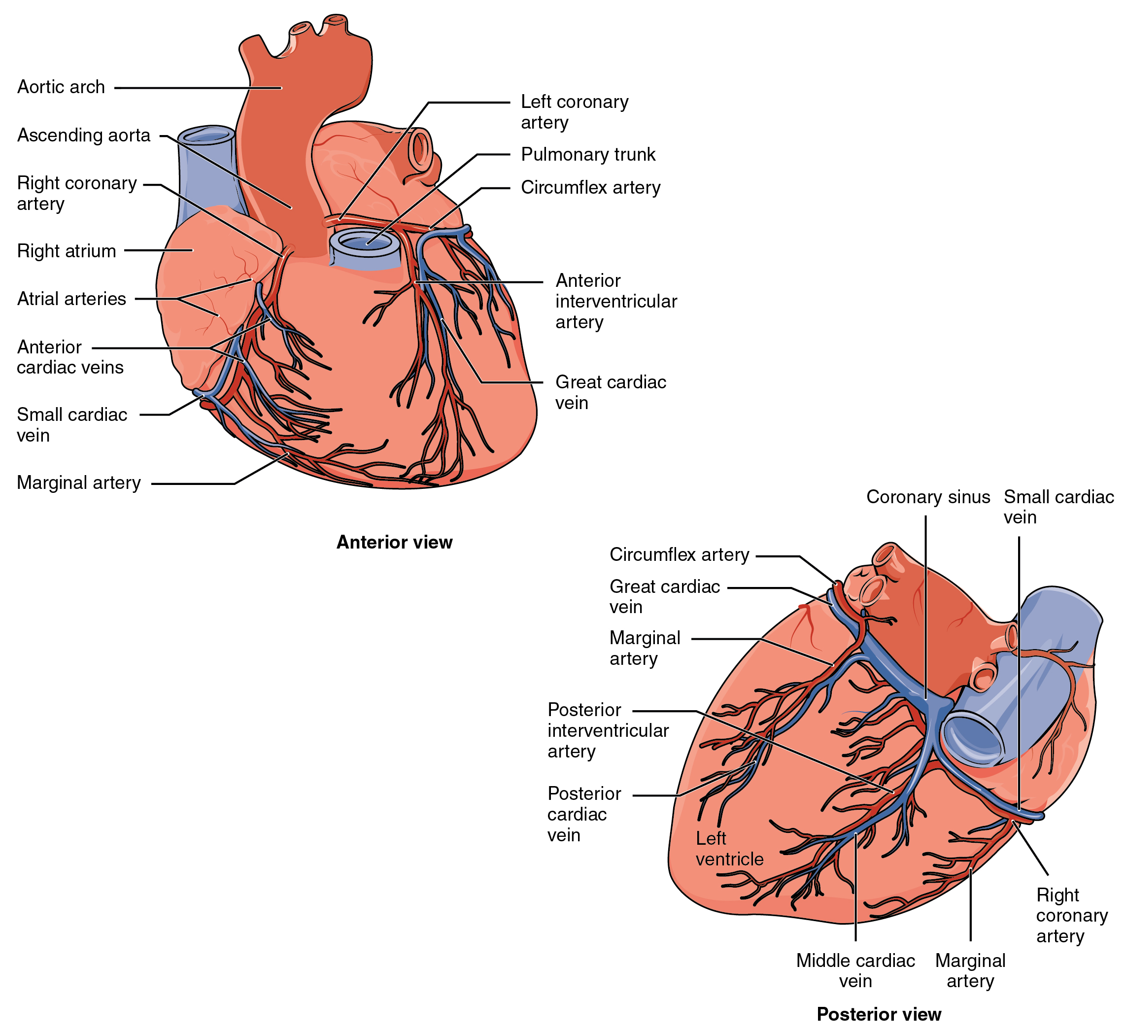
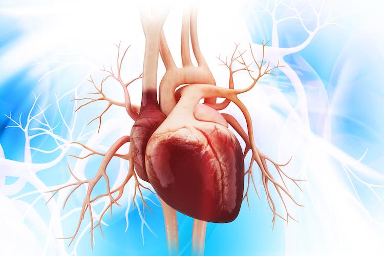
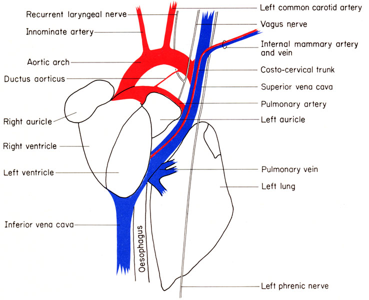
In humans the heart is located between the lungs in the middle compartment of the chest. The heart is a muscular organ in most animals which pumps blood through the blood vessels of the circulatory system. The great veins the superior and inferior venae cavae and the great arteries the aorta and pulmonary trunk are attached to the superior surface of the heart called the base.
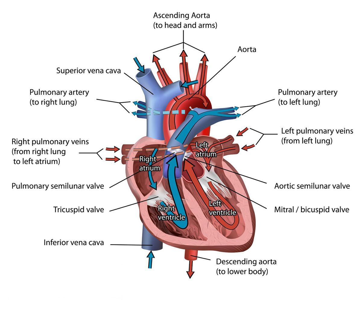
Blood provides the body with oxygen and nutrients as well as assists in the removal of metabolic wastes. Because the heart points to the left about 23 of the hearts mass is found on the left side of the body and the other 13 is on the right. The heart is a muscular organ about the size of a fist located just behind and slightly left of the breastbone.
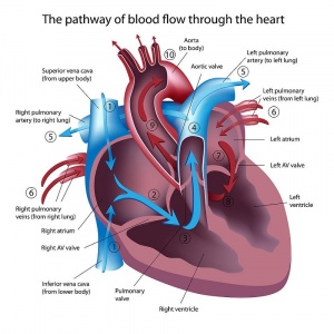
The heart pumps blood through the network of arteries and veins called the. Endocardium lines the inside of the heart and protects the valves and chambers. Location of the heart.
Your heart is located between your lungs in the middle of your chest behind and slightly to the left of your breastbone sternum. Myocardium the muscles of the heart. The heart is situated within the chest cavity and surrounded by a fluid filled sac called the pericardium.
A double layered membrane called the pericardium surrounds your heart like a sac. The heart sits within a fluid filled cavity called the pericardial cavity. Epicardium protective layer mostly made of connective tissue.
The wall of the heart consists of three layers of tissue. The anatomy of the heart. Basic anatomy of the heart.
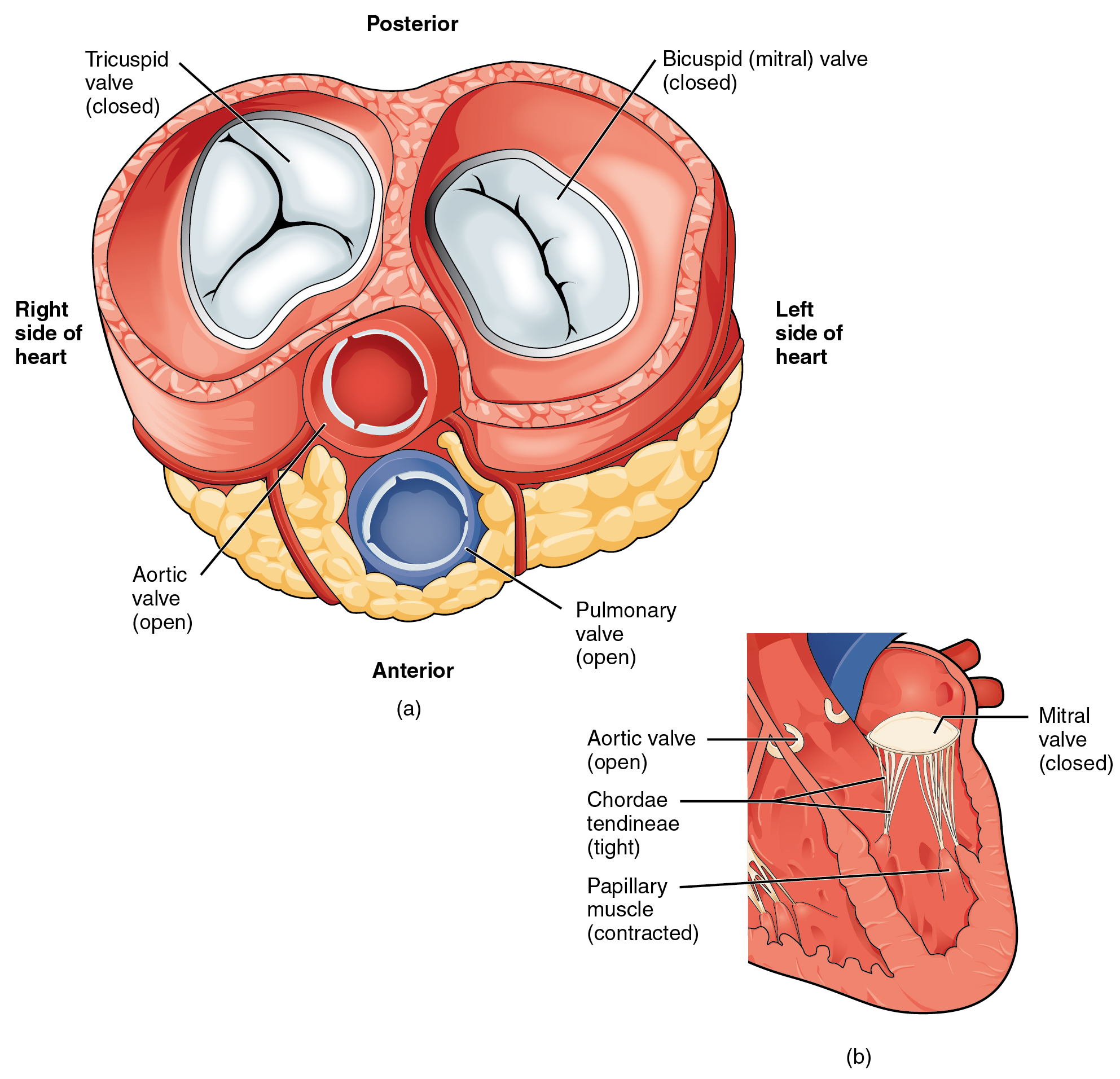
Intraoperatively the anatomy of the heart is viewed from the right side of the supine patient via a median sternotomy incision. It is divided by a partition or septum into two halves and the halves are in turn divided into four chambers.
 19 1 Heart Anatomy Anatomy And Physiology

![]() Heart Anatomy Structure Valves Coronary Vessels Kenhub
Heart Anatomy Structure Valves Coronary Vessels Kenhub
 19 1 Heart Anatomy Anatomy And Physiology
 Pacific Medical Training Acls Bls Pals Certification
 Anatomical Structure Of The Heart Cardiology An
 Anatomy Of The Heart Nurse Adventures
 Cardiac Anatomy And Electrophysiology Thoracic Key
 Human Heart Gross Structure And Anatomy Online Biology Notes
 Anatomy And Physiology Of The Heart
 Anatomy Of The Human Heart Physiopedia
![]() Heart Anatomy Structure Valves Coronary Vessels Kenhub
Heart Anatomy Structure Valves Coronary Vessels Kenhub

 Heart Anatomy Anatomy And Physiology
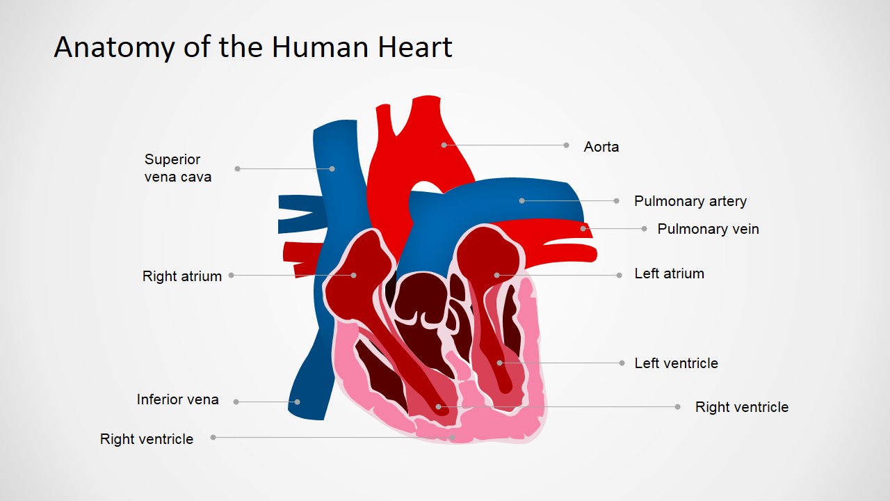 Anatomy Of The Human Heart Powerpoint Shapes
 Heart Chamber Anatomy Pi Uptodate

 Structure And Function Of The Heart
![]() Heart Anatomy Structure Valves Coronary Vessels Kenhub
Heart Anatomy Structure Valves Coronary Vessels Kenhub
 Heart Anatomy 1 Anterior View Quiz By Seattle84
 Free Anatomy Quiz Anatomy Of The Heart Quiz 1
 Heart Anatomy Mydr Com Au
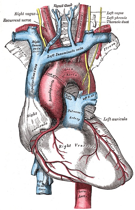 Figure Anatomy Of The Heart Contributed By Gray S Anatomy

 Anatomy Of A Human Heart
 Anatomy Of Heart Interior With Labels Cross Section Blood
 The Anatomy Of The Heart Video Wisc Online Oer
 The Heart Of The Matter National Geographic Society
Belum ada Komentar untuk "The Anatomy Of The Heart"
Posting Komentar