Thyroid Cartilage Anatomy
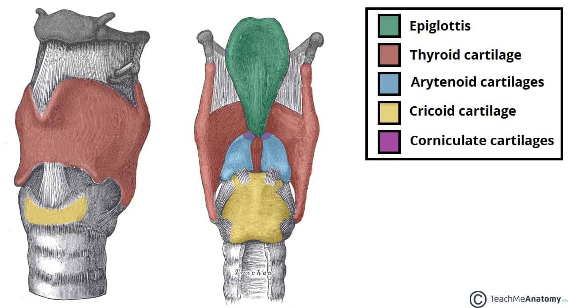
Cartilage of the larynx the thyroid cartilage is made of two plates fused anteriorly in the midline. The lateral surface of the thyroid is covered by the sternothyroid muscle and its attachment to the oblique line of the thyroid cartilage prevents the superior pole from extending superiorly under the thyrohyoid muscle.
 Figure 2 From A Case Of Variant Thyroid Cartilage Anatomy
The thyroid cartilage is located superiorly towards the cricoid cartilage it is another hyaline cartilage.
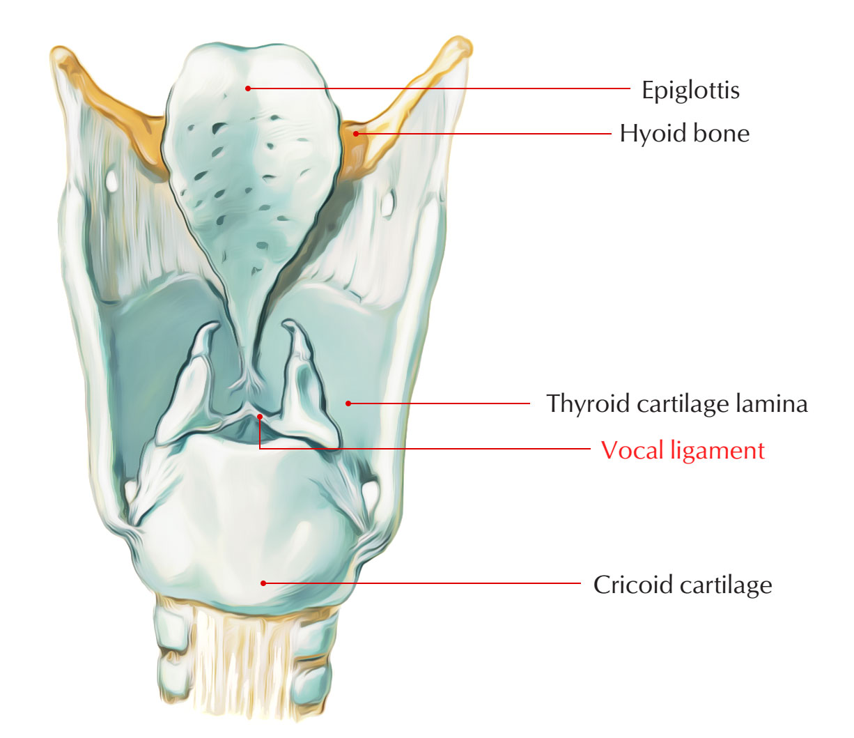
Thyroid cartilage anatomy. Thyroid cartilage anatomy functions and pain. The thyroid cartilage is the largest of the nine cartilages that make up the laryngeal skeleton the cartilage structure in and around the trachea that contains the larynx. At the upper end of the fusion line is an incision the thyroid notch.
Only the cricoid cartilage does. The thyroid cartilage as a whole tended to tilt to the right against the cricoid cartilage. Later morphometric measurements of the laryngeal framework provided valuable information determining the size and extent of the cartilaginous components and human larynx as one unit 4 5.
It plays a role in the production of the human voice providing protection and support for the vocal folds. It does not completely encircle the larynx. The back section of the cartilage that is the farthest also features 2 projections in the downwards and upward directions.
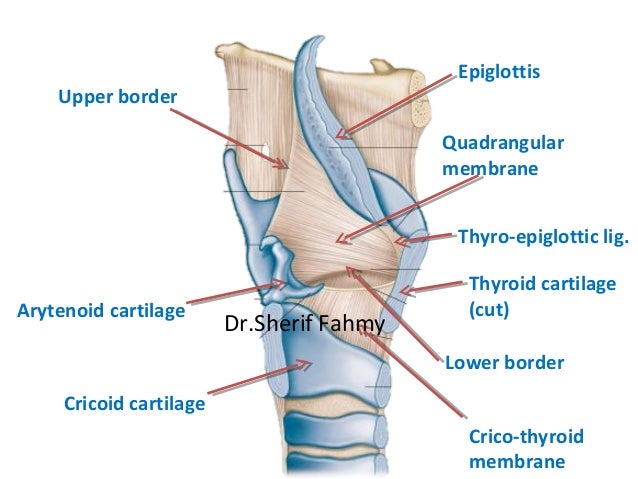
Also the back border of each half of the cartilage communicates inferiorly with the cricoid cartilage at a joint known as the cricothyroid joint. Secreted by the brain tsh regulates thyroid hormone release. Posteriorly its borders are free and project upwards and downwards as the superior and inferior horns.
The muscles of the larynx act on skeletal structures including the thyroid cartilage to produce the vibration of the vocal folds which is necessary to produce vocalization. Below it is a forward projection the laryngeal prominence. Thyroid biopsy is typically done with a needle.
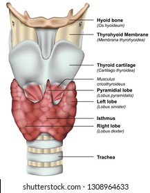
The thyroid cartilage consists of two laminae that are fused anteriorly in the median plane to form the laryngeal prominence. Each laminae possesses an oblique ridge with a tubercle superiorly and inferiorly. Thyroid stimulating hormone tsh.
The cricoid and thyroid cartilages shield the glottis and the entrance towards the trachea. The posterior portion of the cricoid is very stretched out giving support by not involving the thyroid cartilage. A blood test with high tsh indicates low levels of thyroid hormone hypothyroidism and low tsh suggests hyperthyroidism.
 Thyroid Cartilage Earth S Lab
 Surgical Approach To The Thyroarytenoid Branch Of The
 Stock Illustration
![]() Cartilages Of The Larynx Types And Anatomy Kenhub
Cartilages Of The Larynx Types And Anatomy Kenhub
 Illustration Of Thyroid Gland And Cartilage Anatomy Stock
 Laryngeal Cartilages Paired Unpaired Teachmeanatomy
 Thyroid Cartilage Anatomy Illustration License Download
 World S Best Thyroid Cartilage Stock Illustrations Getty
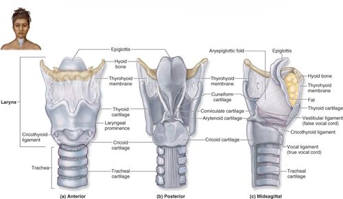
 Review Of Laryngeal Anatomy
 Thyroid Cartilage Anatomy Medical Media Design The
 Neck Atlas Human Anatomy Vertebral Column Thyroid Cartilage
Icm3 2 And Anatomy Icm3 2 And Anatomy Flashcards Memorang
 Cricoid Cartilage Earth S Lab
 Thyroid Cartilage Definition Function Human Anatomy Kenhub
 Thyroid Picture Image On Medicinenet Com
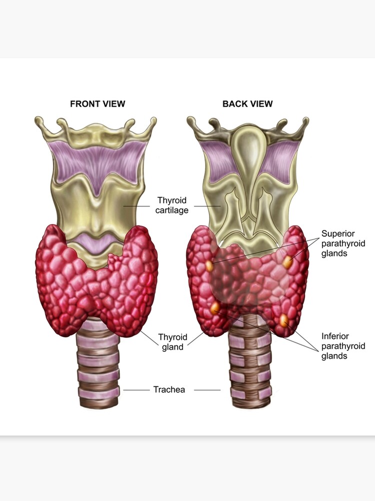 Anatomy Of Thyroid Gland With Larynx Cartilage Canvas Print
 Vocal Cord Dysfunction An Often Misdiagnosed Condition
 Thyroid Cartilage Anatomy Function
 The Larynx Anatomy Of The Neck
 Picture And Anatomy Of The Thyroid Cartilage
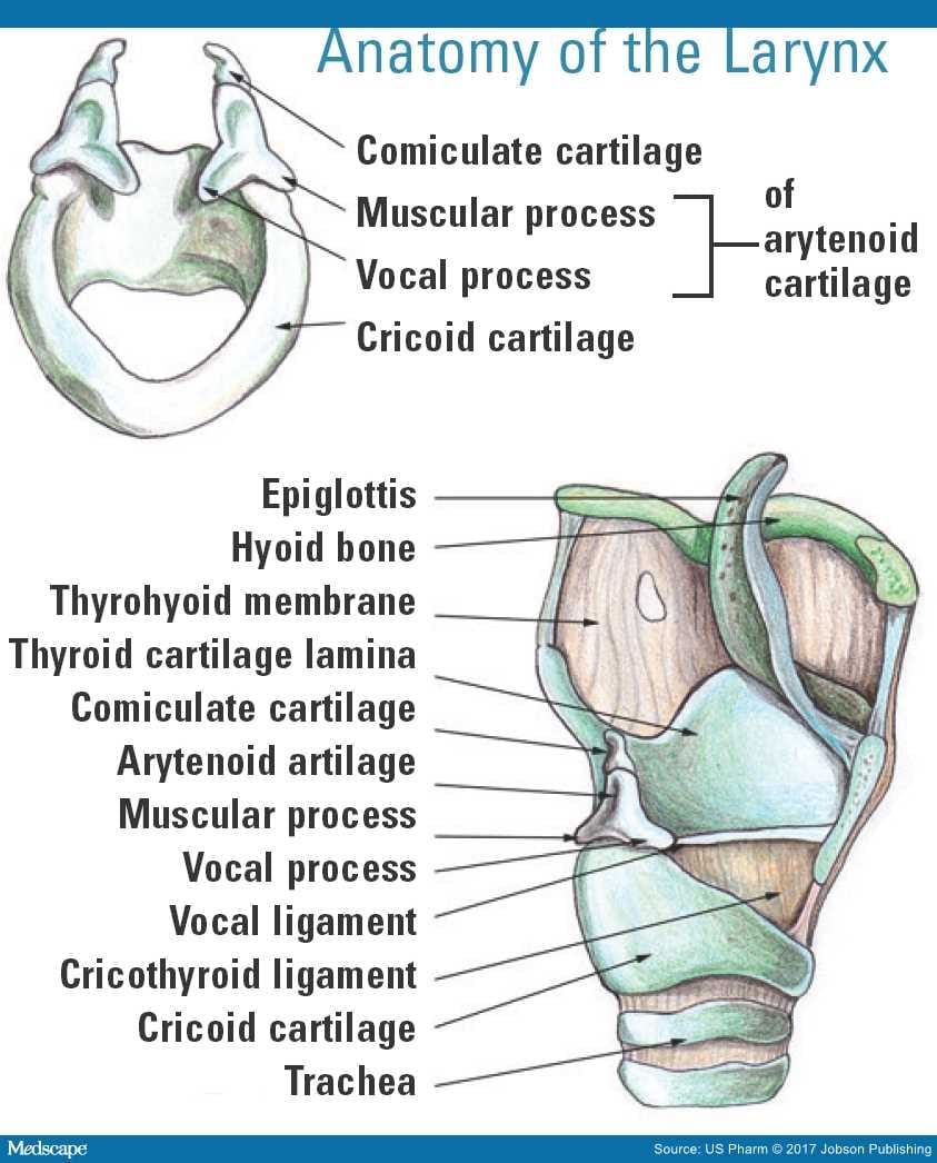
 Larynx Cuneiform Corniculate Arytenoid Cricoid
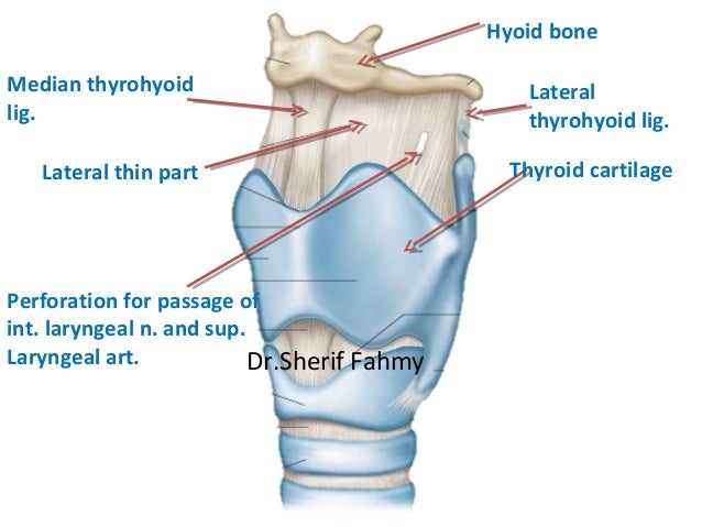
 Oblique Line Of Thyroid Cartilage An Overview
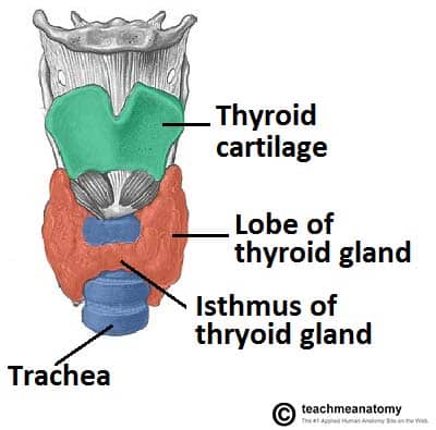 The Thyroid Gland Location Blood Supply Teachmeanatomy
 1000 Thyroid Cartilage Stock Images Photos Vectors
 Thyroid Cartilage Wikipedia
 Larynx Anatomy Of The Respiratory System
 The Larynx Anatomy Of The Neck
 Thyroid Cartilage Horns Anatomy Thyroidissuesinmen

Belum ada Komentar untuk "Thyroid Cartilage Anatomy"
Posting Komentar