Pes Anatomy
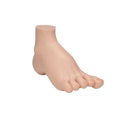
Pes general anatomy definition. Pes the part of the leg of a human being below the ankle joint.
 Outlander Wedding Outlander Anatomy
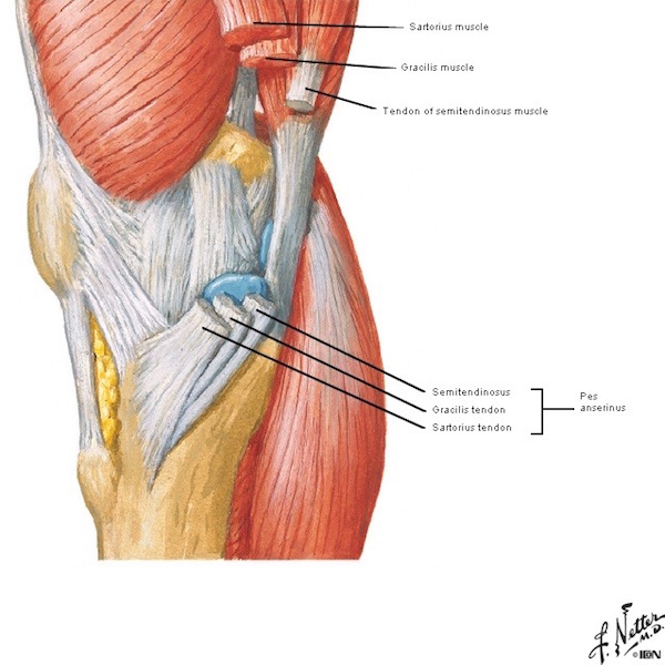
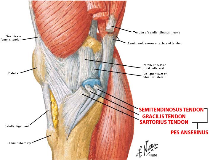
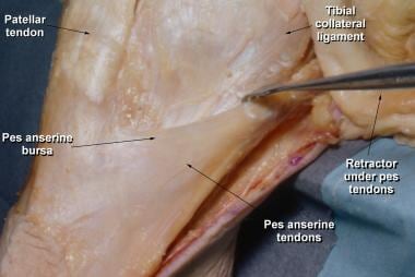
We can locate this at the proximal medial aspect of the knee two inches below the medial knee joint line between the pes anserinus tendons912939139391493.
Pes anatomy. The part of the leg of a human being below the ankle joint. Pes anserine bursitis is when there is inflammation of the pes anserine bursa causing medial knee pain. The name comes from the latin for gooses foot in view of the similarity.
Of course they also need to be equipped to provide leverage. To identify the anatomical basis of imaging mri ct scan and ultrasonography and interpret normal radiological images. Pes anserinus gross anatomy.
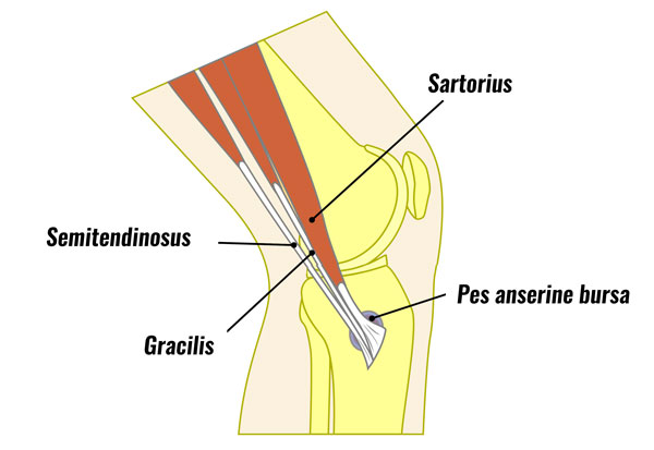
Armored from head to foot. Pes anserine bursitis also known as intertendinous bursa is an inflammatory condition of bursa of the conjoined insertion of the sartorius gracilis and semitendinosus1. Despite their similarities the bones within the foot vary to indicate their ability to take the full weight of the human body.
His bare feet projected from his trousers. Progressive pes planus flatfoot deformity in adults is a common entity that is encountered by orthopedic surgeons. The term pes anserinus may also be used to describe the branching.
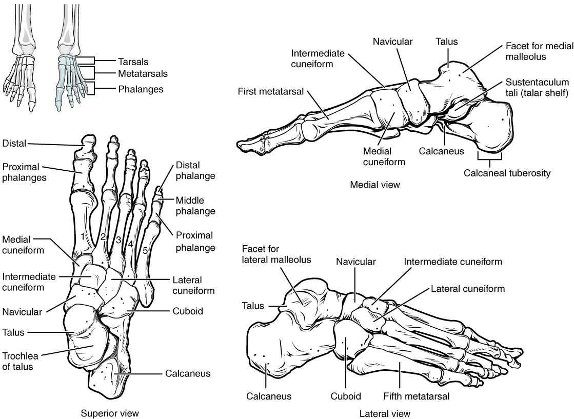
To identify the various normal tissues under microscope differentiate and discuss them. Pes anatomy the pes latin for foot is the zoological term for the distal portion of the hind limb of tetrapod animals. It is the part of the pentadactyl limb that includes the metatarsals and digits phalanges.
The tarsus metatarsus and phalanges are all encompassed within the framework of the foot mildly resembling the framework for the hand. The pes anserinus is an area on the medial inner side of the knee where three muscle tendons attach to the tibia shin bone. A bursa known as the pes anserinus bursa lies between the pes anserinus tendons and.
Human human being homo man any living or extinct member of the family hominidae characterized by superior intelligence articulate speech and erect carriage. A deformity that develops after skeletal maturity is reached is commonly referred to as adult acquired flatfoot deformity aafd. Foot human foot tootsie baby talk.
To identify locate and correlate structures of the body and mark the topography by surface anatomy. During evolution it has taken many forms and served a variety of functions.
 Comparison Of Late Cretaceous Tyrannosauroid Pedal Anatomy
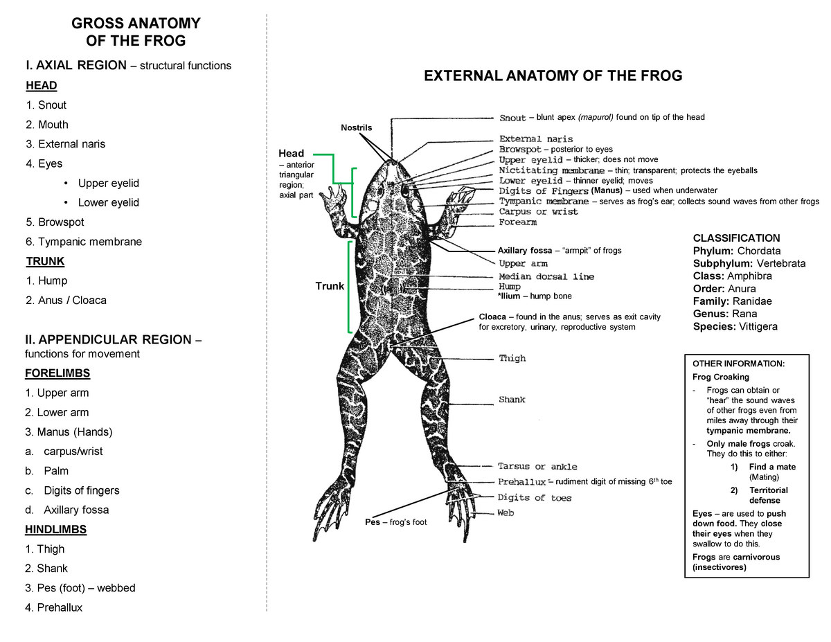 Lecture On Frog Anatomy Bucal Cavity External Biology
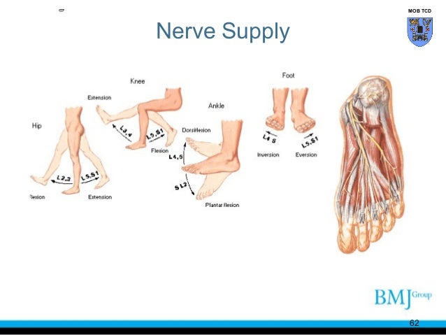
 Hsc 211 Lecture 5 Chapter 5 Lecture Notes Anatomy Oneclass
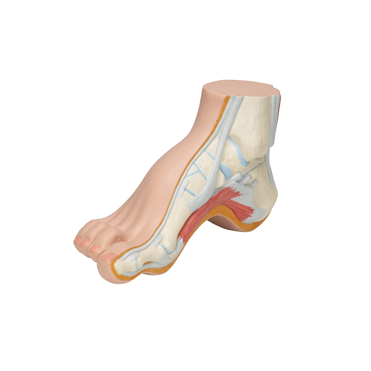
 Hollow Foot Pes Cavus Anatomy Model
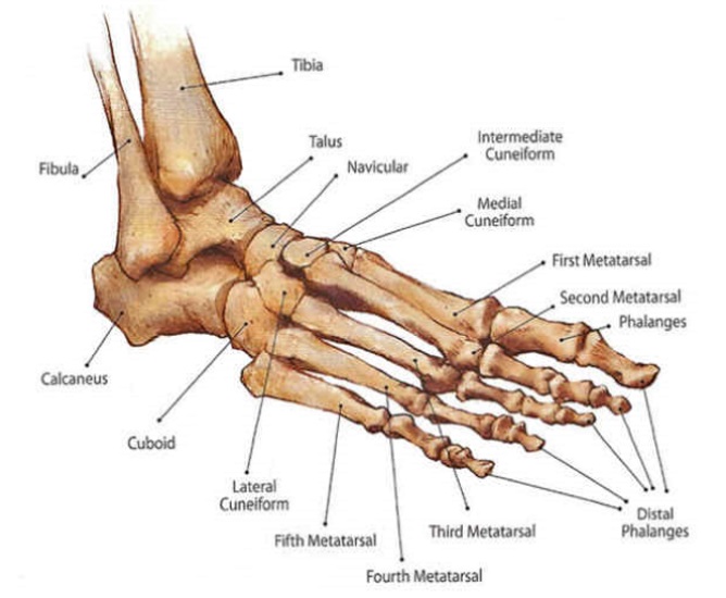
 Anatomy Of Feet
 Videos Matching Pes Anatomy Revolvy
 The Sartorius And The Muscles Of The Pes Anserinus
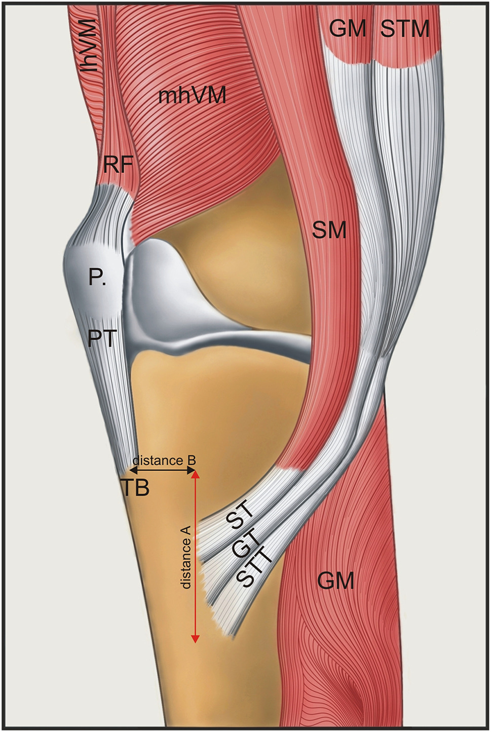 A Proposal For A New Classification Of Pes Anserinus
 Pes Anserine Bursa Kneeguru
 Us 49 0 Free Shipping Pes Arcuatus Model High Arch Foot Model Claw Foot Foot Anatomy Medical In Medical Science From Office School Supplies On
 Leg Anatomy Britannica
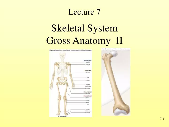 Ppt Skeletal System Gross Anatomy Ii Powerpoint
 Pes Anserine Bursitis Tendinopathy Sportsinjuryclinic Net
 8 4 Bones Of The Lower Limb Anatomy And Physiology
 Okil Anatomy Of Foot And Ankle Poster Anatomical Chart Human Body Educational Home Decor
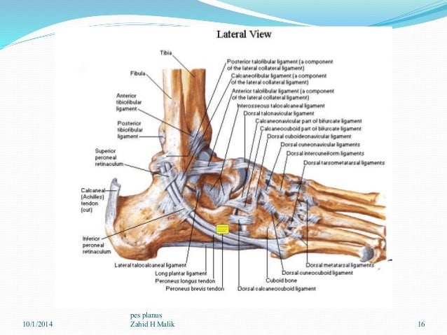 Pes Plenovalgus Dr Zahid H Malik

 Hollow Foot Pes Cavus Model 3b Smart Anatomy
 Pes Anserinus Bursitis Symptoms And Treatment Bone And Spine
 Ao Surgery Reference
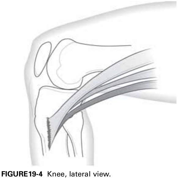 On Figure 19 4 Label The Three Parts Of The Pes Anserine
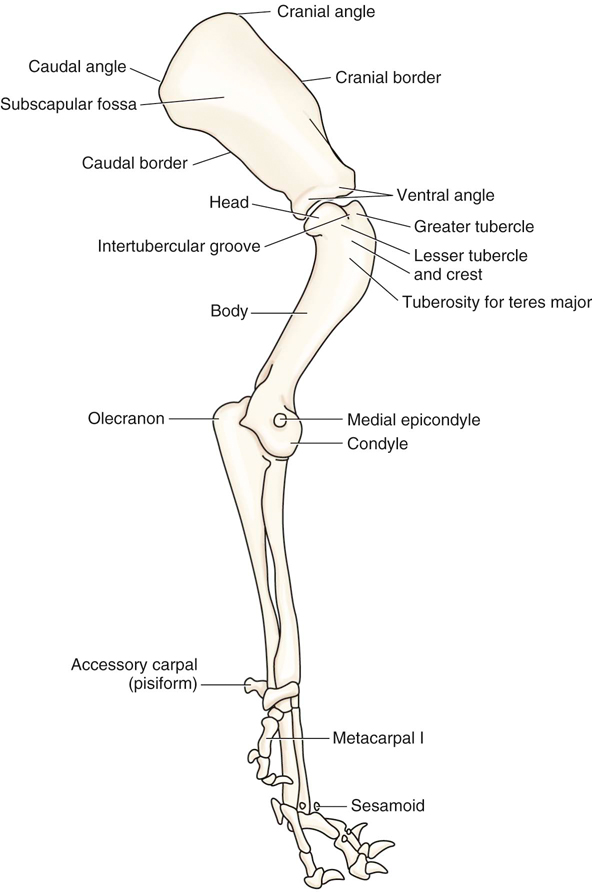 Canine Anatomy Veterian Key
Pes Anatomy 978 613 9 02118 5 6139021189 9786139021185
 Pes Of Eryops Megacephalus Illustration A And Photograph
 Anatomy Of A Rabbit Greeting Card
 Figure 10 From The Pes Of Australovenator Wintonensis
 The Pes Of Australovenator Wintonensis Theropoda
 Vector Illustration Of A Healthy Knee And Unhealthy Knee
 Anatomy And Examination Of The Knee Ligaments Tendons
 Anatomical Teaching Models Plastic Human Joint Models
 Foot Wikipedia
 Pes Anserine Bursitis Background Anatomy Pathophysiology
Belum ada Komentar untuk "Pes Anatomy"
Posting Komentar