Lamina Anatomy
The outermost coat this region is called the lamina cribrosa figure 1. The laminae of the thyroid cartilage.
It is a thin membrane that stretches between the dorsal surface of the optic chiasm to the anterior commissure 1.

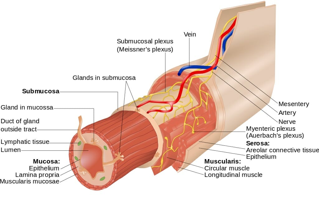
Lamina anatomy. The lamina propria is a large layer of connective tissue which separates the innermost layer of epithelial cells from a layer of smooth muscle tissue called the muscularis mucosa. The lamina terminalis forms the anterior wall of the third ventricle and anterior boundary of the hypothalamus. Zoology a thin scalelike or platelike structure as one of the thin layers of sensitive vascular tissue in the hoof of a horse.
The posterior part of the spinal ring that covers the spinal cord or nerves. Two leaflike plates of cartilage that make up the walls. As with the cornea the innermost layer is a.
Kenhub provides extensive human anatomy learning resources spanning gross clinical and cross sectional anatomy histology and medical imaging. The layers of thalamus tissue. Our dynamic effective and guided approach to learning anatomy is brought to you via a full anatomy atlas in depth articles videos and a variety of quizzes which can be tailored to your level.
The lamina propria is one of three layers which make up the mucosa or mucous membrane. Cytology a thin layer inside the nuclear membrane of cell that is. A thin plate sheet or layer.
Plates of bone that form the posterior walls of each vertebra. Anatomy and physiology chapter 4. The laminae of the thalamus.
Lamina definition spine anatomy overview video the lamina is the flattened or arched part of the vertebral arch forming the roof of the spinal canal. Spinal anatomy the spinal column is a composed of 33 spine bones vertebrae with the lower 9 vertebrae being fused grown together called the sacrum and coccyx. A thin layer of bone membrane or other tissue.
Spine anatomy overview video the pedicle is a stub of bone that connects the lamina to the vertebral body to form the vertebral arch. Two short stout processes extend from the sides of the vertebral body and joins with broad flat plates of bone laminae to form a hollow archway that protects the spinal cord. The blood vessels of the sclera are largely confined to a superficial layer of tissue and these along with the conjunctival vessels are responsible for the bright redness of the inflamed eye.
Consists of cells arranged in continuous sheets in either single or multiple layers cells closely packed together little intercellular space forms coveringslinings throughout the body always has a free surface avascular 1covers structures 2lines spaces 3forms glands 4many times subjected. The vertebrae are stacked on top of each other like building blocks with a cartilage cushion intervertebral disc in between each vertebra.
 Typical Cervical Vertebrae And C7 The Art Of Medicine
 Neuroanatomy Online Lab 4 External And Internal Anatomy
 Lumbar Spine Anatomy Infobarrel
 Lumbar Vertebrae Anatomy And Clinical Aspects Kenhub
 Gate Theory Of Pain Modulation Pain Pathway Kenhub
 Solved 1 Know The Anatomy Involved In Olfaction Recepto
 Spine Anatomy Goodman Campbell Brain And Spine
 Transitional Epithelium Definition And Function Biology
 Patient Education Concord Orthopaedics
 Lamina Propria Definition Function And Structure Biology
 The Grey Matter Of The Spinal Cord Teachmeanatomy
 Thoracic Vertebrae Vs Lumbar Vertebrae Anatomy Kenhub
 Lamina Musculoskeletal Skeletal Anatomyzone
 Spinal Anatomy Usc Spine Center Los Angeles
 Figure Figure A Normal Posterior Vertebral
 The Digestive System Anatomical Chart Poster Lamina Framed
 The Cellular Laminae Facts Functions Role Overview
 Spinal Anatomy Usc Spine Center Los Angeles
 Lamina Anatomy In Hexopetion A B Transverse Section Of
 Anatomy The American Center For Spine And Neurosurgery
 Lamina Anatomy Of Hemionitis Umbrosa A Paradermal Section


Belum ada Komentar untuk "Lamina Anatomy"
Posting Komentar