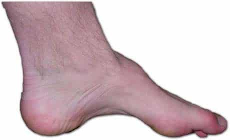
Surface Anatomy Of The Foot
Vastus lateralis muscle. In the study of human anatomy it is easy to become so pre occupied with internal structure that we forget the impor tance of what we can see and feel externally.
 Lower Limb Basicmedical Key
Biceps femoris muscle short head.
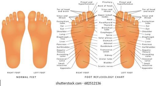
Surface anatomy of the foot. First metatarsalphalangeal joint 11. Broken bones in the foot usually call for rest ice compression and elevation to reduce any swelling. Surface anatomy of the foot for the clinician and operator an exact knowledge of surface anatomy is absolutely essential.
Proximal posterior surface of the tibia a peroneus brevis origin. Foot surface anatomy 1. The opposite side of the foot is called the plantar surface.
The toenail surface anatomy peer review orthopaedicsone peer review workflow is an innovative platform that allows the process of peer review to occur right within an orthopaedicsone article in an open transparent and flexible manner. Tuberos extensor digitorum brevis origin. You may not feel much unless the patient has a heel spur bony overgrowth.
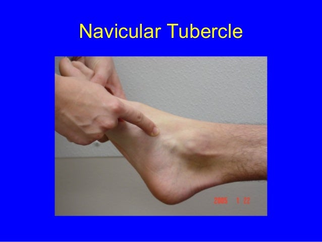
Dont know why you thought that dorsal would be down. Bony palpation medial aspect 3. The medial tubercle of the calcaneus can be palpated by grasping the heel and pressing your thumb into the flesh on the medial plantar surface of the heel.
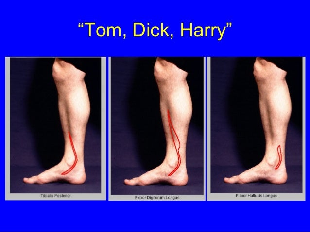
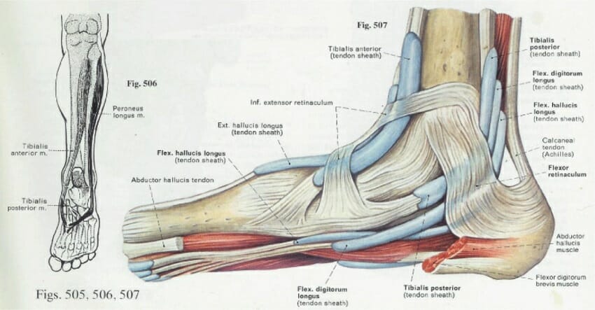
It can readily be acquired because the various bony points and tendons are usually evident both to touch and sight. Dorsal surface of the calcaneus. Extend the second through fifth toes dorsiflex the ankle evert the foot origin of tibialis posterior proximal posterior shafts of tibia and fibula.
A knowledge of the bodys surface landmarks is essential. The opposite side of the hand is the palmar surface. 2nd 4th toe semitendinosus muscle.
Yet external anatomy and appearance are major concerns in giving a physical examination and in many aspects of patient care. Jf the top of both the foot and the hand is the dorsal surface. The most common broken bones in the foot are broken toes which may occur after hitting a toe on a hard or sharp surface while walking running swimming or playing sports.
Distal two thirds of lateral fibula. Foot surface anatomy 2. The tuberosity of the fifth metatarsal is palpable on the lateral side of the foot.
 Duke Anatomy Lab 2 Pre Lab Exercise In 2019 Foot Anatomy
 Lower Limb Surface Anatomy Edinburgh University
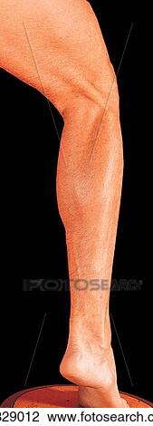 Lateral Aspect Of Leg And Foot A Surface Anatomy
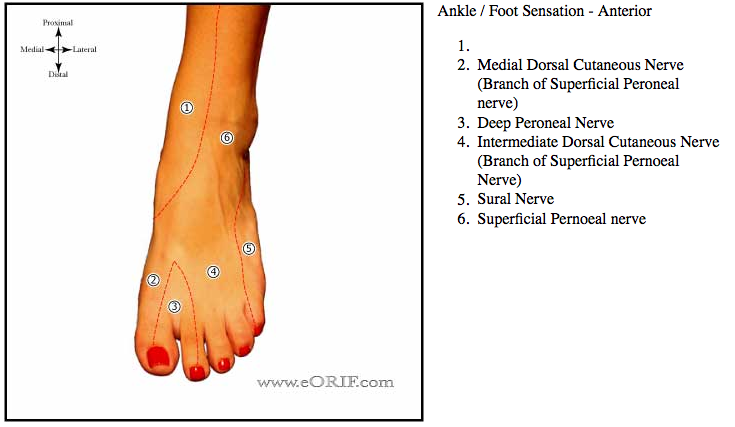
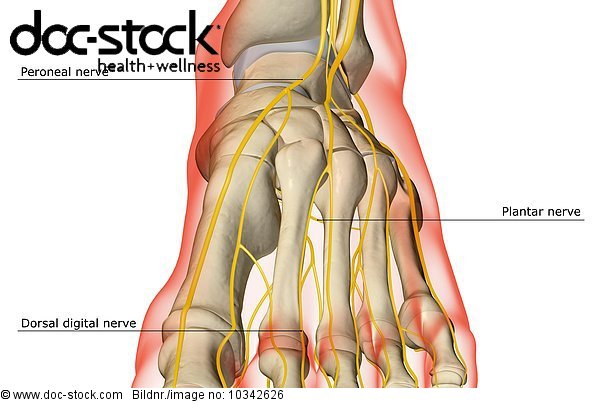 An Anterior View Of The Nerve Supply Of The Foot The
 Human Anatomy For The Artist The Dorsal Foot How Do I Love
 An Exploration Of The Surface Anatomy Of The Anterior Compartment Of The Leg
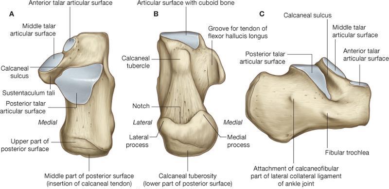 Calcaneus Anatomy And Attachments Bone And Spine
 Talus Bone Wikipedia
 Sole Foot Wikipedia
 Foot Surface Anatomy Bony Palpation Medial Aspect Ppt
 Anatomy Physiology Illustration
 Tib Fib Foot Wikiradiography
 Stock Photo And Image Portfolio By Lotan Shutterstock
 Anatomy Of The Plantar Foot Myfootshop Com
 Patient Education Concord Orthopaedics
 Foot And Ankle Andrea Colten Jessica Tyne Surface
 Pdf Fibularis Tertius Muscle In Women Men A Surface
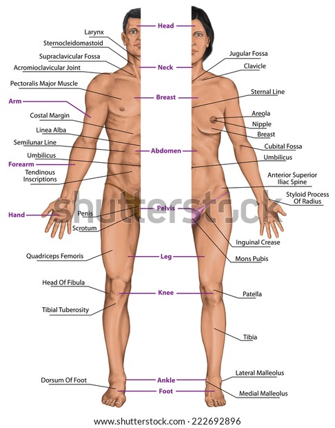 Male Female Anatomical Body Surface Anatomy Stock Image
 Foot Surface Anatomy
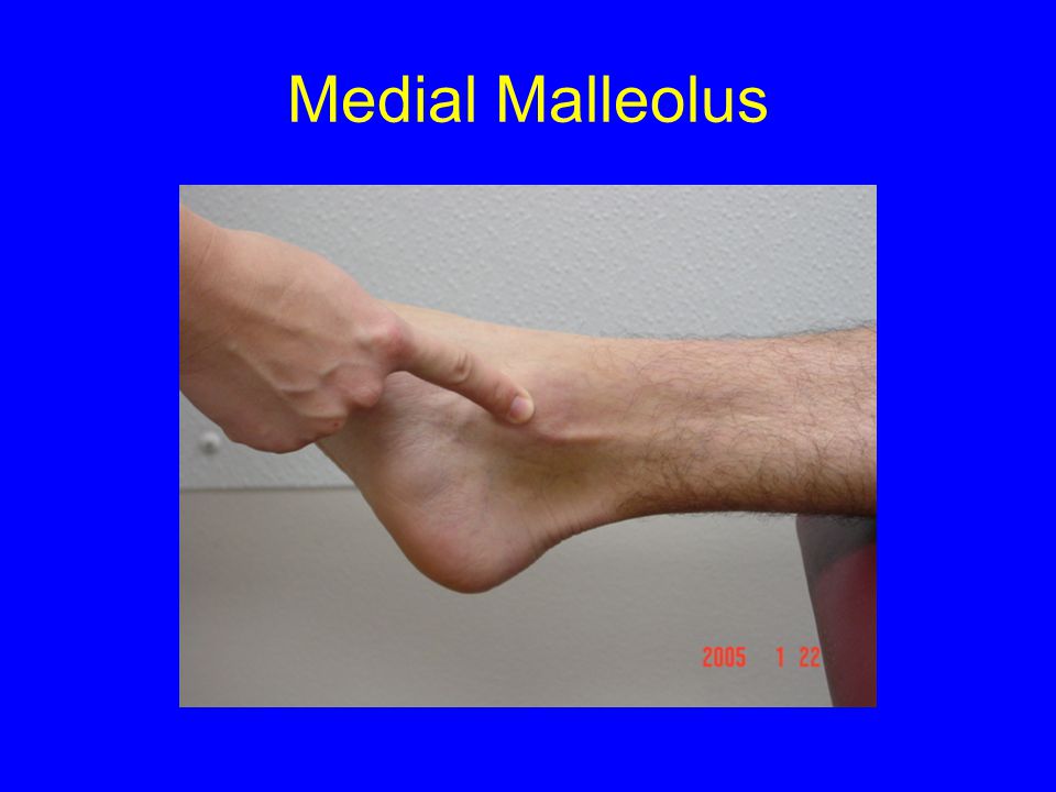
 11 Surface Anatomy Of The Medial Ankle Mm Medial Malleolus
 Ch 12 Diagram Surface Anatomy Anterior Leg Foot
 Ankle Joint Anatomy Overview Lateral Ligament Anatomy And
 Foot Anatomy Eorif
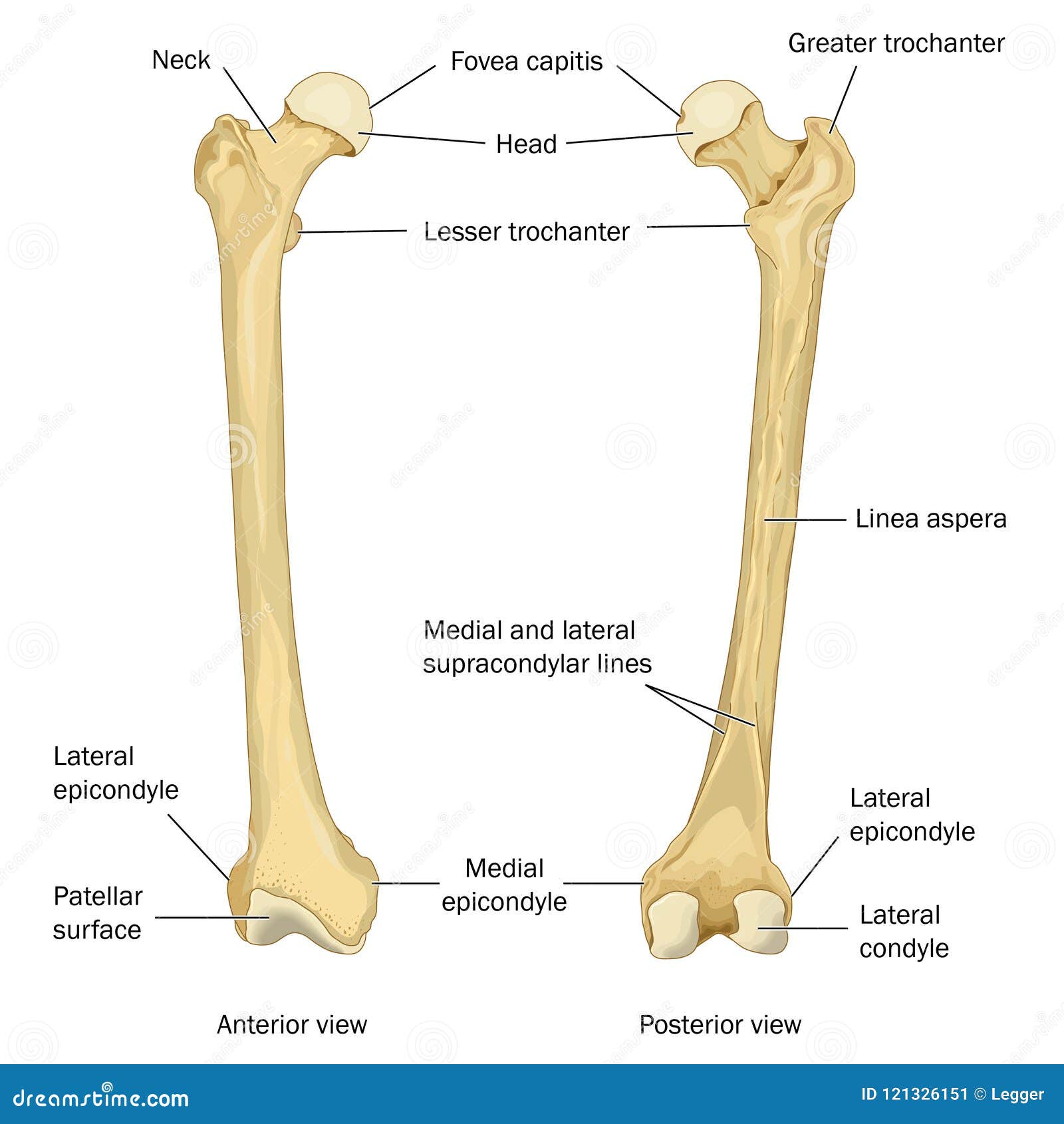 Surface Anatomy Of The Human Femur Stock Illustration
Anatomical Landmarks On The Dorsum Of The Left Foot Showing
 Female Body Front Surface Anatomy Human Body Shapes Anterior
 The Arches Of The Foot Longitudinal Transverse
 Foot Anatomy Bones Ligaments Muscles Tendons Arches
 Surface Anatomy Plantar Aspect Of The Forefoot
Foot Surface Anatomy
Surface Anatomy
 Surface Anatomy Atlas Of Anatomy

Belum ada Komentar untuk "Surface Anatomy Of The Foot"
Posting Komentar