Foot X Ray Anatomy
The human foot has 26 bones and 33 joints. This is a very complex structure due to the need to support your entire body weight.
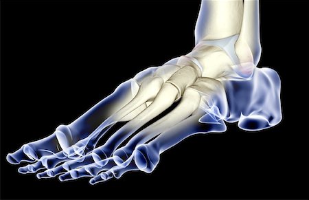
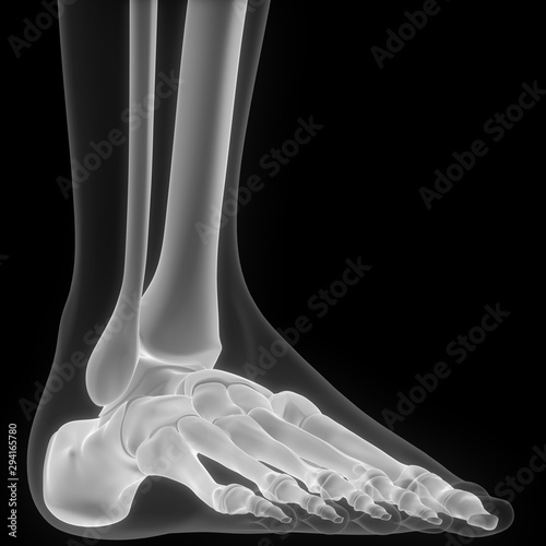 Human Skeleton Anatomy Foot X Ray 3d Rendering Stock Photo
We are pleased to provide you with the picture named foot x ray anatomy.

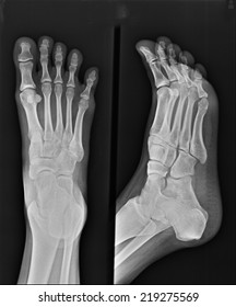
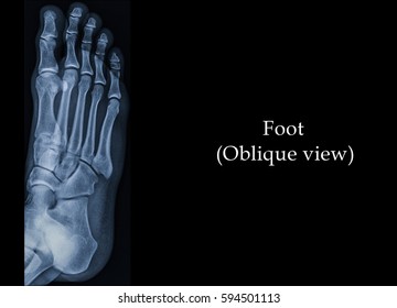
Foot x ray anatomy. Normal radiographic anatomy of the foot. Foot x ray anatomy in this image you will find distal phalanges interphalangeal joint proximal phalanges metatarso phalangeal joints sesamoid bones metatarsals intermediate phalanges in it. When checking any post traumatic foot x ray it is crucial to assess alignment of the bones at the joints.
It is extremely rare and extremely dangerous for the health of the foot if a full break occurs in one of the bones. Check you have the right views. A foot x ray is an x ray conducted to check these various bones of the foot.
Loss of joint alignment can represent severe injury even in the absence of a fracture. Radiographic anatomy foot dp radiographic anatomy radiology. Foot radiographs are commonly performed in emergency departments usually after sport related trauma and often with a clinical request that states lateral border pain.
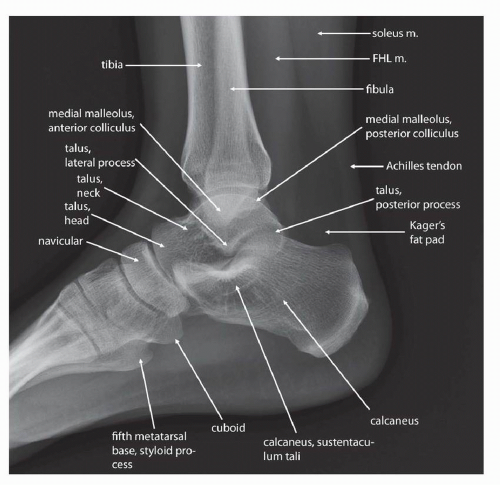
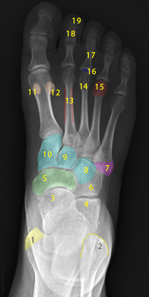
Both should ideally be done when weight bearing if your patient can manage it. 1 calcaneus 2 cuboid. Ankle anatomy sprain clinical anatomy fracture radiology x ray.
Often a foot x ray is also requested for the investigation of osteomyelitis arthritides or a bone lesion. Remember to check the whole film though. Normal radiographic anatomy of the foot.
1 fibula 2 cuboid 3 5th metatarsal bone 4 tibia 5 talus 6 navicular 7 cuneiform 8 1st metatarsal bone 9 proximal phalanx 10 distal phalanx. There are two views in foot x rays dp dorsal plantar and oblique. Detailed anatomy description of foot.
Plain film x ray principles interpretation teachmeanatomy. There are more than a hundred muscles tendons and ligaments. Normal foot and ankle x rays.
Foot fractures are usually in the form of chip injuries or crack injuries. This webpage presents the anatomical structures found on foot radiograph. Ankle is joint that is located between leg and foot a main contributor of stability sunday december 15 2019.
Normal radiographic anatomy of the foot.
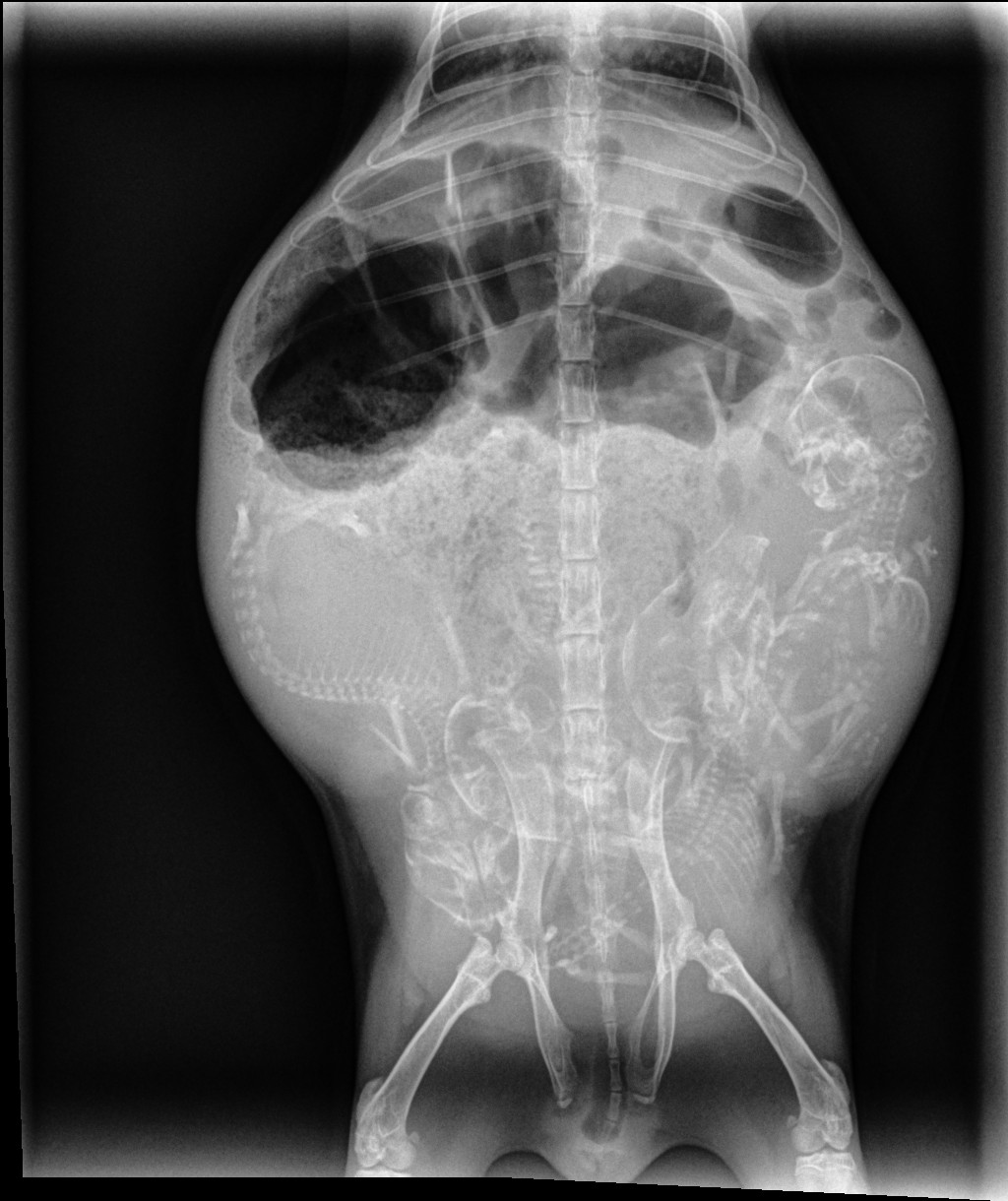 Learn To Read An X Ray Long Beach Animal Hospital
 X Ray Film Collection Of Middle Toe Foot Bone With Red
 E Anatomy Radiologic Anatomy Atlas Of The Human Body
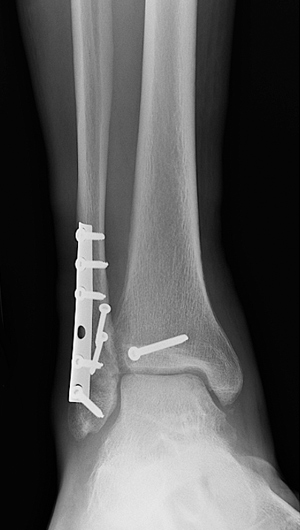 Broken Ankle Types Of Fractures Diagnosis Treatments
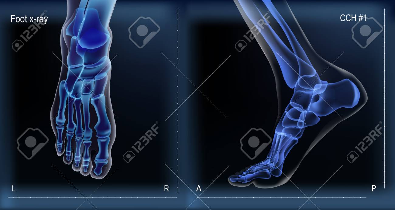 Dark Navy Blue Vector Realistic Medial And Top X Ray Of Skeleton
 Pediatric Ankle And Foot Injuries Sciencedirect
 Royalty Free Foot Xray Stock Images Photos Vectors
 X Ray Of Foot Ankle Wall Mural Anatomy Wallpaper Murals
 X Ray Of The Foot Fracture Of The 5th Metatarsal Bone
 Diagnostic Imaging Techniques Of The Foot And Ankle
 Anatomy Of The Bones Of The Foot The Bmj
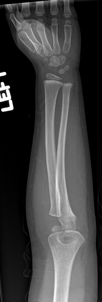
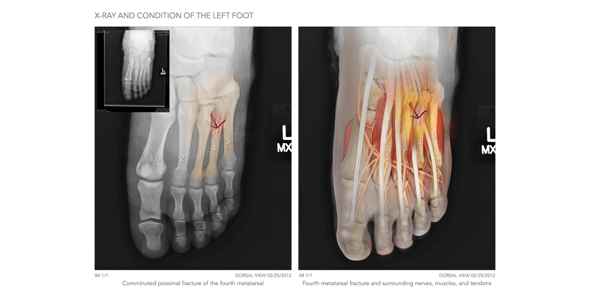 Left Foot X Ray High Impact Visual Litigation Strategies
 X Ray Of The Foot Anterior Posterior View Myfootshop Com
 Foot X Rays
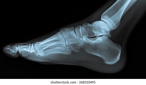
 Foot Ankle X Ray Lateral View
 Sesamoid Bone Wikipedia
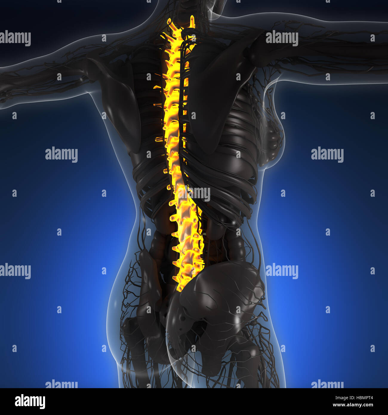 Science Anatomy Of Human Body In X Ray With Glow Back Bones
 X Ray Anatomy Of The Foot Toe
 X Ray Anatomy Foot Ankle Lower Leg Diagram Quizlet
 Lisfranc Injury Tarsometatarsal Fracture Dislocation
 Bone Anatomy Of Foot Frontal Xray Stock Photos Page 1
 Foot Anatomy Ankle X Ray
 Royalty Free Foot Xray Stock Images Photos Vectors
 Foot X Rays
 Royalty Free Foot Xray Stock Images Photos Vectors
 File X Ray Of Normal Right Foot By Oblique Projection Jpg
 Normal Radiographic Anatomy Of The Foot Radiology Case

Startradiology
Belum ada Komentar untuk "Foot X Ray Anatomy"
Posting Komentar