Femoral Region Anatomy
Subacute torsioned femoral hernia it occurs more commonly in the femoral region it is more frequent in women and it usually involves a segment of distal ileum. The anatomical regions shown compartmentalize the human body.
 Femoral Neck Fracture Background Epidemiology Functional
The two sections that meet this requirement are.

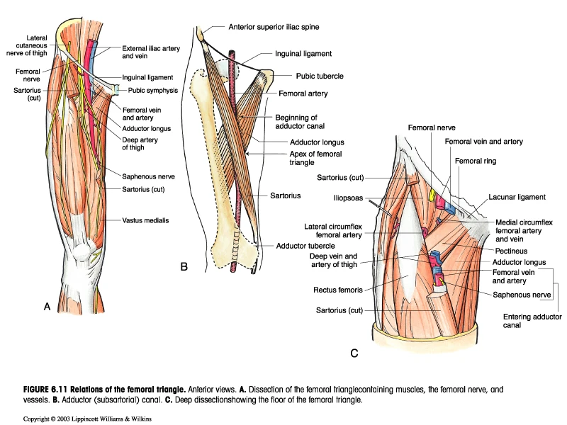
Femoral region anatomy. The anatomical regions of the body. Some regions are combined into larger regions. The adductor canal figure 61 a situated in the lower third of the thigh is delimited anteriorly by the vastus medialis muscle m.
The femoral triangle serves as a crucial anatomical landmark for surgeons when surgery needs to be performed in the region. Just like on a map a region refers to a certain area. Vastus medialis posteriorly by the tendon of the adductor magnus muscle m.
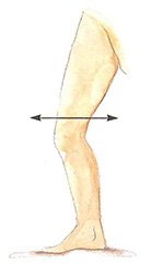
From the abdomen the femoral nerve next passes deep to the inguinal ligament in the groin and crosses the hip joint to enter the femoral region. Embalmers use this artery to supply chemicals to the body to preserve it. The femoral region encompassing the thigh the popliteal region encompassing the back of the knee the sural region encompassing the back of the lower leg the calcaneal region encompassing the heel the plantar region encompassing the sole of the foot.
The axial body runs right down the center axis and consists of everything except the limbs meaning the head neck. Adductor magnus and laterally by the aponeurosis of the adductor canal. A 135 x 90 x 75 mm sized highly intense cystic mass localized in the right inguinal region extending to the femoral region was detected in the ultrasonographic examination.
It is a subfascial space which appears as a triangular depression below the inguinal ligament when the thigh is flexed abducted and laterally rotated. The body is divided into two major portions. Femoral region regio femoris anatomical parts.
In the femoral region the femoral nerve separates into two nerve trunks the anterior and posterior divisions before further dividing into many smaller branches throughout the anterior and medial. Femoral region anatomy boundaries of the gluteal region iliac crest gluteal sulcus fold greater trochanter natal intergluteal cleft. You are told to cut a dissection animal along two planes so that the lungs are observable in both sections.
Illustrated anatomical parts with images from e anatomy and descriptions of anatomical structures. The femoral triangle or scarpas triangle is an anatomical region of the upper third of the thigh.
 Overview Of The Lower Limb Anatomy An Essential Textbook

 Femoral Region Phlebologia
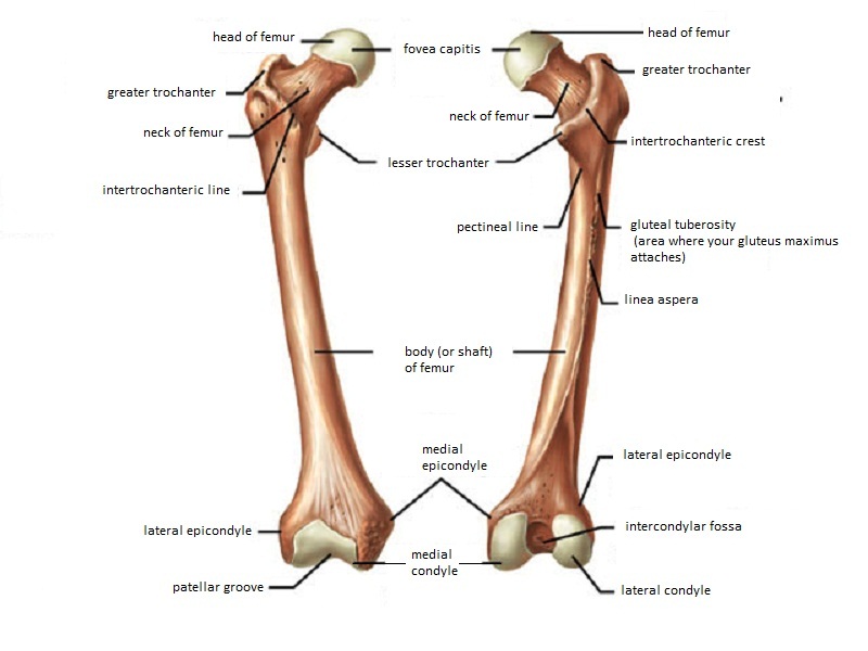
 Femur Wikipedia
 Femoral Triangle And Venous Drainage In The Lower Limg
 Figure 2 From The Surgical Anatomy Of The Lateral Femoral

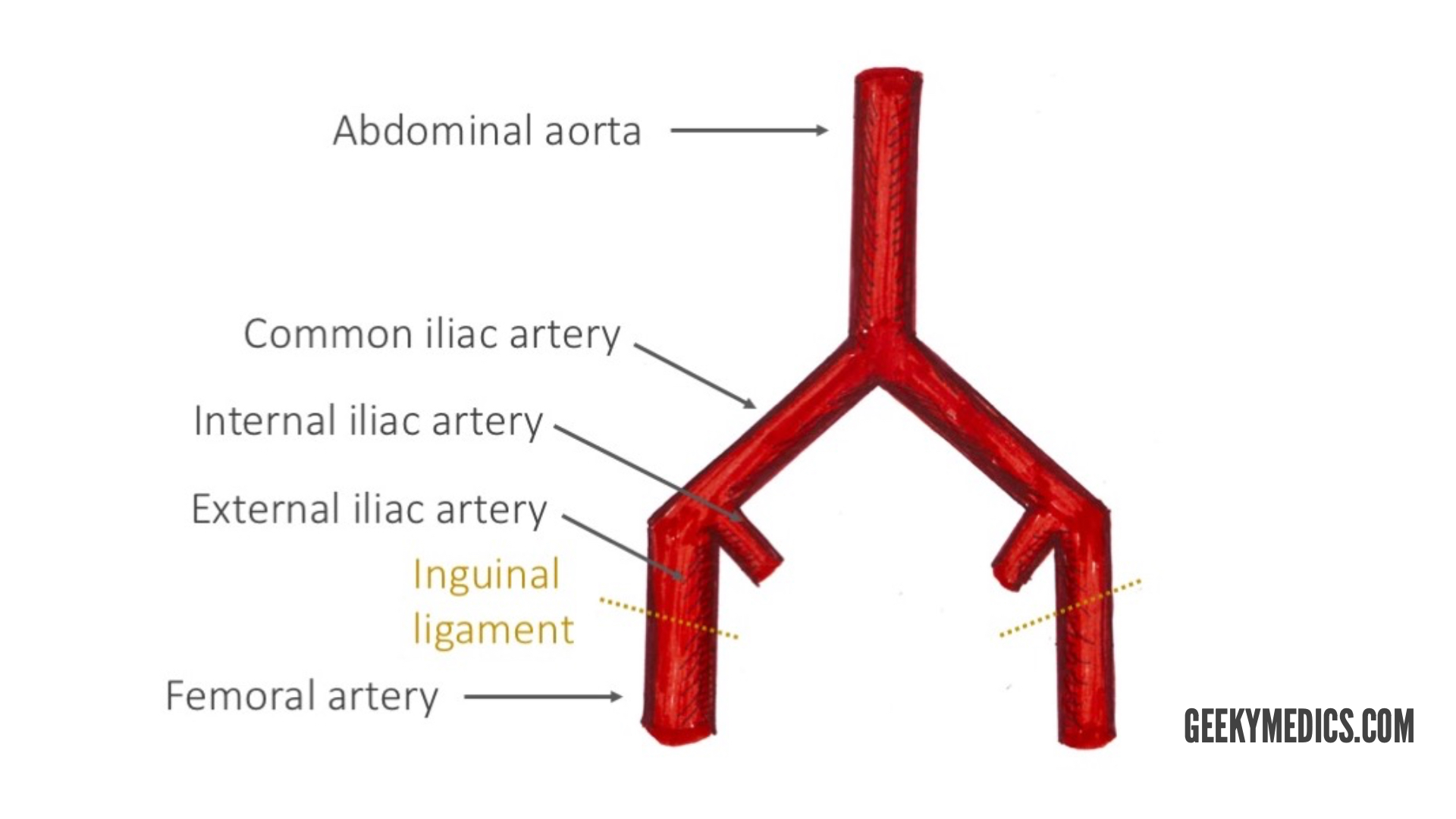
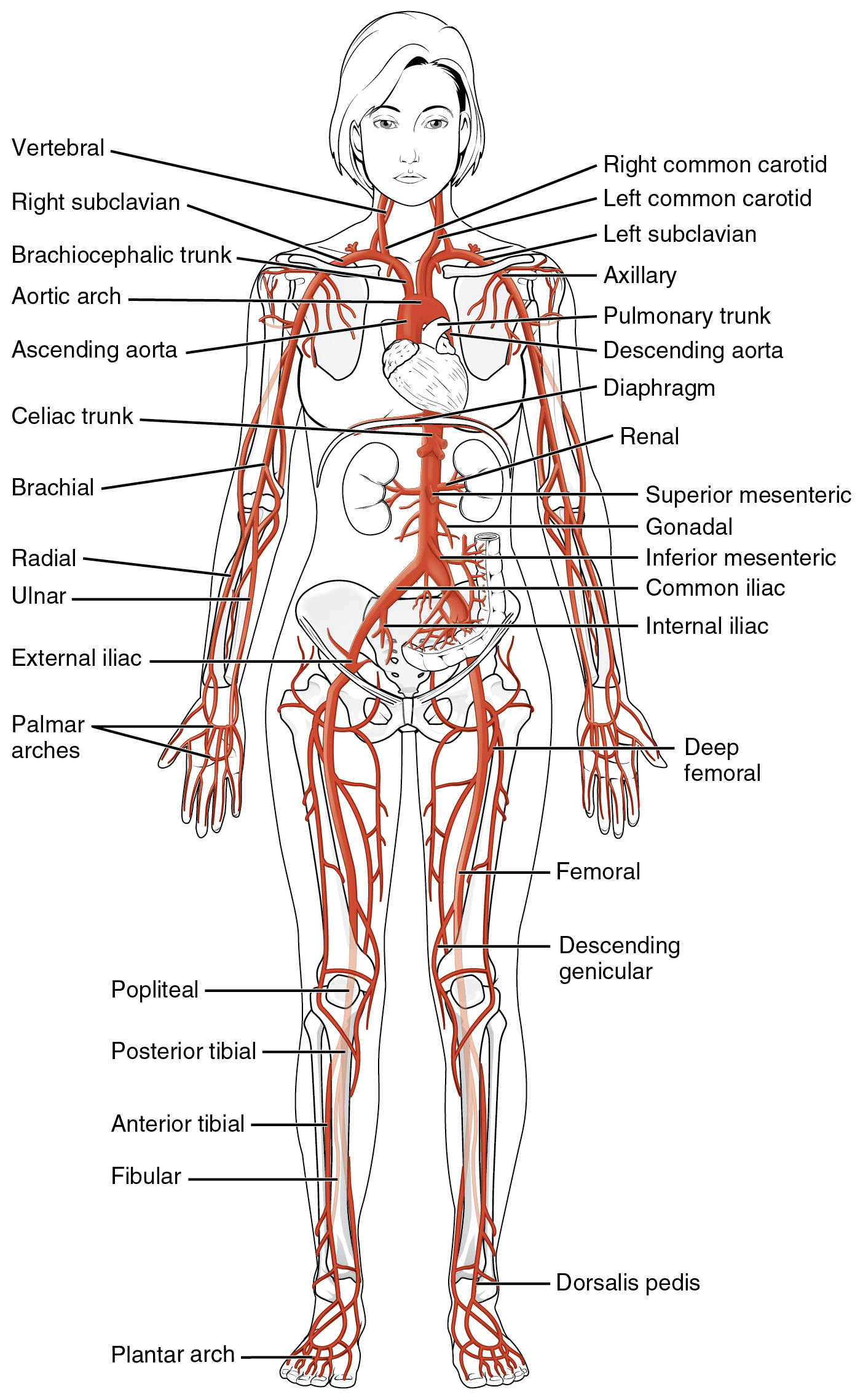 20 5 Circulatory Pathways Anatomy And Physiology
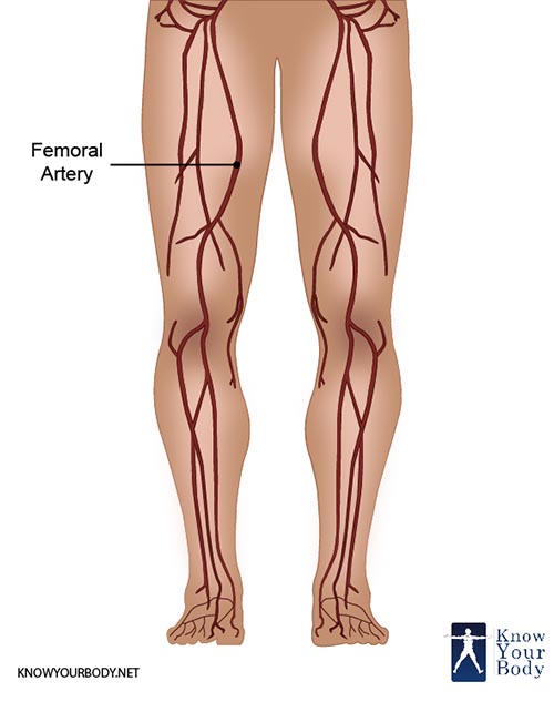 Femoral Artery Location Anatomy Branches Function And Faqs
 Femoral Nerve Block Landmarks And Nerve Stimulator
 Language Of Anatomy Practice
 Lower Limb Clinical Gate
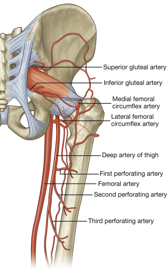
 Arterial Supply Of The Thigh And Gluteal Region Geeky Medics
 Anatomy Of The Femoral Region Springerlink
 Regional Anatomy Ii Proximal Femur At Texas Woman S
 Hip Joint Anatomy Bone And Spine
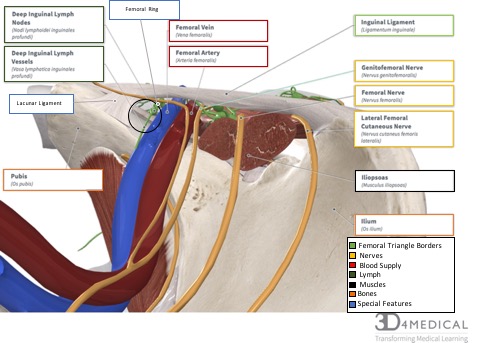
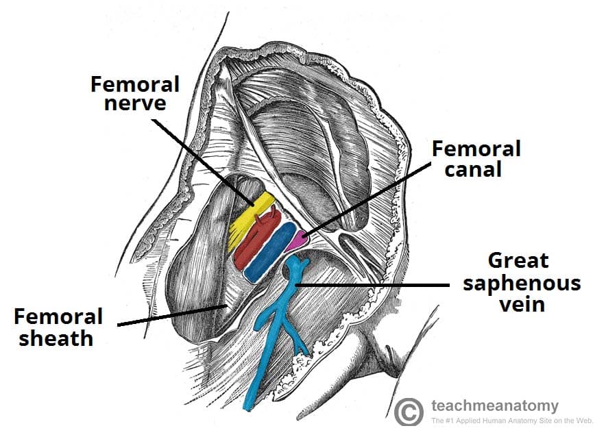 The Femoral Triangle Borders Contents Teachmeanatomy

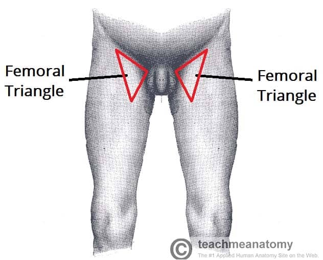 The Femoral Triangle Borders Contents Teachmeanatomy
 Leg Knee Anatomy
 The Femur Proximal Distal Shaft Teachmeanatomy
 Ucsd S Practical Guide To Clinical Medicine
 Special Anatomical Regions Advanced Anatomy 2nd Ed
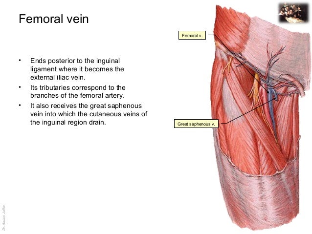
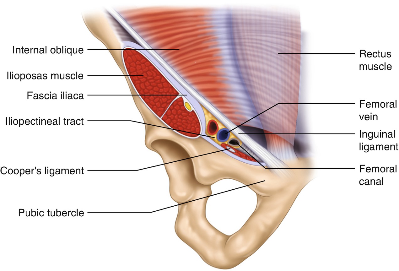
Femoral Vein Wikipedia
 Regions Anterior Femoral Triangle Boundary Contents
 Atlas Of Human Anatomy 1st Edition
 Femoral Region Gastrointestinal Medbullets Step 1
Belum ada Komentar untuk "Femoral Region Anatomy"
Posting Komentar