Anatomy Of The Dermis
The dermis contains the following. The papillary layer the upper layer of the dermis.
 Anatomy Chapter 5 Anatomy Physiology 2100c With Nguyen
The fin rays of fishes are dermal derivatives as are many types of pigment cells.

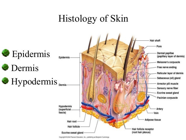
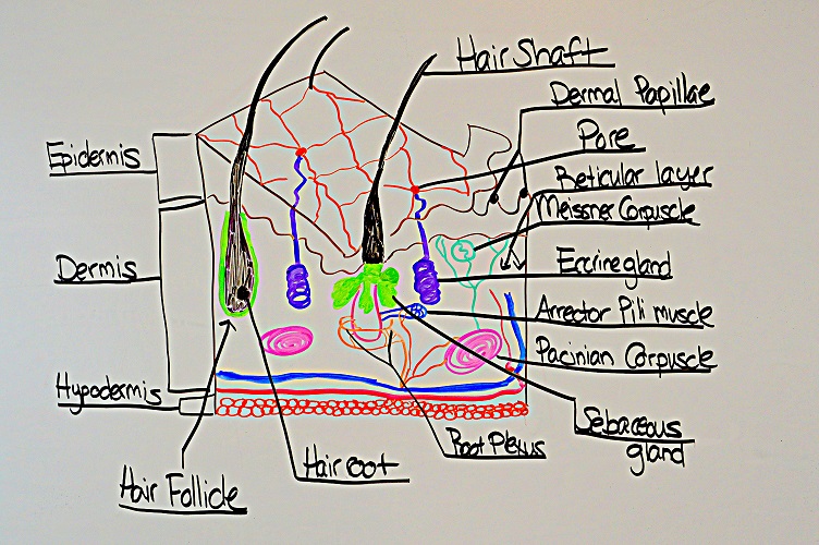
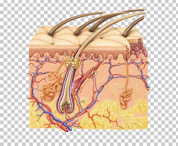
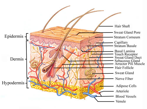
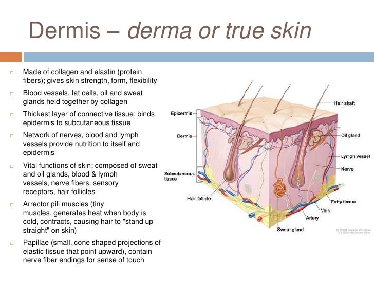
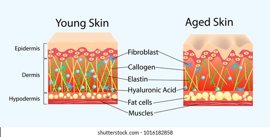
Anatomy of the dermis. The dermis beneath the epidermis contains tough connective tissue hair follicles and sweat glands. It contains connective tissue blood capillaries oil and sweat glands nerve endings and hair follicles. Interwoven within these layers are numerous elastin and collagenous fibers produced by fibroblasts figure 56.
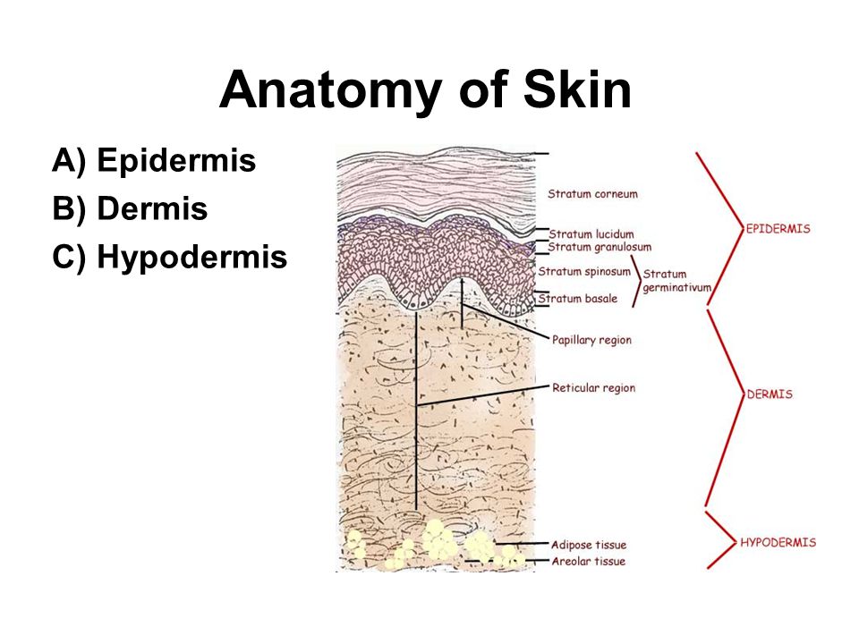
The dermis from its earliest evolutionary appearance has been a depository of bone as expressed in dermal armour primitive fishes scales fishes and certain amphibians and plates crocodile lizard turtle armadillo. The dermis is mostly composed of dense irregular connective tissue that is divided to two layers. The papillary layer and reticular layer.
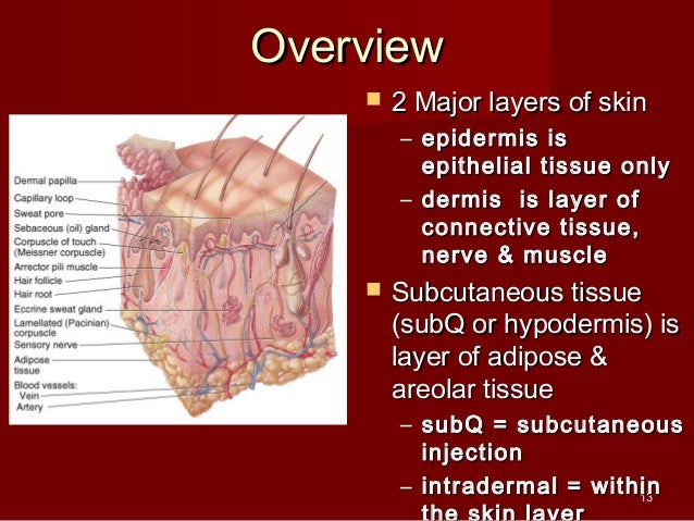
The second layer of the skin the dermis consists of various connective tissues. The structure provides strength extensibility the ability to be stretched and elasticity. The deeper subcutaneous tissue hypodermis is made of fat and connective tissue.
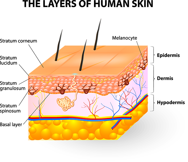
The papillary dermis which lies superficially the recticular dermis which lies deeper. Broadly the dermis can be divided into two layers. The dense inner layer of skin beneath the epidermis composed of connective tissue blood and lymph vessels sweat glands hair follicles and an elaborate sensory nerve network.
As connective tissue it contains fibroblasts and macrophages within a gelatinous matrix containing collagen elastic and reticular fibers. The dermis has two parts. Its located between the epidermis and the subcutaneous tissue.
The dermis is the middle layer of the three layers of skin. A thin upper layer known as the papillary dermis. The dermis is the thickest layer of skin and arguably the most.
Anatomy and function of the dermis anatomy and structure. It is not strictly a part of the skin although the border between the hypodermis and dermis can be difficult to distinguish. The hypodermis also called the subcutaneous layer or superficial fascia is a layer directly below the dermis and serves to connect the skin to the underlying fascia fibrous tissue of the bones and muscles.
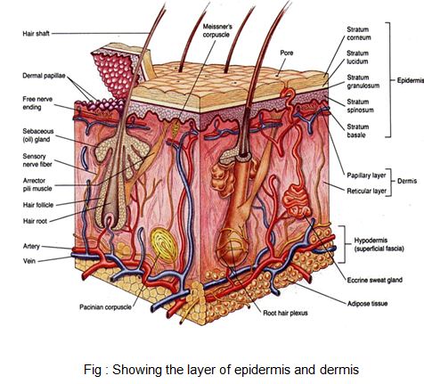
 Figure Anatomy Of The Skin Showing The Epidermis Dermis
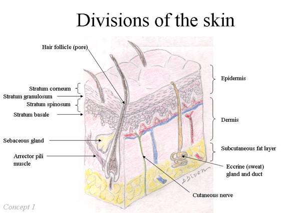
 Skin Anatomy
 Skin Integumentary System Review For Anatomy Physiology
 Integumentary System Facts Now Let S Talk About The Dermis
 Anatomy Quiz Dermis And Co Diagram Quizlet
 Skin Anatomy And Wound Healing Dermatology Medbullets Step 1
:max_bytes(150000):strip_icc()/skin-anatomy-1068880_review-01-9adf9daebac8464eb693274a960bd850.png) Skin Anatomy The Layers Of Skin And Their Functions
Skin Anatomy The Layers Of Skin And Their Functions

 Anatomy Of The Skin
 Anatomy Gross Anatomy Physiology Cells Cytology Cell
 Hair And Nails Anatomy And Physiology I
 Human Skin Anatomy Image Photo Free Trial Bigstock
 Subcutaneous Tissue Human Skin Integumentary System Dermis
 Figure Anatomy Of The Skin Showing Pdq Cancer
 Skin Anatomy Chart Dermis
 Intro To Anatomy 7 The Integumentary System Freethought Forum
 Mblex Review Integumentary System Anatomy And Physiology
 Human Body Skin Anatomy Vector Illustration With Parts Vein Artery
 5 1 Layers Of The Skin Anatomy And Physiology
 Dermis Anatomy Britannica
 Wound Healing Anatomy Of Skin A Epidermis B Dermis C
 Structure Of The Skin Course Hero
 Anatomy Of The Skin Lecture
 Dermis Definition Anatomy And Function
 Skin Physiology Griffin Row
 The Skin Ross And Wilson Anatomy And Physiology In Health
 Week 2 Skin Anatomy And Function 15171 Sp Uw
 Anatomy Of Your Skin Dermatology Associates Savannah Ga
 Anatomy Lab 2 Integumentary Sys
Dermis Wikipedia
 Anatomy Of Epidermis And Dermis Images Stock Photos
 Skin Structure Epidermis Dermis Subcutis Subcutaneous
Belum ada Komentar untuk "Anatomy Of The Dermis"
Posting Komentar