Thoracic Aorta Anatomy
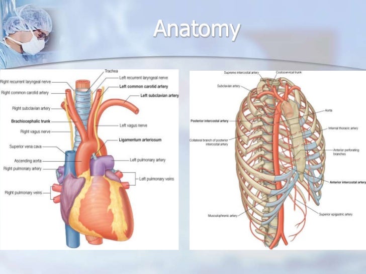
The thoracic aorta is the portion of the descending aorta contained within the posterior mediastinal cavity that is continuous with the aortic arch. In this tutorial we will explore the anatomy of the thoracic aorta and tell you about its arterial branches.
The aortic arch curves over the heart giving rise to branches that bring blood to the head neck.
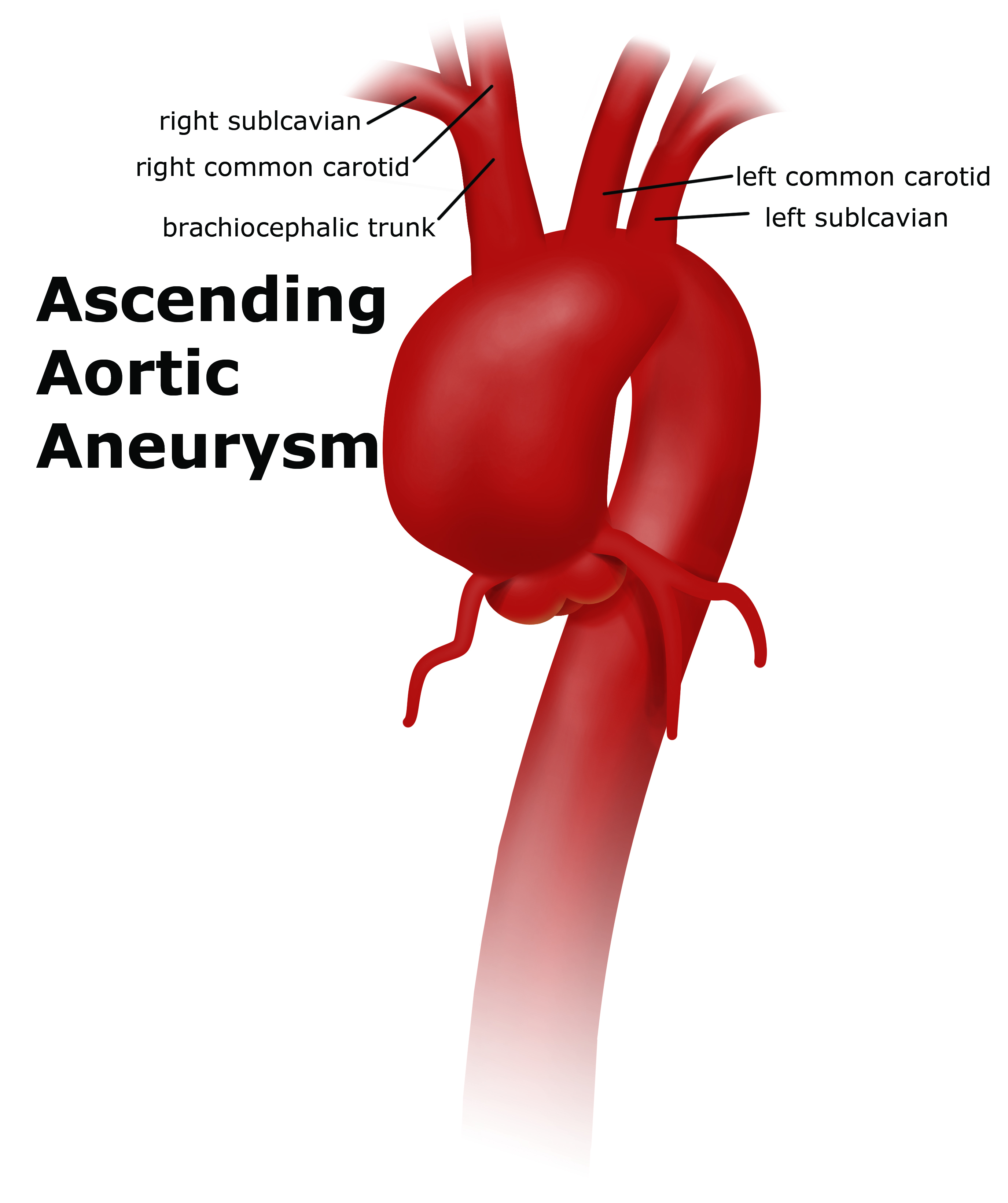
Thoracic aorta anatomy. Symptoms including diminished or absent pulses in the arms are related to narrowing and obstruction of these vessels. The branches of the thoracic aorta run to various organs esophagus lungs adrenal. The ascending aorta begins at the sinotubular junction.
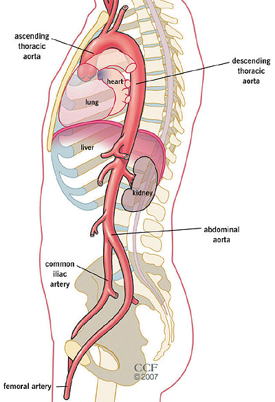
It terminates at the level of l4 by bifurcating into the left and right common iliac arteries. Takayasu arteritis is most common in young asian women. The descending thoracic aorta travels down through the chest.
The diagnosis and extent of vascular. Aortic root ascending aorta proximal aortic arch distal aortic arch and descending thoracic aorta. At the origination point it is on the left side of the vertebrae.
The aortic arch is the portion of the aorta that is in the shape. As it descends it winds around the vertebrae and ends in front. The aorta involves principally the thoracic aorta chest portion and the adjacent segments of its large branches.
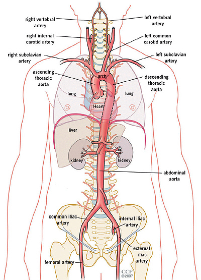
The descending thoracic aorta is a part of the aorta located in the thorax. The thoracic aorta is divided into five segments. The descending thoracic aorta begins at the lower border of the fourth thoracic vertebra where it is continuous with the aortic arch.
Picture of the aorta the ascending aorta rises up from the heart and is about 2 inches long. The diameter of the artery is 232 centimeters. The aorta classified as a large elastic artery and more information on its internal structure can be found here.
The aortic root is the portion of the aorta that is attached to the heart. The ascending aorta the aortic arch the thoracic descending aorta and the abdominal aorta. The abdominal aorta begins at.
At its origin this portion of the aorta lies just to the left of the vertebral column and drifts medially as it continues to move downward through the chest until it crosses the diaphragm. Anatomy of the aorta aortic root. This proximal descending portion of aorta gives rise to the visceral and the parietal branches above the aortic hiatus at the diaphragm.
Normal anatomy of the aorta. It is a continuation of the descending aorta and contained in the posterior mediastinal cavity. Descending aorta thoracic the descending aorta thoracic aorta is between the arch of the aorta and the diaphragm muscle below the ribs.
The aorta can be divided into four sections. An abnormal balloon or sac like dilatation in the wall of the thoracic aorta.
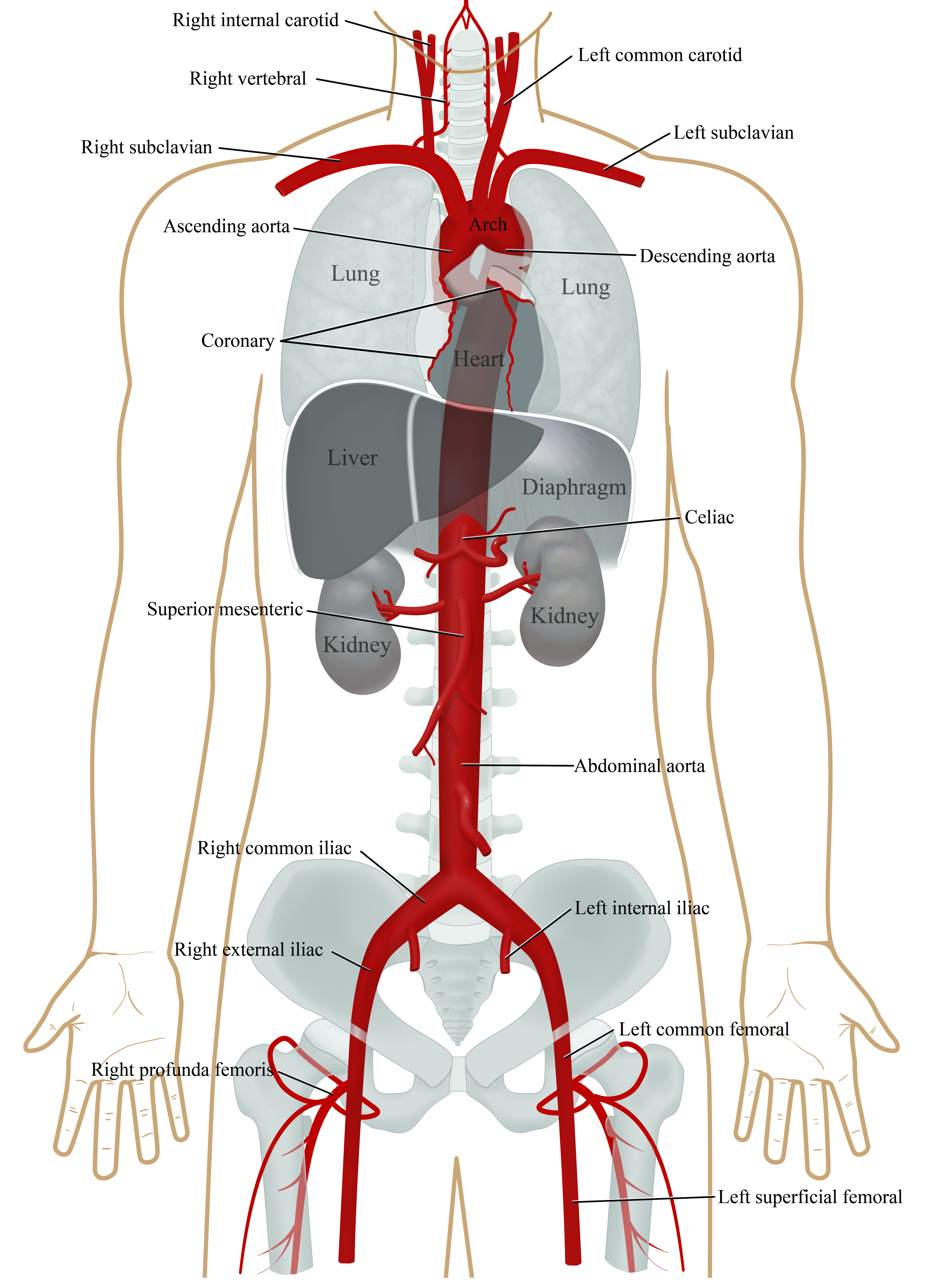 Aorta Anatomy Uf Health Aortic Disease Center Overview
 A Magnetic Resonance Angiography Mra Image Of A Moderate
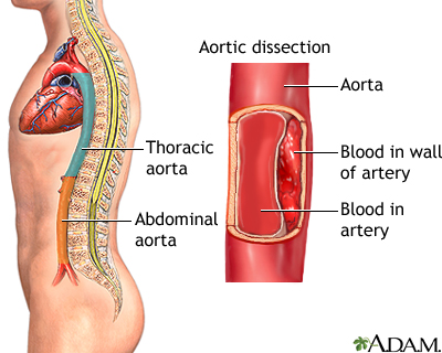 Aortic Dissection Medlineplus Medical Encyclopedia
 Thoracic Aorta Radiology Reference Article Radiopaedia Org
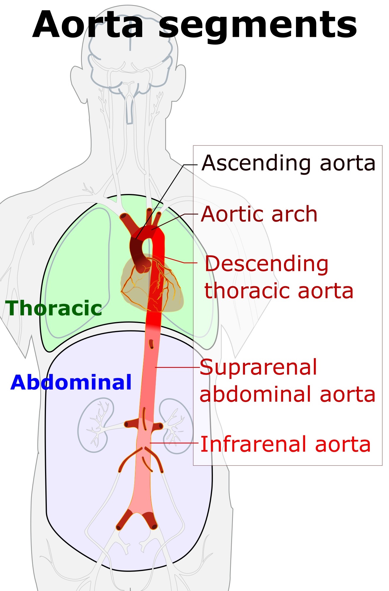 Descending Thoracic Aorta Wikipedia
 Thoracic Aorta An Overview Sciencedirect Topics
 Thoracic Aortic Aneurysm Uf Health Aortic Disease Center
:watermark(/images/logo_url.png,-10,-10,0):format(jpeg)/images/anatomy_term/aorta-thoracica-2/O7k9d5b146USmjo8yfTAoA_Aorta_pars_descendens_01.png) Aorta Anatomy Branches Course Divisions Kenhub
Aorta Anatomy Branches Course Divisions Kenhub
 Thoracic Aortic Aneurysm Society For Vascular Surgery
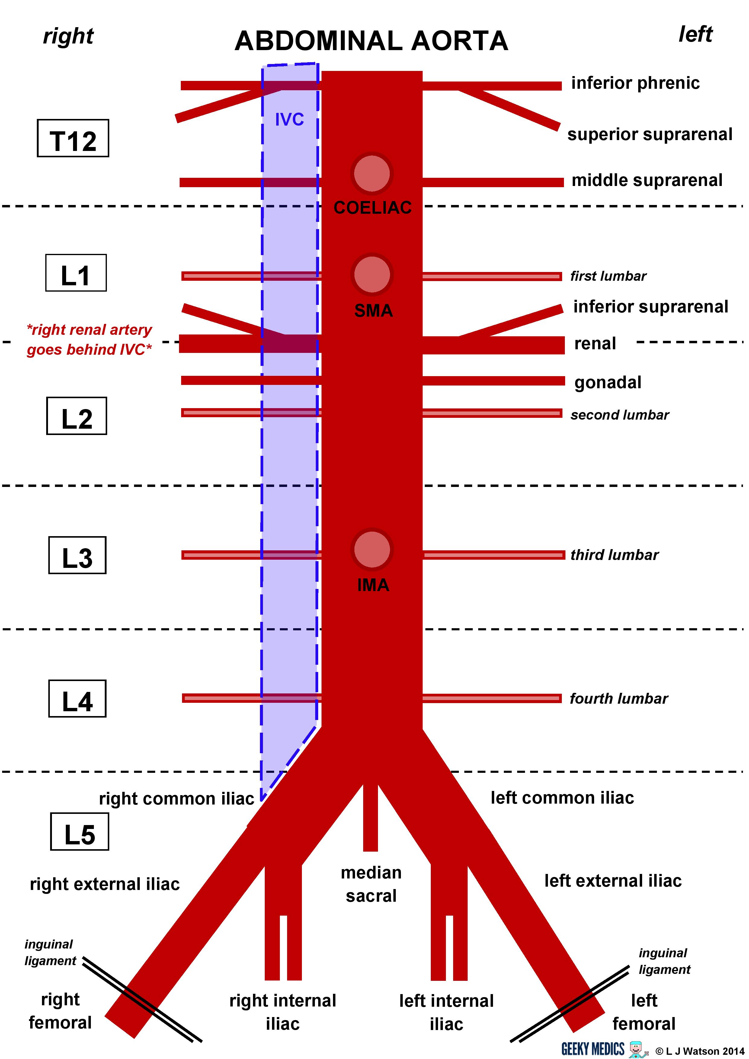 Abdominal Aorta Anatomy Geeky Medics
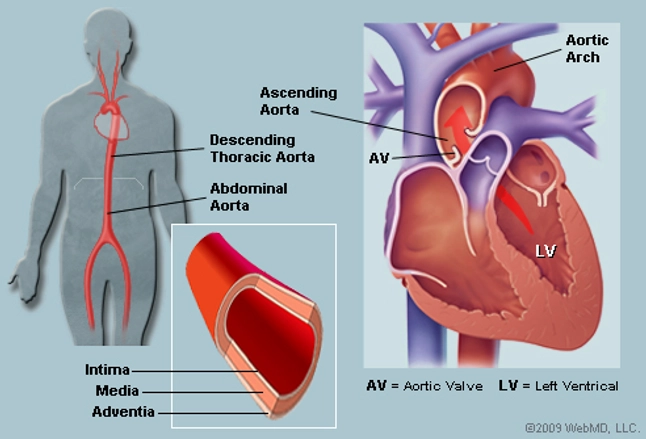 The Aorta Human Anatomy Picture Function Location And
 Aorta And Its Branches Anatomy
 1 Thoracic Aortic Anatomy C Massachusetts General Hospital
Thoracic Aortic Aneurysm Repair
 Racgp Aortic Aneurysms Screening Surveillance And Referral
 Aortic Anatomy Massachusetts General Hospital Boston Ma
 Thoracic Aortic Aneurysm Wikipedia
 Aortic Replacement In Cardiac Surgery Cleveland Clinic
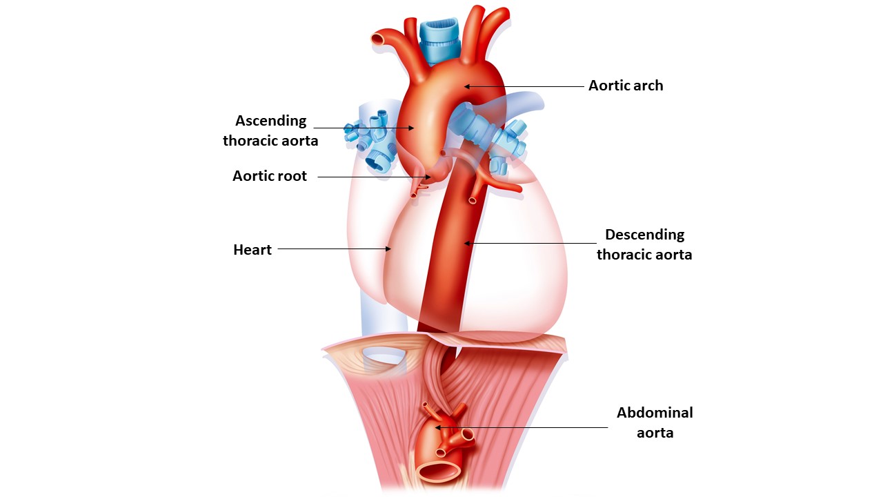 The Aorta Taad Genetic Aortic Disorders Association Canada
 Thoracic Aortic Endografts Massachusetts General Hospital
 Thoracic Aortic Aneurysm Medecine
 Thoracic Aorta An Overview Sciencedirect Topics



Belum ada Komentar untuk "Thoracic Aorta Anatomy"
Posting Komentar