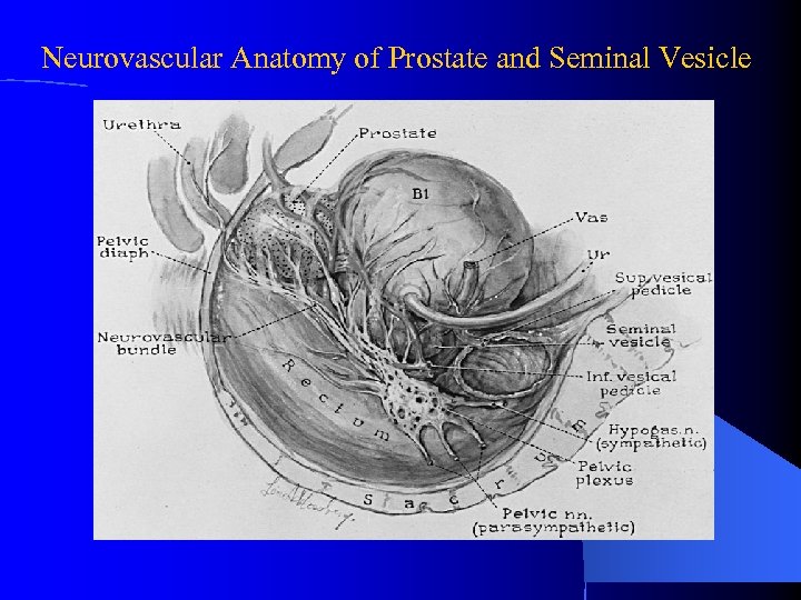
Seminal Vesicle Anatomy
The seminal vesicles latin. Glandulae vesiculosae vesicular glands or seminal glands are a pair of simple tubular glands posteroinferior to the urinary bladder of some male mammals.

The size and activity of the seminal vesicles are controlled by hormones.
Seminal vesicle anatomy. The vesicles are blind pouches and are rounded on their most superior aspects and taper to their inferior aspects where they constrict to ultimately form short ducts. Seminal vesicles are located within the pelvis. In the absence of this hormone the seminal vesicles will degenerate atrophy.
The prostate produces a thin milky white fluid containing calcium citrate and phosphate ions. Unlike what their name suggests. An individual seminal vesicle consists of a single coiled tube off of which several pouches branch.
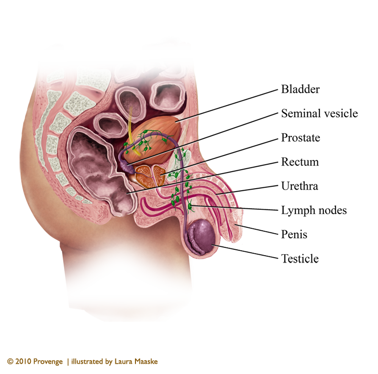
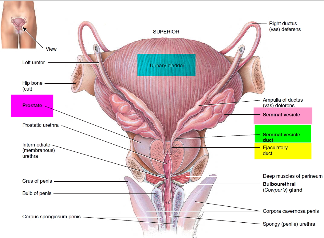
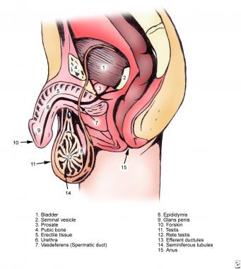
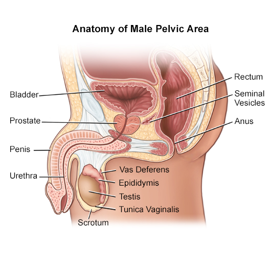
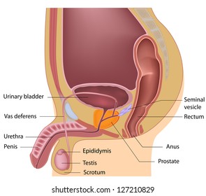
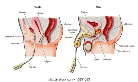
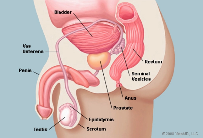
The seminal vesicles are bilateral lobulated glands see the following image. They are a pair of contorted or twisted tubes which are located between the bladder and the rectum. The seminal vesicles are accessory glands of the male reproductive system.
Production of androgen the major hormone that influences the growth and activity of the seminal vesicles begins at puberty and starts to decline at about the age of 30. The prostate and seminal vesicles contribute the following constituents to the semen. At puberty the seminal vesicles form sacs and contribute up to 85 of the seminal fluid.
The seminal vesicles contribute an alkaline viscous yellowish fluid. The seminal vesicles are located below the bladder and above the prostate gland. They are soft and approximately 5 7 cm long.
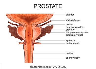
Coronal Diagram Posterior View Illustrates The Anatomy Of
 Royalty Free Seminal Vesicles Stock Images Photos Vectors
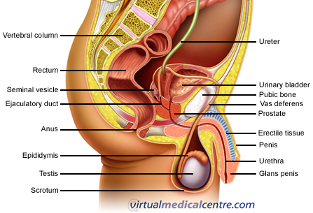 Male Reproductive System Urogenital System Anatomy
 Royalty Free Seminal Vesicles Stock Images Photos Vectors
 Ejaculatory Duct Wikipedia
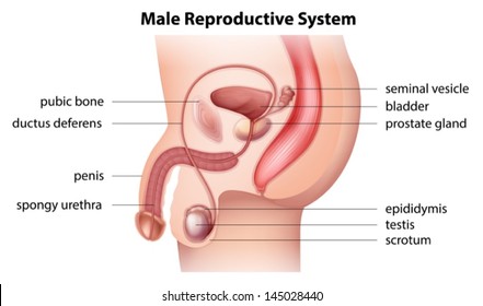
 Structure Of The Male Reproductive System Men S Health
 Prostate Picture Image On Medicinenet Com
 Prostate And Seminal Vesicle Normal Development Function
 Seminal Vesicle Wikipedia
 Seminal Vesicle Anatomy Location Function Inflammation
 The Seminal Vesicles Structure Function Lymphatics
 Prostate Functions Diseases And Tests


 Vas Deferens And Ejaculatory Duct Anatomy
 Prostate And Seminal Vesicle Ultrasonography And Biopsy
 Royalty Free Seminal Vesicles Stock Images Photos Vectors
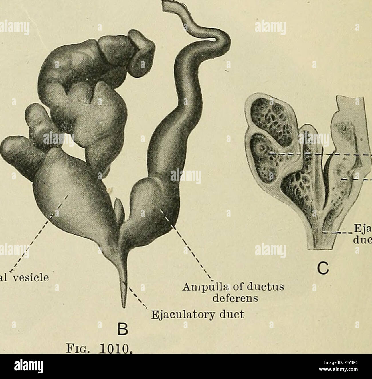 Cunningham S Text Book Of Anatomy Anatomy Seminal Vesicle
 Seminal Vesicle น กเร ยนพยาบาล
 Royalty Free Seminal Vesicles Stock Images Photos Vectors

 35 The Anatomy Histology And Development Of The Seminal

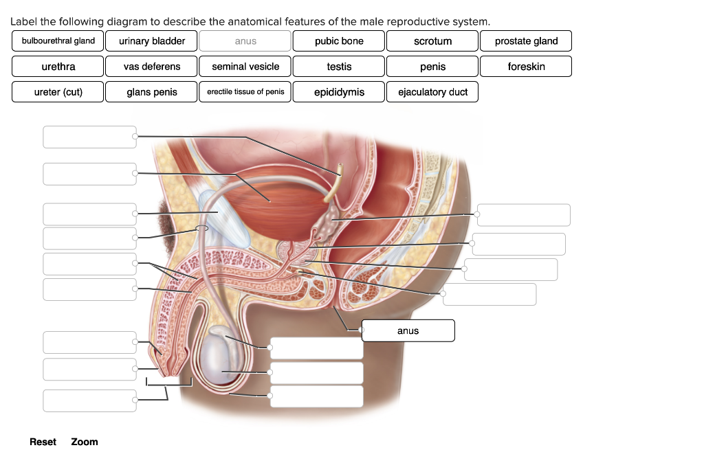 Solved Label The Following Diagram To Describe The Anatom
 Prostate Gland Human Anatomy Prostate Picture Definition
Belum ada Komentar untuk "Seminal Vesicle Anatomy"
Posting Komentar