Female Cervical Anatomy
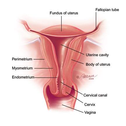
The canal is formed by and often almost occluded by mucosal folds. The lumen of the cervix is the cervical canal.
The cervical canal opens cranially into the body of the uterus at the internal uterine ostium.
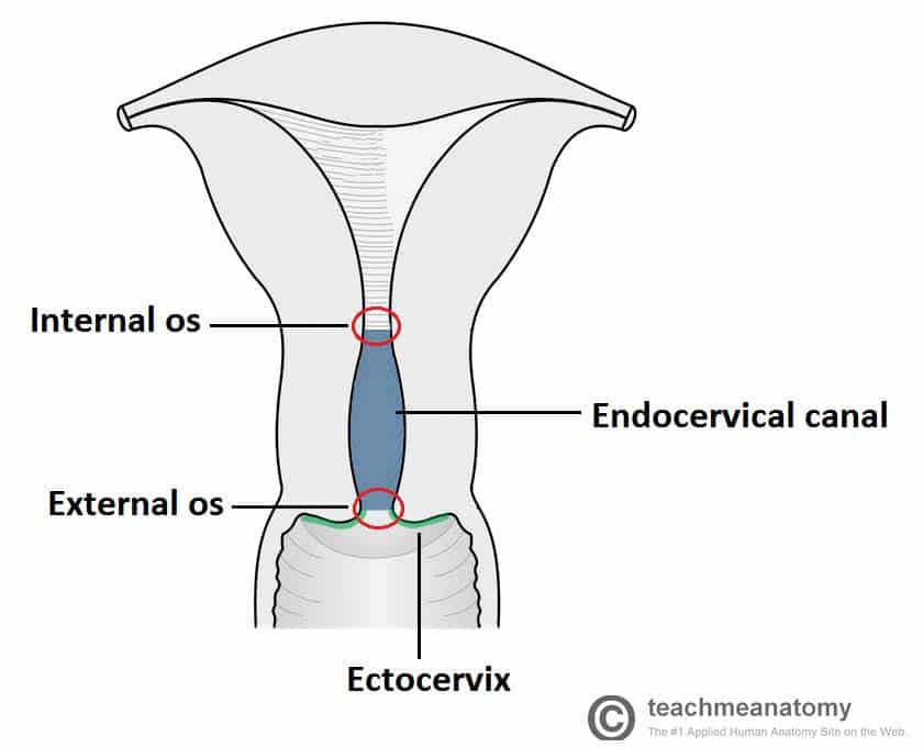
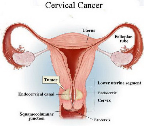
Female cervical anatomy. This can prevent early opening of the cervix during pregnancy which can cause premature delivery. During childbirth the cervix thins out. There are two main classifications of cervical cancer.
Cervical spine and intervertebral disc anatomy female version medical animation this 3d medical animation depicts the anatomy of the cervical spine and a typical intervertebral disc from c3. Implication for scaling injury criteria. The cervical canal is the hollow orifice through the cervix that connects the uterine cavity to the hollow lumen of the vagina.
When the woman isnt ovulating the cervical mucus thickens and serves as a barrier to keep sperm out of the uterus. The cervical spine has 7 stacked bones called vertebrae labeled c1 through c7. As viewed from the side the cervical spine forms a lordotic curve by gently curving toward the front of the body and then back.
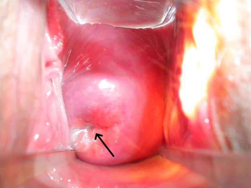
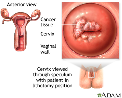
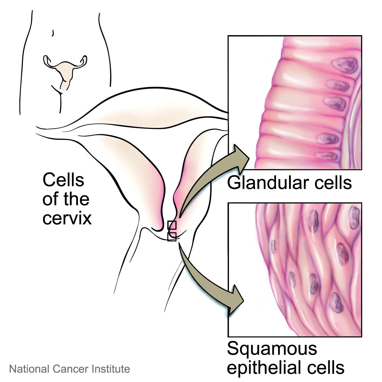
Cancer of the cervix is the most common cancer affecting the female reproductive tract. Single fold and smooth surface in the queen and bitch multiple folds protruding into the cervical canal in the cow ewe sow and mare. Squamous cell carcinoma cancer of the epithelial lining of the ectocervix.
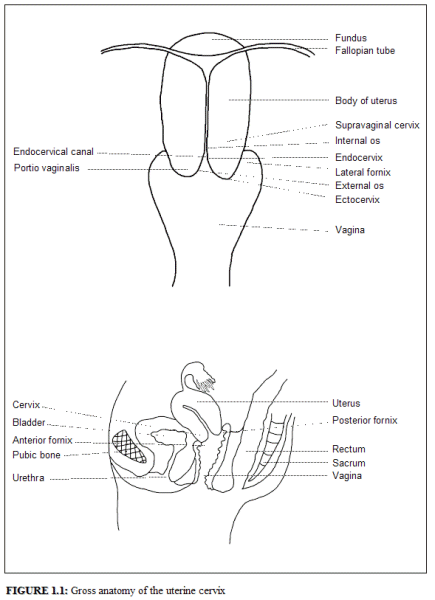
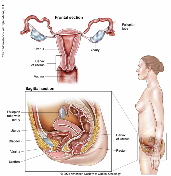
The female upper genital tract consists of the cervix uterine corpus fallopian tubes and ovaries. Lining the inside of the cervix is a thin layer of endometrium containing the epithelial cells that constantly produce cervical mucus. The cervix can be broken down into several anatomically distinct regions.
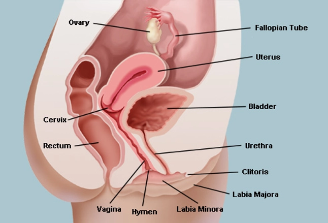
Yoganandan n1 bass cr2 voo l3 pintar fa4. The anatomy of the lower genital tract comprised of the vulva and vagina is discussed separately. The vagina connects the uterus to the outside world.
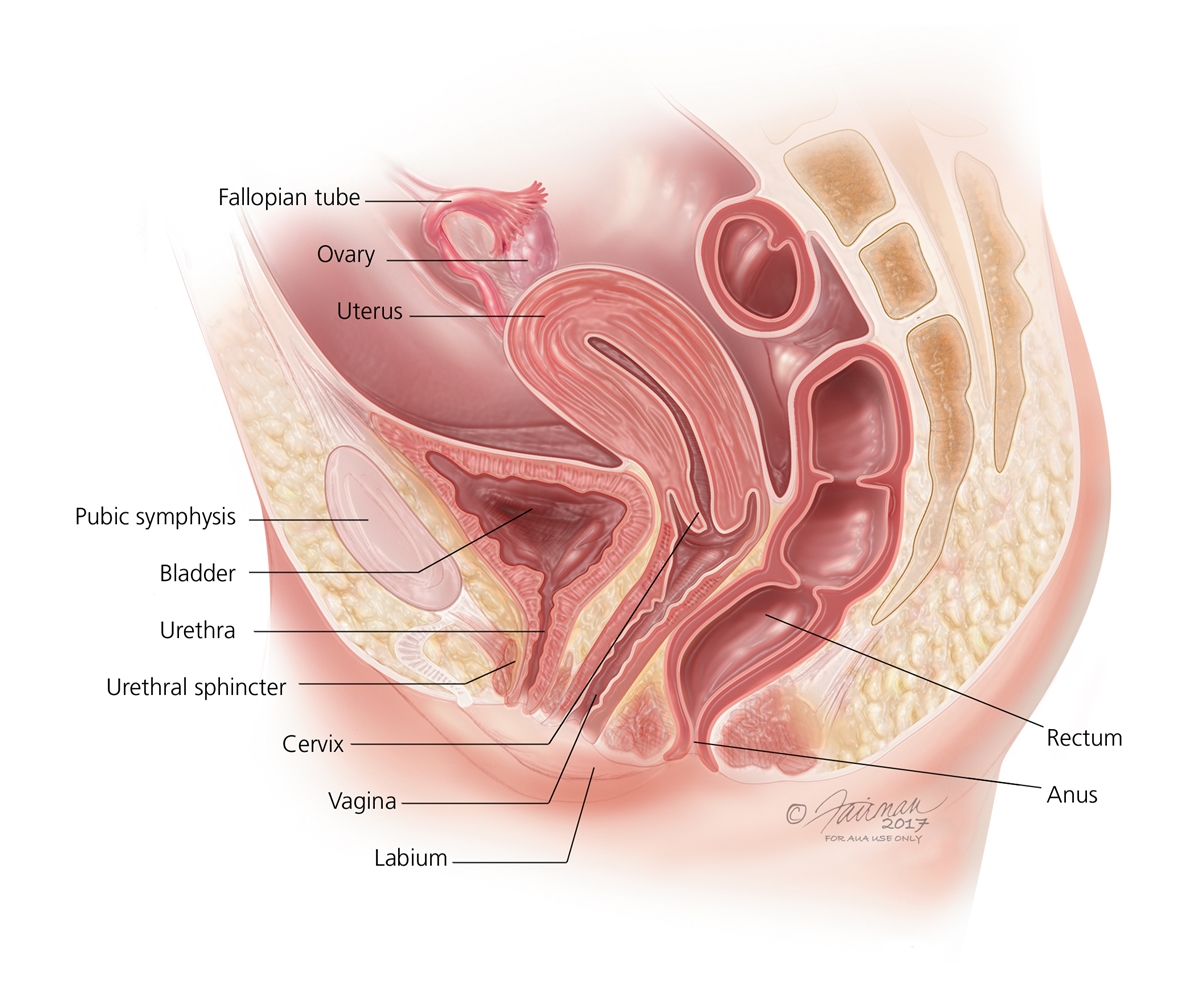
In women with cervical incompetence the cervix can be sewn closed. A sagittal view of the female pelvis is shown in the figure figure 1. The uterine cervix produces a mucus that aids in carrying sperm from the vagina to the uterus where it can fertilize an egg if the woman is ovulating.
Cervical spine anatomy video. Male and female cervical spine biomechanics and anatomy. The vulva and labia form the entrance and the cervix of the uterus protrudes into the vagina forming the interior end.
The top of the cervical spine connects to the skull and the bottom connects to the upper back at about shoulder level. Adenocarcinoma cancer of the glands found within the lining of the cervix.
 About Brachytherapy Introduction To Cervical Cancer
 Pelvis Clinical Anatomy A Case Study Approach
 Anatomical And Pathological Basis Of Visual Inspection With
 The Vagina Vulva Female Anatomy Pictures Parts
 Vaginal Cancer Vanderbilt Ingram Cancer Center
 The Female Reproductive System Boundless Anatomy And
 Female Reproductive System The Big Picture Gross Anatomy
 The Cervix Structure Function Vascular Supply
 About Cervical Cancer At Home Hpv Test Gynaehealth Uk
 Cervical Cancer Medlineplus Medical Encyclopedia
 Cervix Definition Function Location Diagram Facts
Human Papilloma Virus Hpv Cleveland Clinic
Cervical Cancer Causes Diagnosis And Symptoms Nccc
 Cervix Of Uterus Anatomy Pictures And Information
 Uterine Cancer General Information Symptoms Signs Of
 Vaginal Abnormalities Fusion And Duplication Symptoms
 Figure Hysterectomy The Uterus Is Surgically Pdq
 Uterus Radiology Reference Article Radiopaedia Org
Raman Spectroscopy In Cervical Cancers An Update Rubina S
Barrier Methods Of Birth Control Series
 Cervical Cancer Medical Illustrations Cancer Net
 The Cervix Structure Function Vascular Supply
 Royalty Free Cervix Stock Images Photos Vectors




Belum ada Komentar untuk "Female Cervical Anatomy"
Posting Komentar