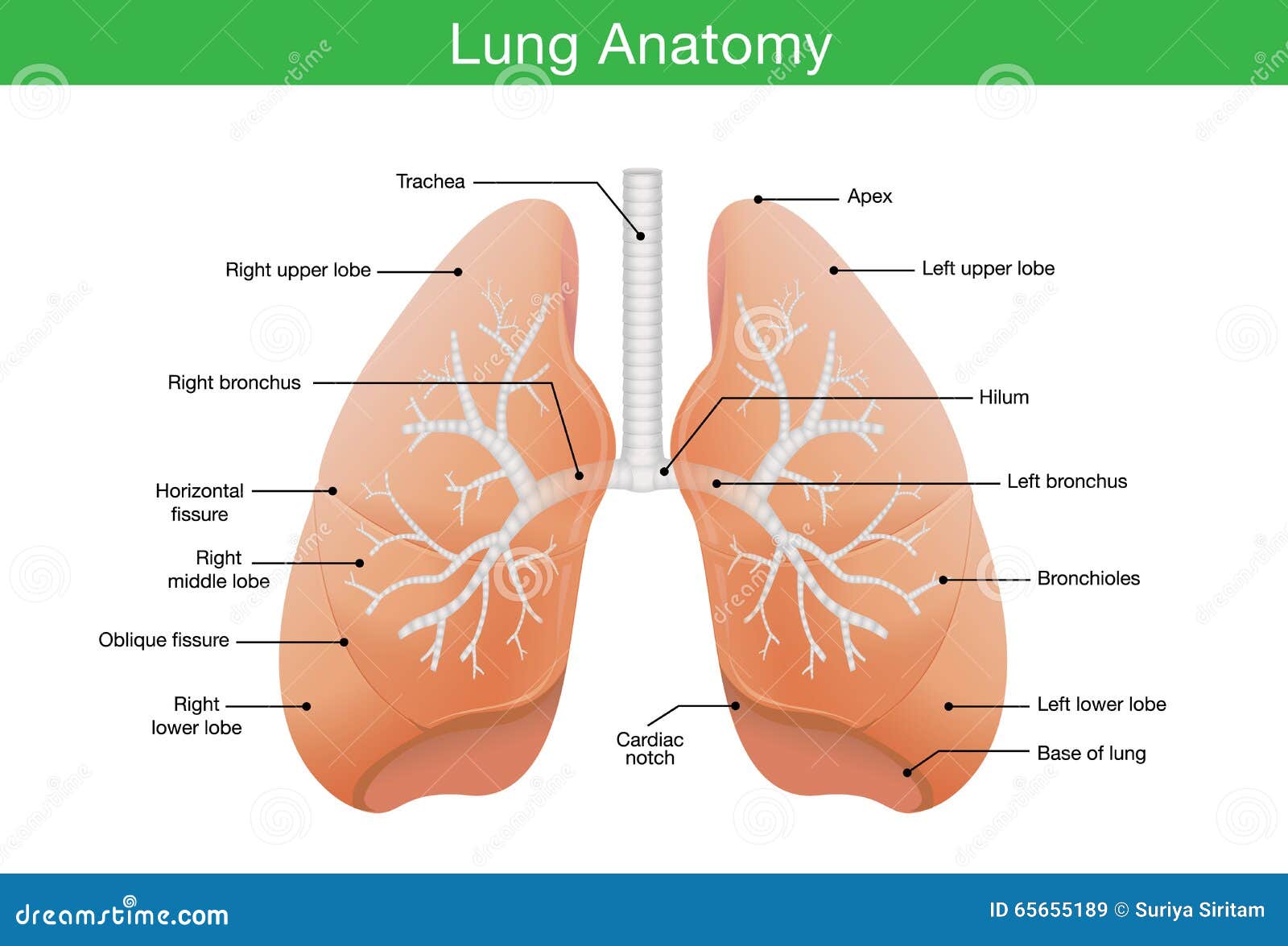
Lung Anatomy Diagram
Lung alveolus structure lung alveoli anatomy the alveolar sac contains two types of cells type 1 and type 2 cells. Surfaces and borders of lungs 3.
 Lungs Facts Function And Diseases Live Science
Human lip anatomy diagrams abdomen sketch with organs abdomen sketch with organs 7 photos of the abdomen sketch with organs abdominal organs chart diagram of abdominal organs diagram of abdominal organs and ribs diagram of stomach organs where is your abdomen inner body abdominal organs chart diagram of abdominal organs diagram of abdominal organs and ribs diagram of stomach organs where is your.

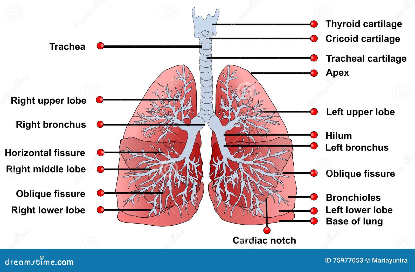
Lung anatomy diagram. Apex the blunt superior end of the lung. A small piece of tissue is taken from the lungs either through bronchoscopy or surgery. The animation also deals with lung cancer and the role of lymph in transporting bacteria allergens and cancer cells away from.
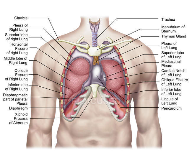
Fissures and lobes of lungs 5. Hilum and root of lungs 4. Surfaces three these correspond to the area of the thorax that they face.
Type 1 cells secrete pulmonary surfactant keeping the alveolar walls from sticking together as they deflate in exhalation. Structure and anatomy of the lungs. Gross anatomy of lungs 2.
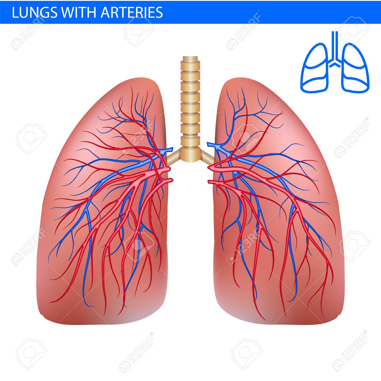
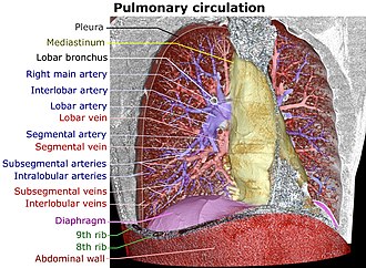
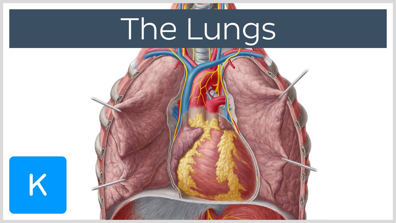
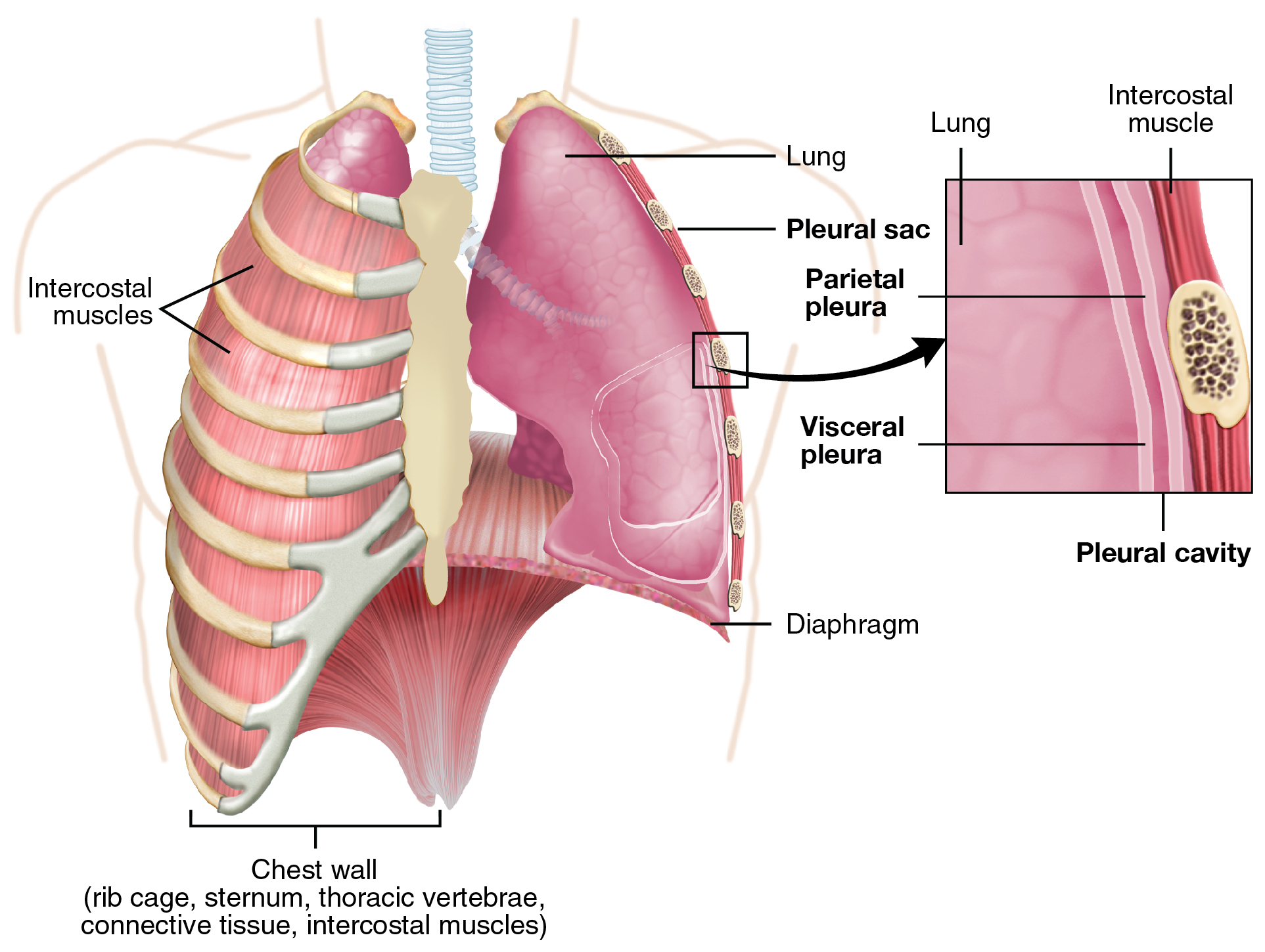
Blood supply of lungs 9. The left lung shares space with the heart and has an indentation in its border called the cardiac notch of the left lung to accommodate this. Base the inferior surface of the lung which sits on the diaphragm.
Nerve supply of lungs 11. Histopathology of alveoli 7. Lymphatics of lungs 10.
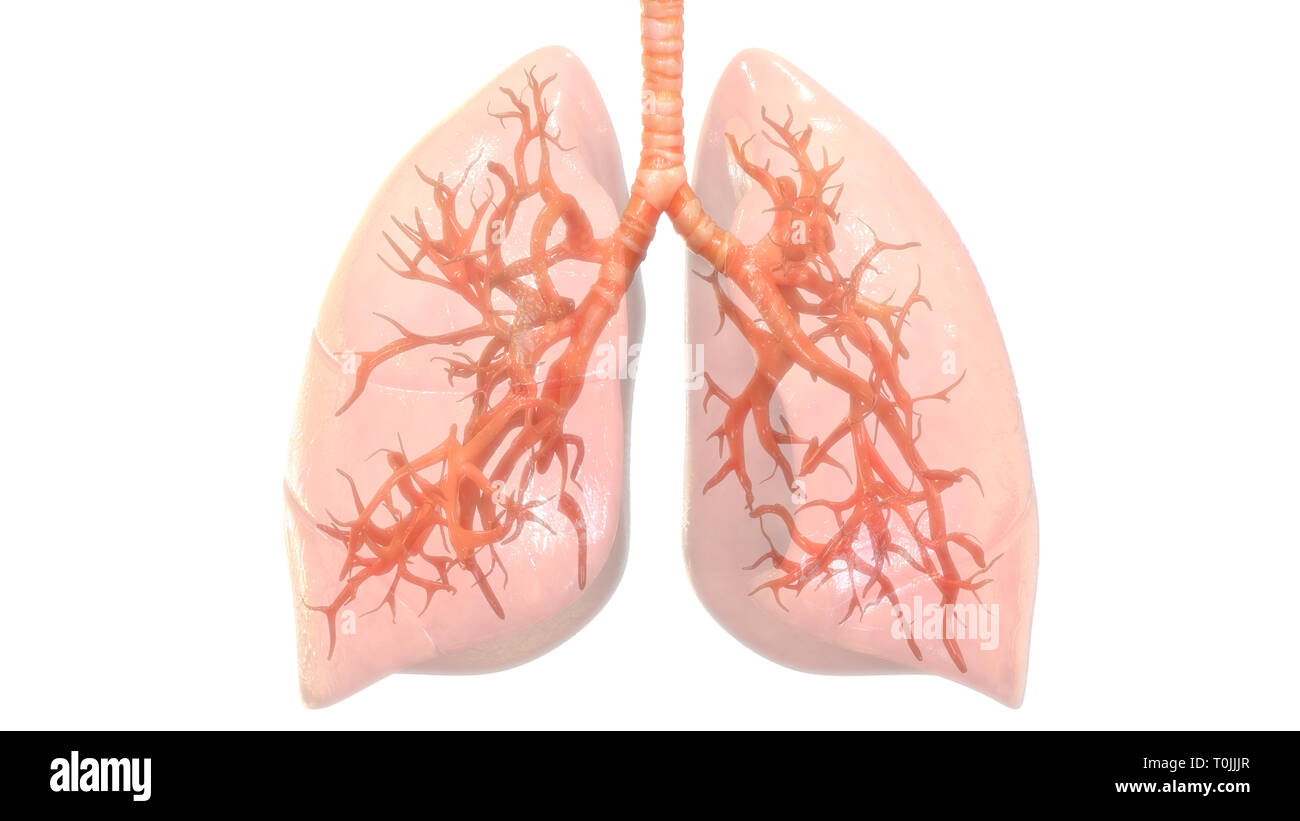
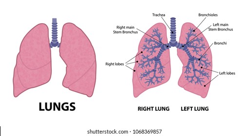
Each lung consists of. Lobes two or three these are separated by fissures within the lung. Here is how lungs work as the center of your breathing the path a full breath takes in your body and a 3 d model of lung anatomy.
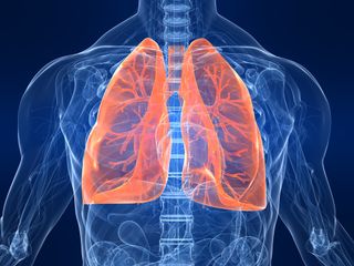
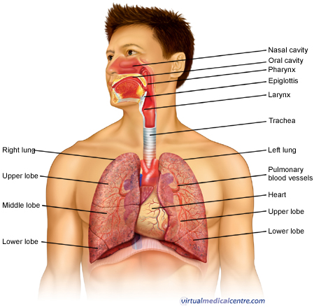
The lungs stretch from close to the backbone in the rib cage to the front of the chest and downwards from the lower part of the trachea to the diaphragm. The apex is the superior part of the lungs with its highest point located above the first rib extending through the superior opening of the thoracic cavity into the inferior floor of where the neck starts 8. Lung anatomy anatomy and physiology human anatomy respiratory therapy respiratory system heart and lungs runny nose radiology lung cancer webmds lungs anatomy page provides a detailed image and definition of the lungs.
The lungs are the main part of your respiratory system. Examining the biopsied tissue under a microscope can help diagnose lung conditions. It describes the pleura and diaphragm which aid in lung expansion.

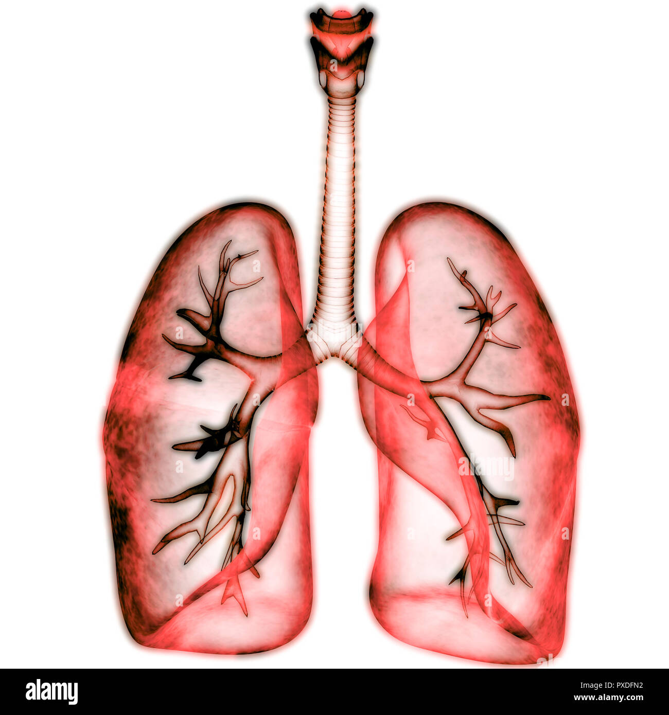
 Cancer Lungs Anatomy
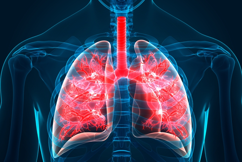
 Human Lungs Anatomy With Artery Circulatory System Realistic
 Anatomy Lung
 Lung Wikipedia
![]() Lung Anatomy Blood Supply Innervation Functions Kenhub
Lung Anatomy Blood Supply Innervation Functions Kenhub
 Human Respiratory System Lungs Anatomy Stock Photo
How Do We Breathe Lungs And Pleura Interactive Biology
 Lung Wikipedia
 Respiratory System Pulmonary System Anatomy Healthengine
 The Lungs Human Anatomy
 Label Lungs Diagram Printout Enchantedlearning Com
 Lung Human Icon Vector Photo Free Trial Bigstock
 Human Lung Anatomy Respiratory System Process Function
 Ilustraciones Imagenes Y Vectores De Stock Sobre Human
 Lungs Definition Location Structure Human Anatomy Kenhub
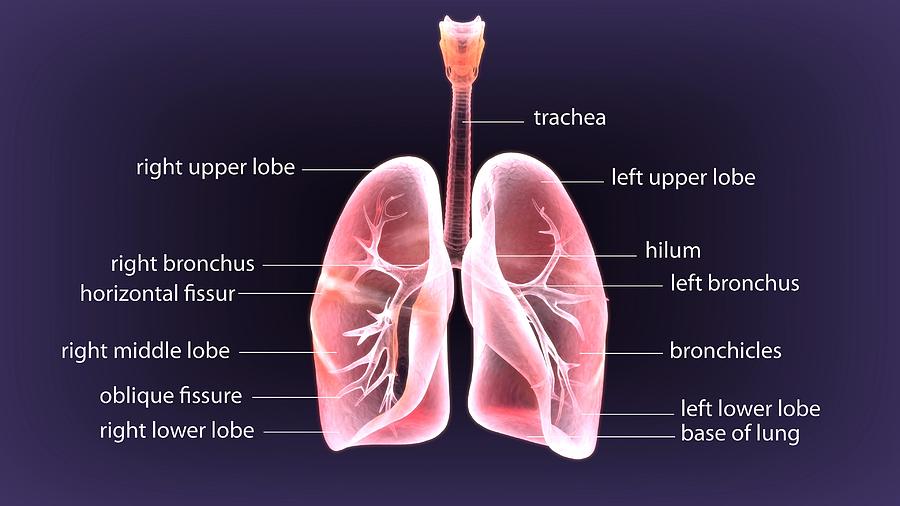
 Medical Illustration Of Human Lungs Anatomy With Labels
 Human Respiratory System Lungs Anatomy Stock Photo
![]() Lung Human Icon Respiratory System Healthy Lungs Anatomy Flat
Lung Human Icon Respiratory System Healthy Lungs Anatomy Flat
 3d Illustration Of Human Body Lungs Anatomy By Palmihelp

 A Step Closer To Understanding Idiopathic Pulmonary Fibrosis
 22 2 The Lungs Anatomy And Physiology
 Lungs Anatomy Stock Illustration Illustration Of
 Components Of Human Lung Stock Vector Illustration Of

 Drawing Anatomy Of Human Lungs Clipart Drawing Gg70248592
 Human Lung Anatomy Diagram
Belum ada Komentar untuk "Lung Anatomy Diagram"
Posting Komentar