Foot Anatomy Top
The cuneiform bones the navicularis and the cuboid all of which function to give your foot. Instability and tenderness to touch.
The dorsal fin of the shark is on his back.

Foot anatomy top. These all work together to bear weight allow movement and provide a stable base for us to stand and move on. The foot is an extremely complex anatomic structure made up of 26 bones and 33 joints that must work together with 19 muscles and 107 ligaments to execute highly precise movements. Measure your shoe size.
The forefoot contains the five toes phalanges and the five longer bones metatarsals. Posted by yclipse at 332 pm on march 18 2007. The opposite side of the foot is called the plantar surface.
The metatarsals which run through the flat part of your foot. The hindfoot forms the heel and ankle. Hinge joints typically allow for only one direction of motion much like a door hinge.
The phalanges which are the bones in your toes. The ankle joint is both a synovial joint and a hinge joint. Ankle stiffness in the morning which eases with movement.
The ankle joint or tibiotalar joint is formed where the top of the talus the uppermost bone in the foot and the tibia shin bone and fibula meet. The talus which is the. Tarsals five irregularly shaped bones of the midfoot that form the foots arch.
Ice exercises joint mobilisations stability training. Foot pain on top and outside of the ankle which gets better with rest and worse with activity. Foot and ankle anatomy is quite complex.
The other bones of the foot that create the ankle and connecting bones include. The foot consists of thirty three bones twenty six joints and over a hundred muscles ligaments and tendons. The opposite side of the hand is the palmar surface.
The feet are divided into three sections. Calcaneus the largest bone of the foot which lies beneath the talus to form the heel bone. Jf the top of both the foot and the hand is the dorsal surface.
Many of the muscles that affect larger foot movements are located in the lower leg. These make up the toes and broad section of the feet. At the same time the foot must be strong to support more than 100000 pounds of pressure for every mile walked.
The worst shoes to wear. The bones of the feet are. The bones of the foot are organized into rows named tarsal bones metatarsal bones and phalanges.
Talus the bone on top of the foot that forms a joint with the two bones of the lower leg. Dont know why you thought that dorsal would be down. The calcaneus which is the bone in your heel.
The talus bone supports the leg bones. The midfoot is a pyramid like collection of bones that form the arches of the feet. Top of foot pain.
 Foot Ankle Anatomy Pictures Function Treatment Sprain Pain
 Anatomy 101 Strengthen Your Big Toes To Build Stability
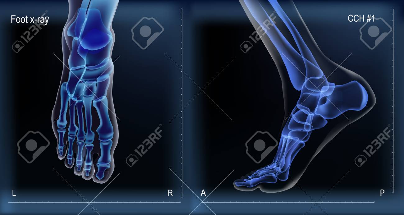 Dark Navy Blue Vector Realistic Medial And Top X Ray Of Skeleton
 Top Of The Foot Pain And Swelling Treatment Your Health
Patient Education Concord Orthopaedics
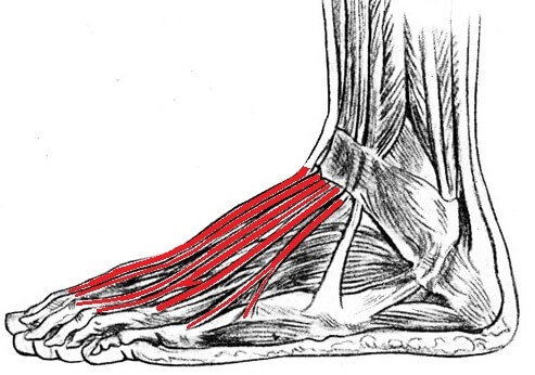 Extensor Tendonitis Causes Symptoms Treatment
 Foot Anatomy Detail Picture Image On Medicinenet Com
 Aliexpress Com Buy J0091 Foot Muscles And Bones Anatomy Pop 14x21 24x36 Inches Silk Art Poster Top Fabric Print Home Wall Decor From Reliable Art
 Foot Skeleton With Ligaments And Muscles Model Clinicalposters
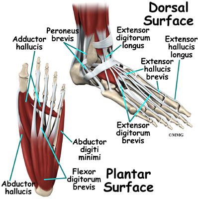
:background_color(FFFFFF):format(jpeg)/images/library/9528/muscles-foot_english.jpg) Dorsal Muscles Of The Foot Anatomy And Function Kenhub
Dorsal Muscles Of The Foot Anatomy And Function Kenhub
 Pain On Top Of The Foot Causes And Treatment
 The Foot Advanced Anatomy 2nd Ed
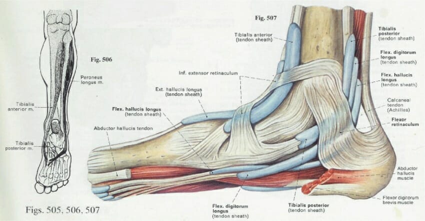 Foot Anatomy Bones Ligaments Muscles Tendons Arches
 Bones The Of Foot Top And Medial View Stock Vector
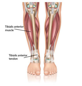 Ankle Tendonitis Anterior Tibial Tendonitis

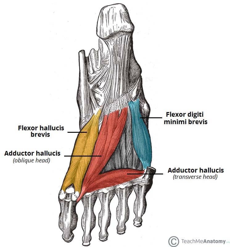 Muscles Of The Foot Dorsal Plantar Teachmeanatomy
Foot Anatomy Orthopedic Surgery Algonquin Il Barrington
 Foot Anatomy Pictures Model Body Maps
 Top View Of Human Foot Muscles Anatomy Isolated With Clipping Path
 10 Common Foot Problems Everyday Health


Belum ada Komentar untuk "Foot Anatomy Top"
Posting Komentar