Zygomatic Arch Anatomy
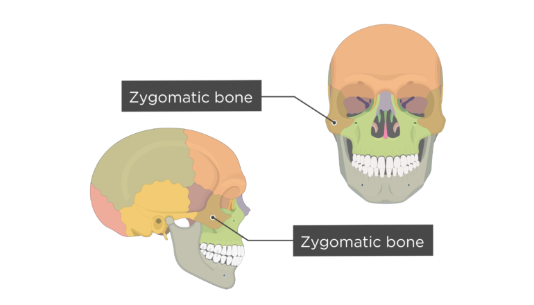
They are also commonly referred to a as the cheekbones or malar bones l mala the cheek. The zygomatic bone forms in membrane ie.

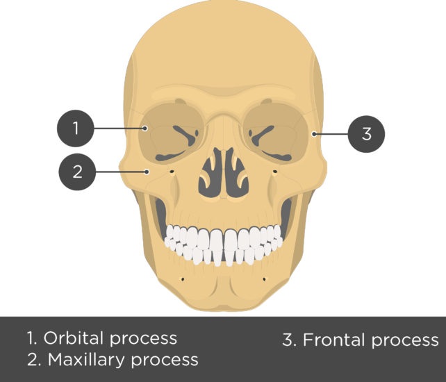
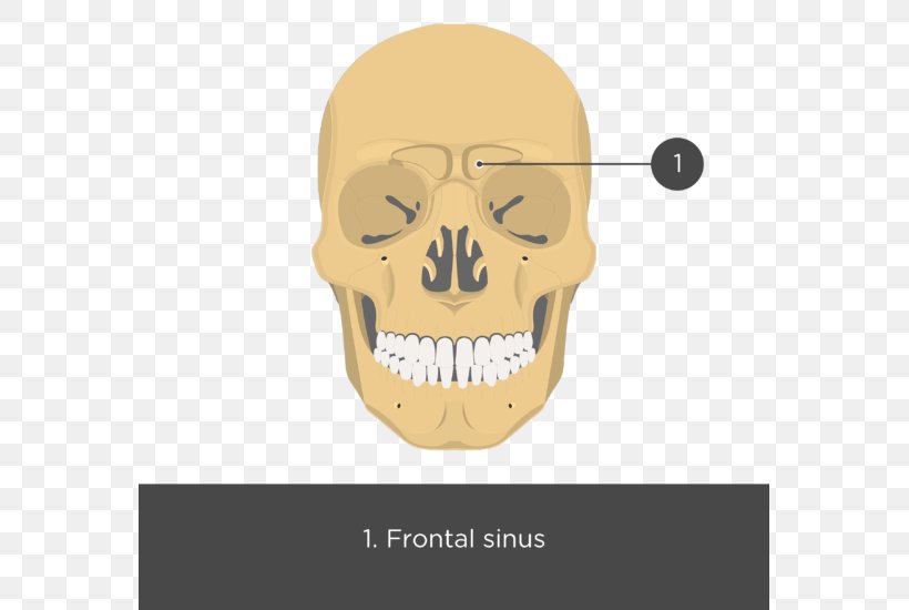
It adjoins the frontal bone at the outer edge of the orbit and the sphenoid and maxilla within the orbit.
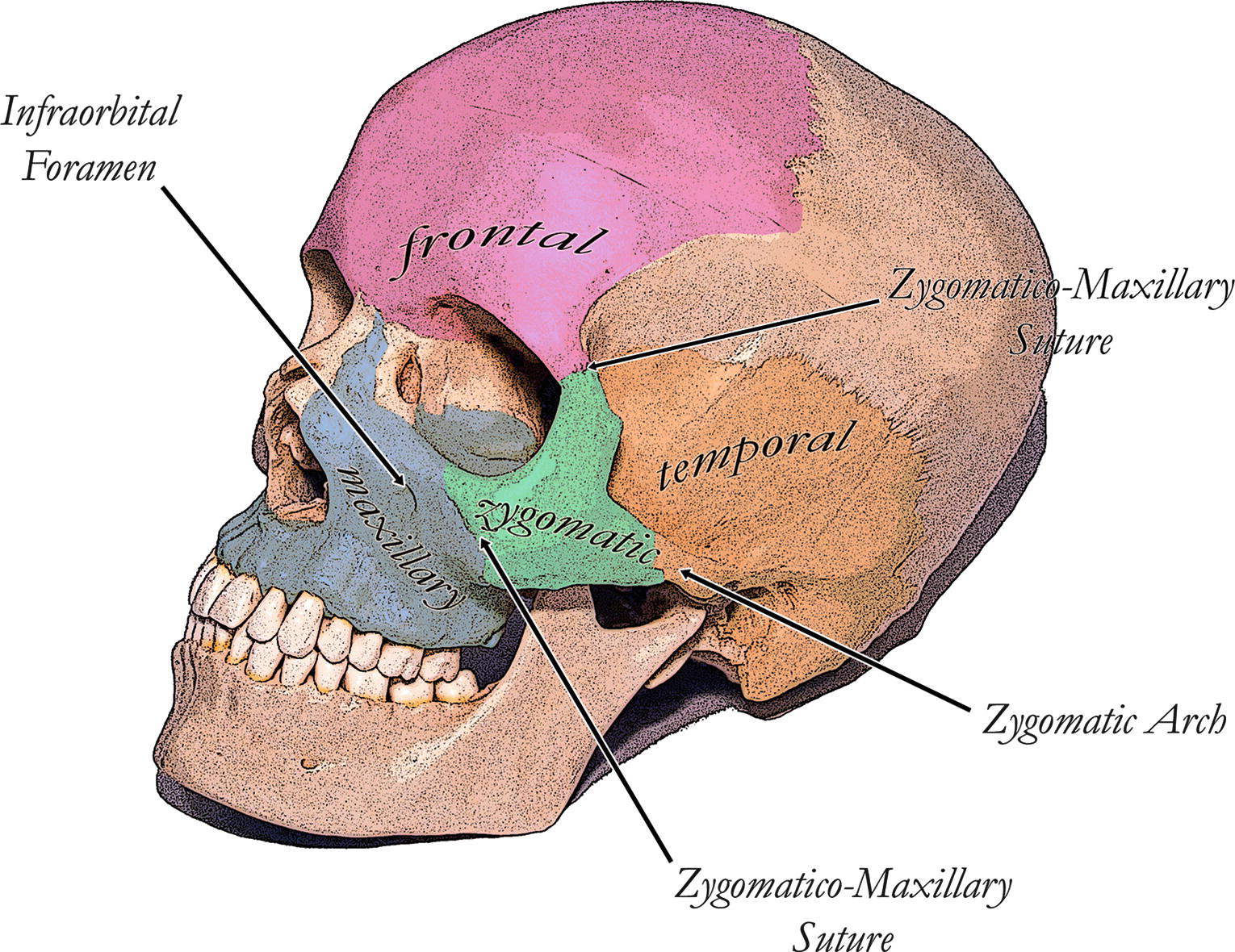
Zygomatic arch anatomy. The zygomatic bones gr zygoma yoke are two facial bones that form the cheeks and the lateral walls of the orbits. A posterior which runs backward above the external acoustic meatus and is continuous with the supramastoid crest. Zygomatic arch bridge of bone extending from the temporal bone at the side of the head around to the maxilla upper jawbone in front and including the zygomatic cheek bone as a major portion.
It forms the prominence of the cheek part of the lateral wall and floor of the orbit and parts of the temporal and infratemporal fossæ fig. Musculoskeletal and neurologic diseases. The zygomatic bone is small and quadrangular and is situated at the upper and lateral part of the face.
The anatomy of the common marmoset. Myofascial trigger point treatment for headache and tmd. Introduction to temporal bone anatomy.
The zygomatic process of the temporal arises by two roots. Frontal bone via the frontozygomatic suture which creates the rounded form of the bony orbit. Zygomatic arch temporomandibular joint dysplasia.
An anterior directed inward in front of the mandibular fossa where it expands to form the articular tubercle. Zygomatic process of the temporal bone linked by the temporozygomatic suture. The bony arch at the outer border of the eye socket formed by the union of the cheekbone and the zygomatic process of the temporal bone.
Zygomatic process of the maxillary bone articulated by the. Another major chewing muscle the temporalis passes through the arch. Disorders of the eye and vision.
Le fort type 3 fracture. It forms the central part of the zygomatic arch by its attachments to the maxilla in front and to the zygomatic process of the temporal bone at the side. Zygomatic arch noun anatomy.
The zygomatic arch is formed by the union of the temporal process of the zygomatic bone and the zygomatic process of the temporal bone at the zygomaticotemporal suture. Surgery of the orbit. The masseter muscle important in chewing arises from the lower edge of the arch.
Each zygomatic bone articulates with the temporal bone frontal bone maxilla and sphenoid bones. Several bones and joints surround the zygoma including the.
 Zygomatic Bone Anatomy
Alloplastic Contouring Of The Orbital Maxillary And
 Zygomatic Process Of The Temporal Bone Zygomatic Arch

 Zygomatic Arch Fracture Google Search Russell Westbrook
 Zygomatic Process Of Temporal Bone Zygomatic Bone Zygomatic
 Zygomatic Process Wikipedia
 Zygomatic Bone Anatomy
 The Zygomatic Bone Human Anatomy
Search Zygomatic Arches
 Zygomaticus Major O Zygomatic Arch Ins Angle Of Mouth
 Depression In Zygomatic Arch Region Download Scientific
 Gray Henry 1918 Anatomy Of The Human Body Page 1292
 Ancestral Variations In The Shape And Size Of The Zygoma
Does The Zygoma Arch Have Any Muscles Major Veins Or Major
 Zygomatic Bone Maxilla Zygomatic Arch Zygomatic Process
Is The Cheekbone In A Rabbit Called The Maxilla Or The
 Anp 1107 Lecture Notes Winter 2017 Lecture 3 Zygomatic

Zygomatic Arch

 Ontogeny Of Bone Strain The Zygomatic Arch In Pigs

 Zygomatic Arch An Overview Sciencedirect Topics
![]() Zygomatic Plate Wikipedia
Zygomatic Plate Wikipedia
 Zygomatic And Nasal Injury Rcemlearning
 Stunning Zygomatic Arch Art Fine Art America
 Zygomatic Arch Wikipedia
Belum ada Komentar untuk "Zygomatic Arch Anatomy"
Posting Komentar