Anatomy Of The Knee Ligaments
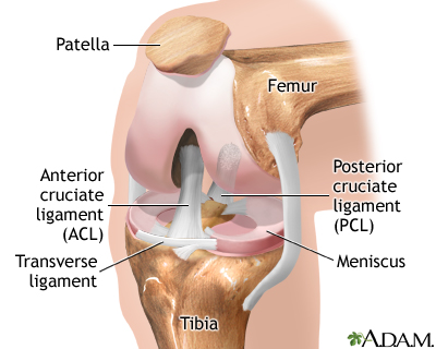
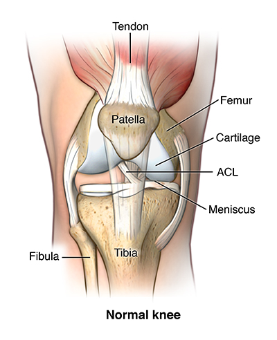
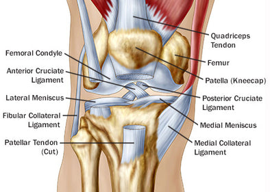
There are four knee ligaments thick bands of tough tissue that serve to maintain the stability of the knee joint. Another bone the patella kneecap is at the center of the knee.
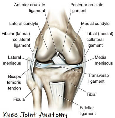
There is also a patellar ligament that attaches the kneecap to the tibia and aids in stability.
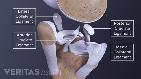
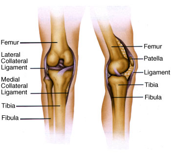
Anatomy of the knee ligaments. The largest joint in the body the knee moves like a hinge allowing you to sit squat walk or jump. There are also several key ligaments a type of fibrous connective tissue that connect these bones. The knee consists of three bones.
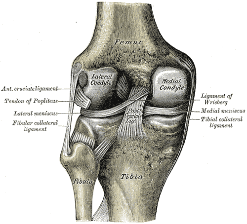
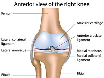
In knee joint anatomy they are the main stabilising structures of the knee acl pcl mcl and lcl preventing excessive movements and instability. On the sides of the knee are the medial collateral ligament mcl and the lateral collateral ligament lcl. Ligaments of the knee.
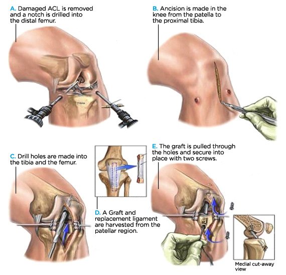
Ligaments in the knee. The knee is designed to fulfill a number of functions. The most common ligament injuries are acl tears mcl tears pcl tears and knee sprains which occur when the ligaments are overstretched.
It consists of bones meniscus ligaments and tendons. The knee includes four important ligaments all of which connect the femur to the tibia. The anterior cruciate ligament and posterior cruciate ligament provide front and back anterior and.
These are called the cruciate ligaments and consist of the anterior cruciate ligament and the posterior cruciate ligament. These two prevent sideways sliding of the knee joint ad also brace it against unusual movement. Two concave pads of cartilage strong flexible tissue called menisci minimize the friction created at the meeting of the ends of the tibia and femur.
A belt of fascia called the iliotibial band runs along the outside of the leg from the hip down to the knee and helps limit the lateral movement of the knee. Knee ligament impose limitations on the movement of the knee allowing it to concentrate forces of the muscles on extension and flexion. The medial collateral ligament on the inner side and the lateral collateral ligament on the outer side.
One ligament is on each side of the knee joint. The knee is a hinge joint that is responsible for weight bearing and movement. The anterior cruciate ligament prevents the femur from sliding backward on the tibia or the tibia sliding forward on the femur.
Ligaments join the knee bones and provide stability to the knee. Femur the upper leg bone or thigh bone tibia the bone at the front of the lower leg or shin bone patella the thick triangular bone that sits over the other bones at the front of the knee or kneecap.
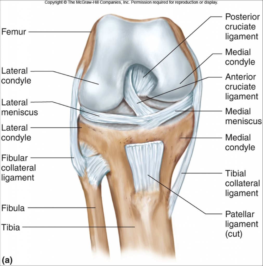 Anterior Cruciate Ligament Acl Injuries Core Em
 Knee Ligament Injury Anatomy Ligament Injury Knee
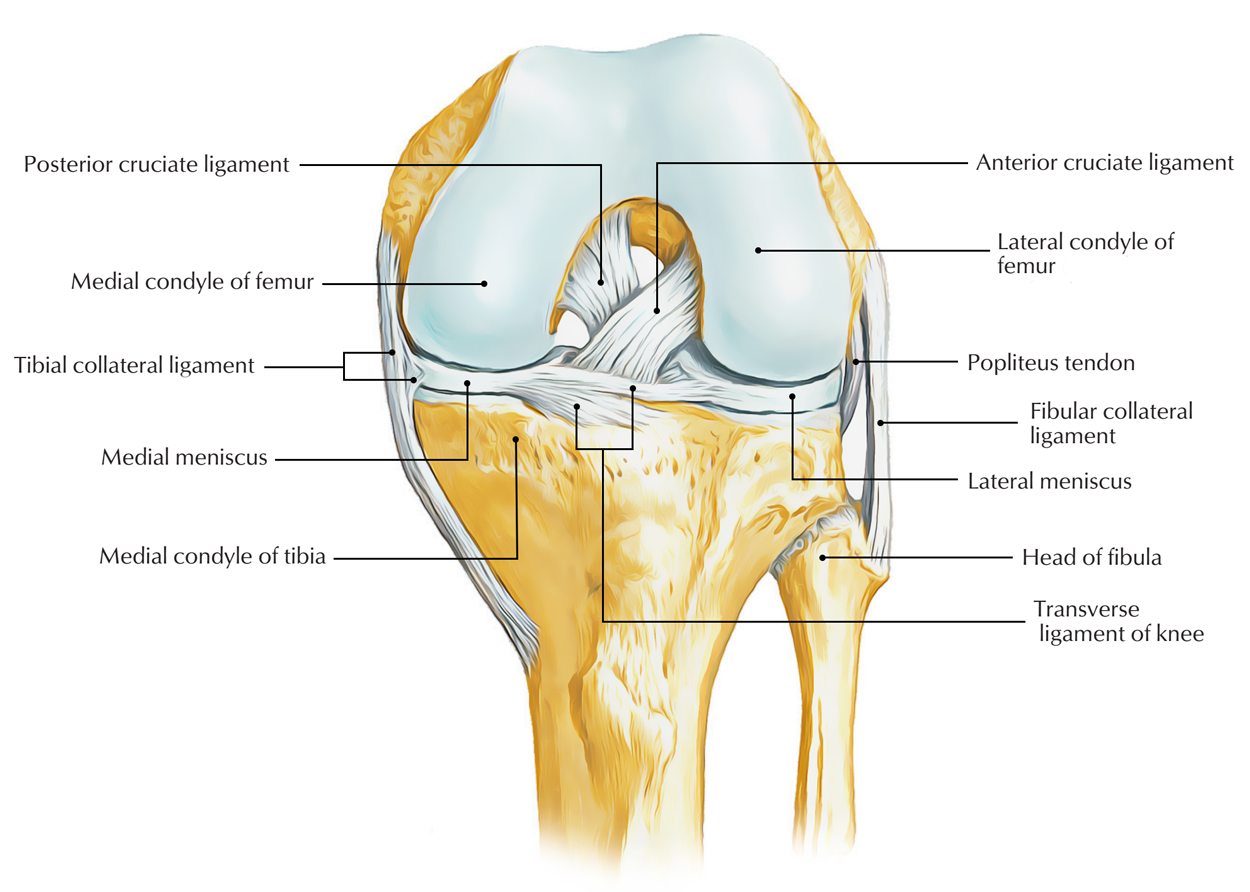 Easy Notes On Ligaments Of The Knee Joint Learn In Just 3
 10093 02x Ligaments Of Left Knee Anatomy Exhibits
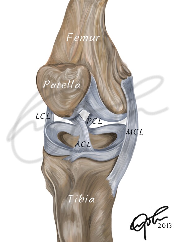 Human Knee Ligaments Printable Download Digital Illustration Medical Drawing Human Anatomy Knee Joint Knee Ligaments Anatomy Joint
 Knee Pain The Center For Physical Rehabilitation
 Knee Arthroscopy Series Normal Anatomy Medlineplus
 Acl Solutions Acl Knee Anatomy And Diagram Images
 Knee Joint Anatomy Motion Knee Pain Explained
 Posterolateral Knee Injuries Anatomy Evaluation And
 Coronary Ligament Of The Knee Wikipedia
 Knee Anatomy Including Ligaments Cartilage And Meniscus
 Guide Knee Injuries In Bjj Fighters Market
Common Knee Injuries Orthoinfo Aaos
Anterior Cruciate Ligament Acl Injuries Orthoinfo Aaos
 Knee Anatomy Wilmington Health
 Anatomy Of The Knee How The Knee Works Knee Anatomy
 Understanding The Anatomy Of The Knee Bodyheal
 Knee Anatomy The Orthopedic Sports Medicine Institute In
 The Knee Anatomy Injury Care And Prevention Soccer
 Knee Ligament Injuries Spanish
 Anatomy Of An Injury Acl Anterior Cruciate Ligament Tear
Knee Ligament Injuries Singapore Tears In Acl Mcl Pcl
 Anatomy Of The Knee For Dancers Dance Work Balance
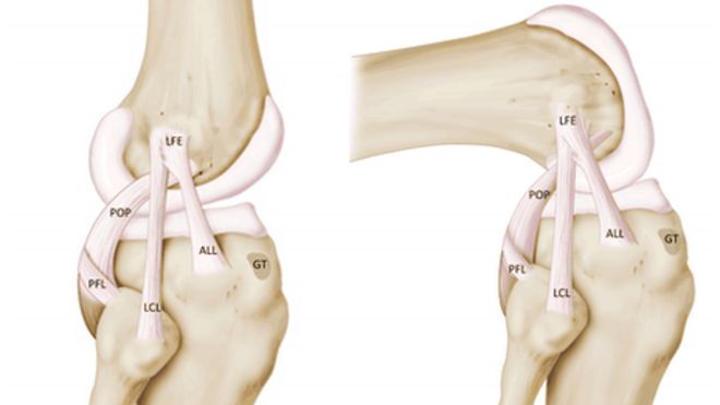 New Ligament Discovered In Knee Belgian Surgeons Say Bbc News


Belum ada Komentar untuk "Anatomy Of The Knee Ligaments"
Posting Komentar