Anatomy Of Ear Canal
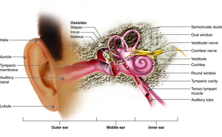
When sounds enter the middle ear they are transmitted to tiny bones called the ossicles. The eustachian tube connects the middle ear to the back of the nose.

External auditory canal or tube.
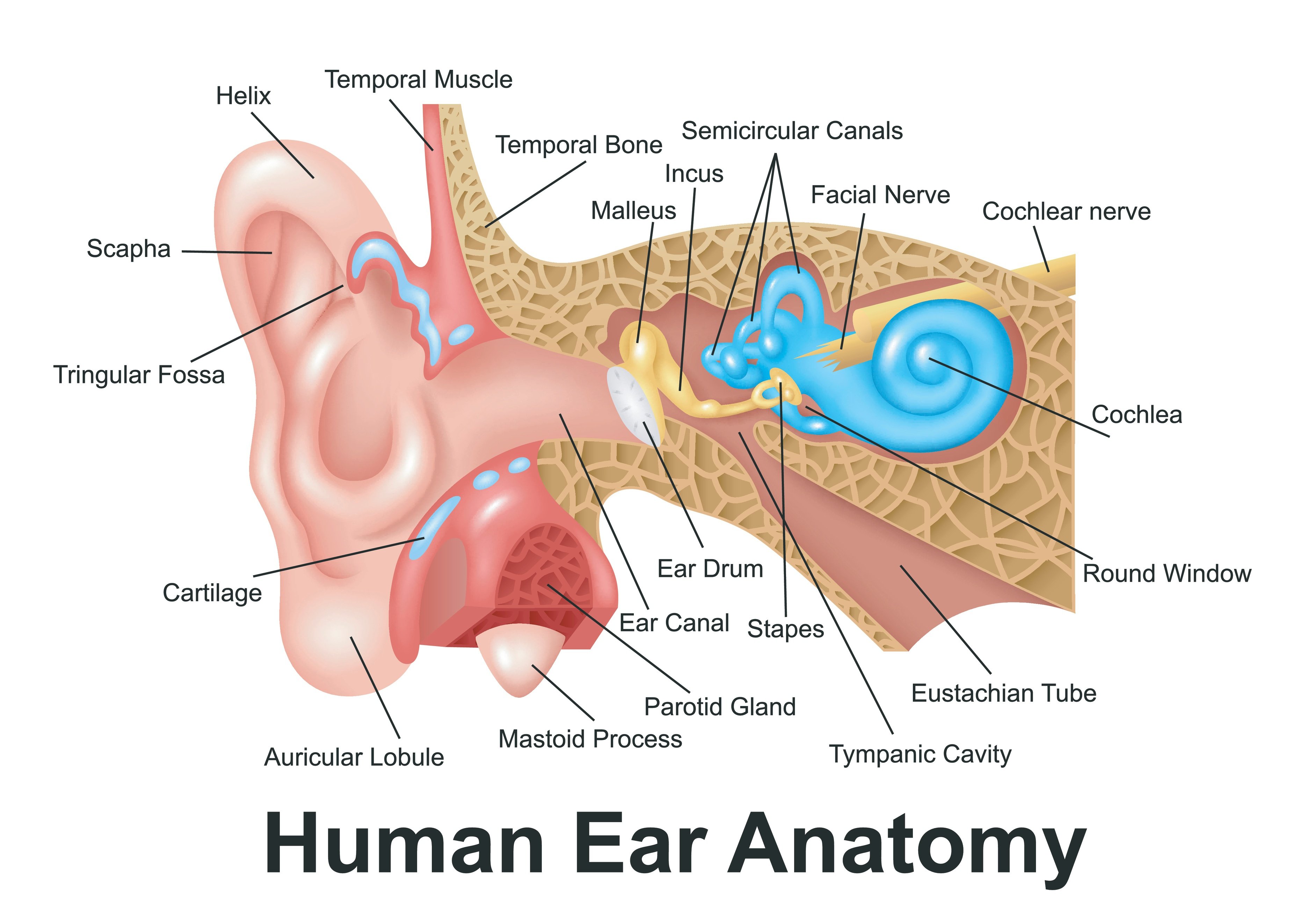
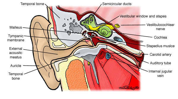
Anatomy of ear canal. External auditory canal or tube. The skin of the ear canal is very sensitive to pain and pressure. It contains two glands.
Sebaceous gland and ceruminous gland. The outer ear is called the pinna and is made of ridged cartilage covered by skin. The three bones are called the malleus the incus and the stapes.
The outer ear includes. The ear canal contains protective hairs and ear wax. Sound causes the eardrum and its tiny attached bones in the middle portion of the ear to vibrate.
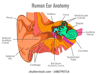
This is the outside part of the ear. The meatus is lined with skin continuous with auricle. Sound travels through the auricle and the auditory canal a short tube that ends at the eardrum.
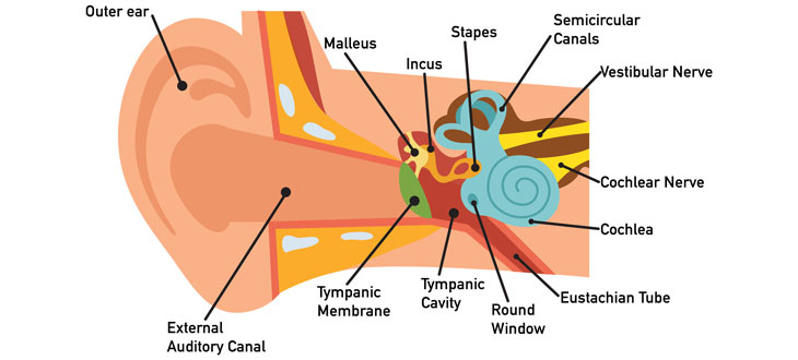
The ear is comprised of the ear canal also known as the outer ear the middle ear and the inner ear. The ear canal functions as an entryway for sound waves which get propelled toward the tympanic membrane known as the eardrum. The ear canal external acoustic meatus external auditory meatus eam is a pathway running from the outer ear to the middle ear.
The adult human ear canal extends from the pinna to the eardrum and is about 25 centimetres 1 in in length and 07 centimetres 03 in in diameter. The tympanic membrane is covered by epidermis and lined by simple cuboidal epithelium. Ear canal the ear canal starts at the outer ear and ends at the ear drum.
Anatomy and physiology of the ear what is the ear. The external auditory canal is a curved tube about 25 cm 1 in long that lies in the temporal bone and leads to the eardrum. Ceruminous gland are modified sweat gland that secretes cerumen wax.
Tympanic membrane also called the eardrum. The tympanic membrane or ear drum is a thin semitransparent partition between the external auditory canal and middle ear. Picture of the ear.
External or outer ear consisting of. This is the tube that connects the outer ear to the inside or middle ear. The eardrum separates the outer ear from the middle ear.
The canal is approximately an inch in length. Ear anatomy outer ear. The middle ear tympanic cavity lies behind the eardrum.
Auricle cartilage covered by skin placed on opposite sides of the head auditory canal also called the ear canal eardrum outer layer also called the tympanic membrane the outer part of the ear collects sound. The ear is the organ of hearing and balance. This is the tube that connects the outer ear to the inside or middle ear.
The ear is the organ of hearing and balance. The parts of the ear include. External or outer ear consisting of.
The middle ear contains three small bones ossicles that transmit sound waves from the eardrum to the inner ear. This is the outside part of the ear. Under the skin the outer one third of the canal is cartilage and inner two thirds is bone.
Sound funnels through the pinna into the external auditory canal a short tube that ends at the eardrum tympanic membrane. The parts of the ear include. External auditory meatus is slightly curved canal of about 25 cm ling extending from floor of concha to tympanic membrane ear drum.
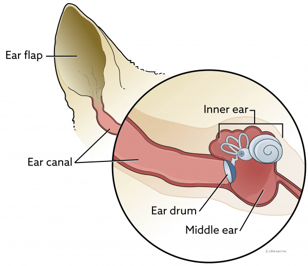 Instructions For Ear Cleaning In Cats Vca Animal Hospital
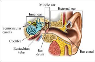 Do You Hear What I Hear Signs And Symptoms Of Tinnitus
How Hearing Works Anatomy Of The Ear
 Anatomy Of The Ear Inner Ear Middle Ear Outer Ear
Ear Anatomy Causes Of Hearing Loss Hearing Aids Audiology
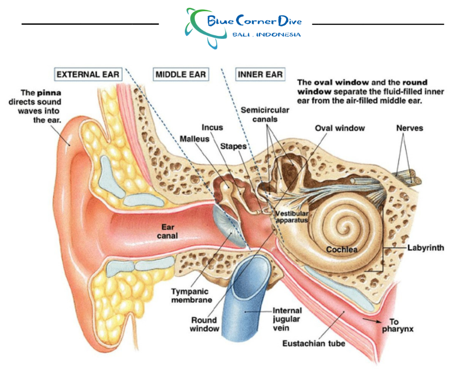 What Happens To My Ears When I Scuba Dive Blue Corner
 Acute Otitis Externa The Successful First Opinion Ear
Ear Disorders Problems And Treatment Ent Florida
 Q How Can I Safely Remove Ear Wax
How Hearing Works How We Hear Hearnet Online
 Anatomy Atlases Anatomy Of First Aid A Case Study Approach
 Can Ear Infections Cause Hearing Loss
 Hearing And Equilibrium Anatomy And Physiology
 Eardrum Images Stock Photos Vectors Shutterstock
Hearing Loss Annapolis Sensorineural Conductive Hearing Loss
 Human Ear Art Print Human Ear Canal Vintage Illustration Wall Art Decor Medical Human Anatomy Art Print Ear Anatomy Drawing Print Ear
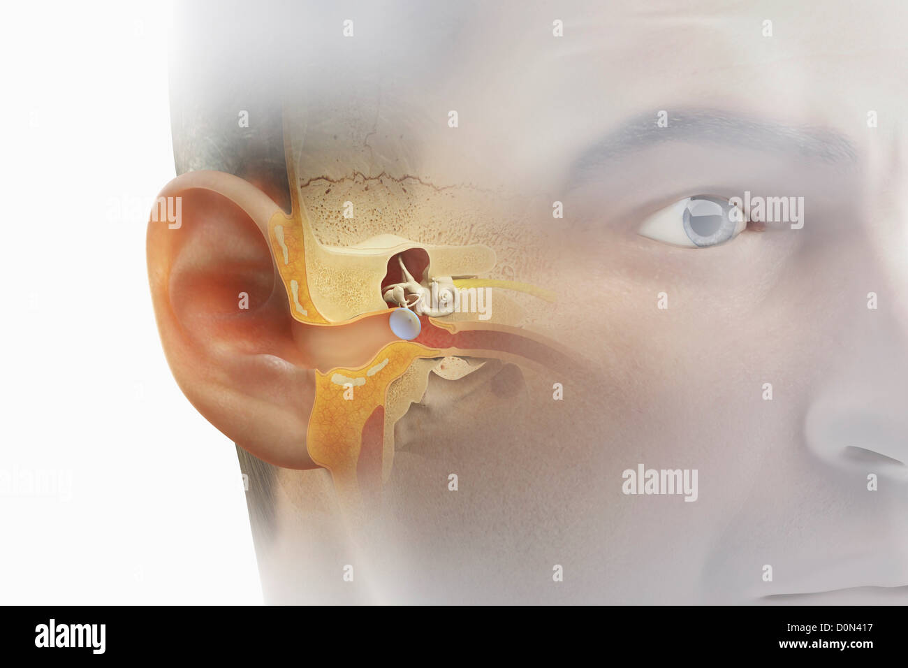 A Sectional View Of The Human Head Revealing The Anatomy Of
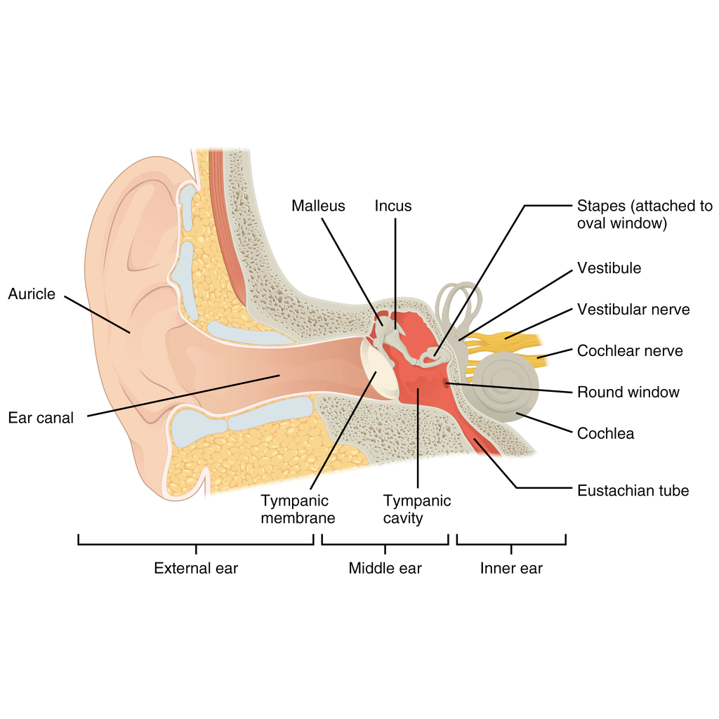 Inner Ear Illustrations Radiology Case Radiopaedia Org
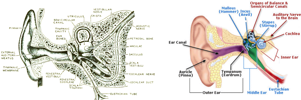 The Moovin Groovin Outer Ear Lydia Gregoret Hearing Aids
 10 Interesting Facts About Ears Hearing
 It S Better Hearing And Speech Month Hough Ear Institute
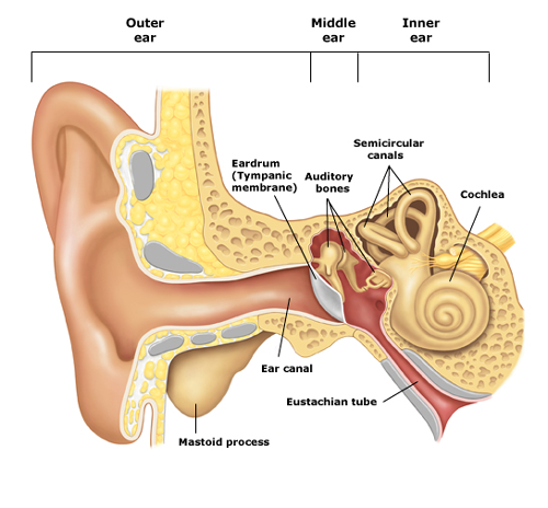 Ear Education Tampa Bay Hearing
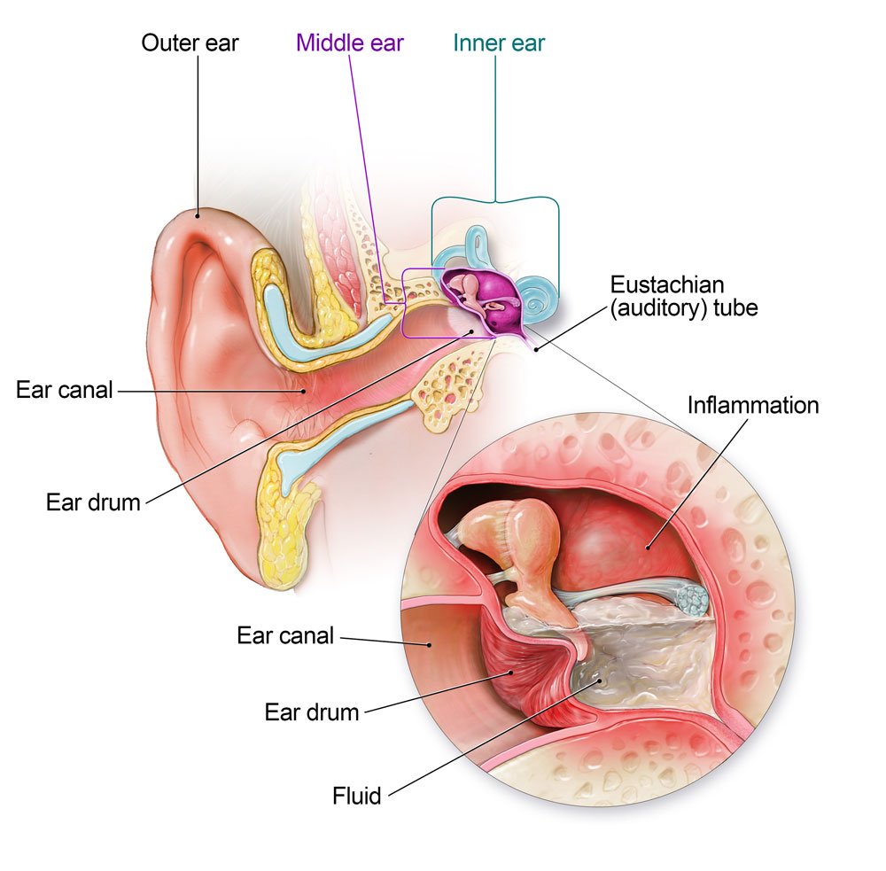 Ear Infection Community Antibiotic Use Cdc
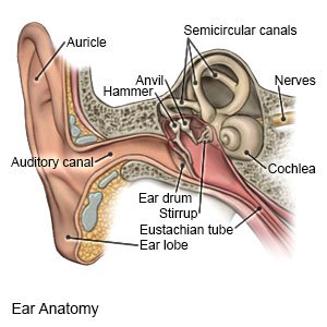 Tympanoplasty Precare What You Need To Know


Belum ada Komentar untuk "Anatomy Of Ear Canal"
Posting Komentar