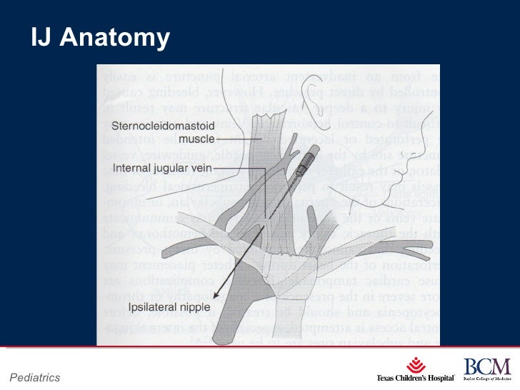
Ij Anatomy
There are numerous described approaches to the ij. Infected skin site in patients with higher risks for pneumothorax or inability to tolerate pneumothorax the ij or femoral sites may also be preferred.
The ij vein is a readily compressible vessel.

Ij anatomy. It exits the cranium via the jugular foramen and then courses through the anterior neck lateral to the carotid. Located besides the common carotid artery in the neck the internal jugular veins are primarily responsible for carrying deoxygenated blood from the brain as well as superficial parts of the face and the neck to the heart. The internal jugular vein ijv is the major venous return from the brain upper face and neck.
Anatomically there are two of these veins that lie. Anatomy the ij vein begins in the cranium at the conclusion of the sigmoid sinus. The internal jugular veins lie deeper and are larger than the external jugular veins which lie closer to the surface.
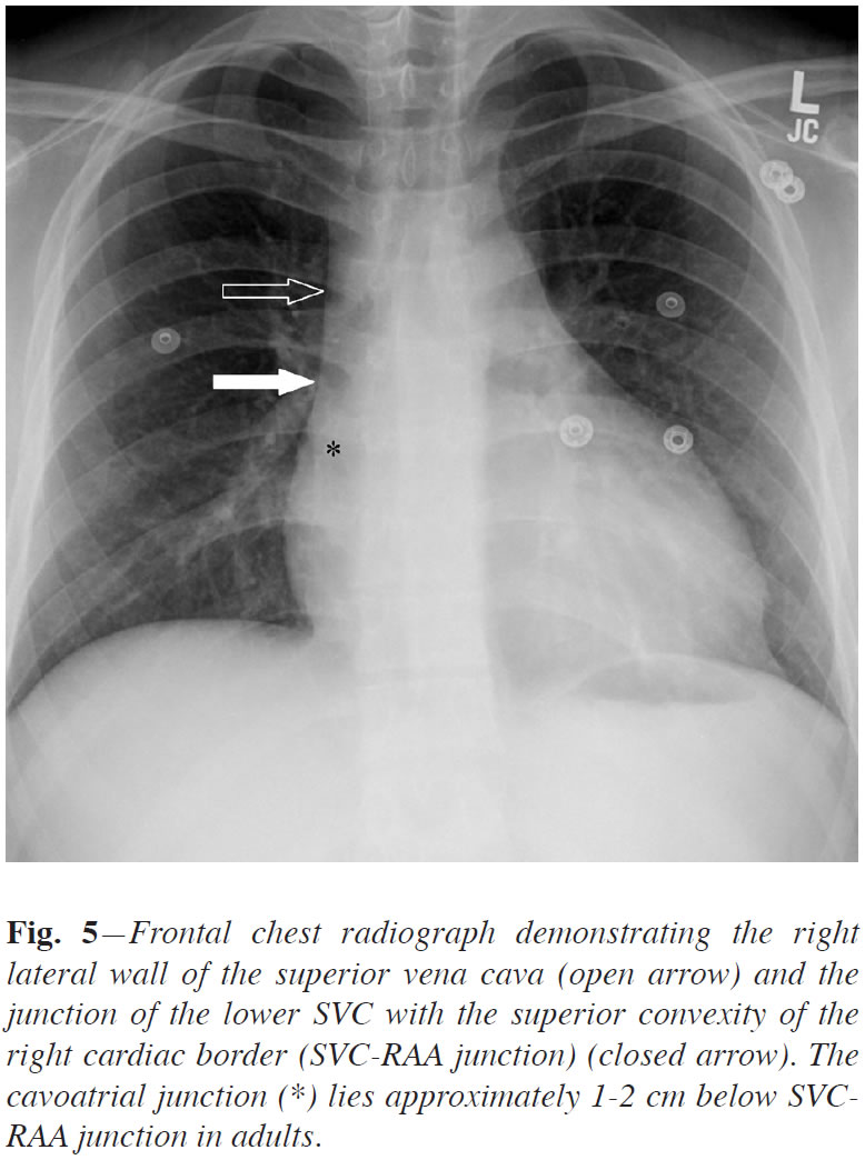
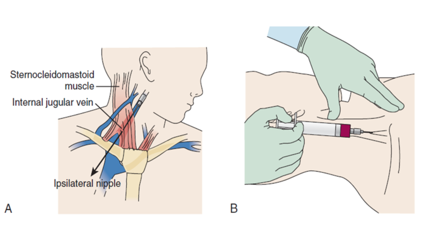
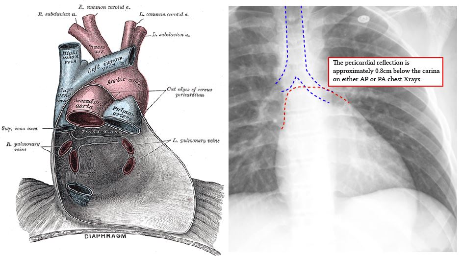
The brachiocephalic veins innominate vein on either side then join one another to form the superior vena cava to bring blood into the right atrium of the heart. The landmark approach most widely used is between the medial and lateral heads of the sternocleidomastoid muscle and lateral to the carotid artery in most cases. Local anatomy and ij vein internal jugular cvc insertion.
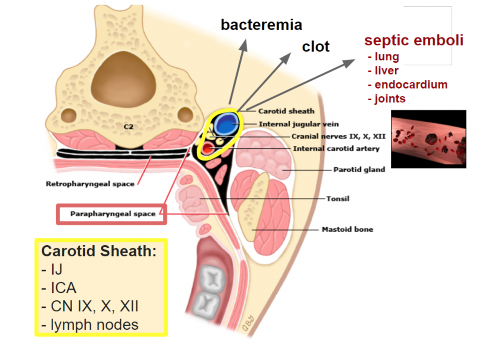
The internal jugular vein is a major blood vessel that drains blood from important body organs and parts such as the brain face and neck. Internal jugular vein the external jugular vein drains into the subclavian vein. The vein runs in the carotid sheath with the common carotid artery and vagus nerve.
Anatomical terminology edit on wikidata the internal jugular vein is a paired jugular vein that collects blood from the brain and the superficial parts of the face and neck. The internal jugular vein joins the subclavian vein to form the brachiocephalic vein. The femoral or ij site is preferred with a coagulopathy or anticoagulation due to the ability to compress the vein in the event of serious hemorrhage.
Gross anatomy origin and course it is formed by the union of inferior petrosal and sigmoid dural venous sinuses in or just distal to the jugular fo.
 Ij Cannula Circuit Surfers
 Ultrasound Leadership Academy Central Line Placement Em
 2012 Vascular Access Pt 1
 Vascular Internal Jugular Highland Em Ultrasound Fueled
 Thread By Edgarvlermamd Hemodialysis 101 Venous Catheter
 Gross Anatomy Picture
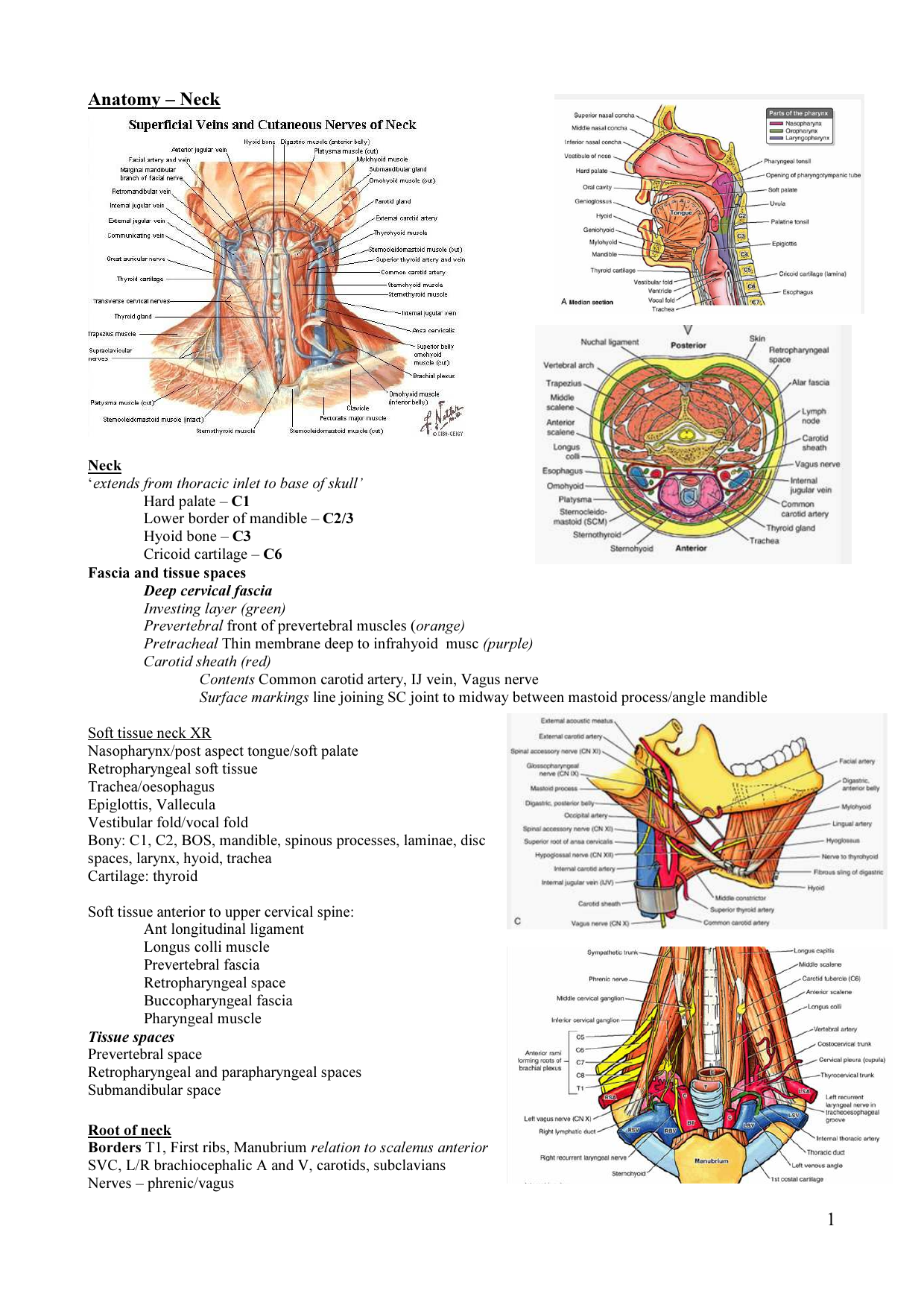 1 Anatomy Neck

 The Central Line Part 1 The Basics Brown Emergency Medicine
 Ultrasound Guided Internal Jugular Vein Catheterization
 Central Venous Access Device Insertion Deranged Physiology
 Anatome Testudinis Europaeae Turtles Anatomy R Io
 Color Atlas Of Human Anatomy Vol 1 Locomotor System By
 The Blind Ij Central Catheter Em Curious
 Introduction Of Ultrasound Into Gross Anatomy Curriculum
 Anatomy Of Studied Cypselas G H Zinnia Pauciflora G
 The Central Line Part 1 The Basics Brown Emergency Medicine
 Central Line Placement Crashing Patient
 Biology Of The Vertebrates A Comparative Study Of Man And
 Anatome Testudinis Europaeae Turtles Anatomy 3 Fio V
 Central Venous Access Device Insertion Deranged Physiology
 The Anatomical Record Anatomy Anatomy 288 Charles F W
 Insertion Of The Dual Lumen Cannula Into The Ij Vein Via


 Internal Jugular Central Line Ultrasound Manikin
 The Comparative Anatomy Of The Domesticated Animals Horses
Belum ada Komentar untuk "Ij Anatomy"
Posting Komentar