Anatomy Of Conjunctiva
The conjunctiva is highly vascularised with many microvessels easily accessible for imaging studies. The clear tissue covering the white part of your eye and the inside of your eyelids.
Conjunctiva American Academy Of Ophthalmology
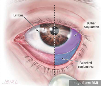
The conjunctiva is the mucous membrane that lines the eyelid and covers the visible portion.

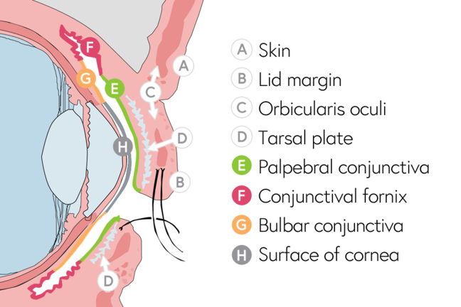
Anatomy of conjunctiva. This portion of the conjunctiva covers the anterior part of the sclera the white of the eye. Palpebral conjunctiva marginal tarsal orbital. Extends from the lid.
Recessed in the eyelids the conjunctiva forms a cul de sac which is open in front at the palpebral fissure and only closed when the eyes are shut. The conjunctiva is the clear thin membrane that covers part of the front surface of the eye and the inner surface of the eyelids. Eye anatomy in eyelid the normal functioning of the conjunctiva and cornea.
It has two segments. The potential space between tenons capsule and the sclera is frequently used for local anesthesia. Dry eye retinal detachment.
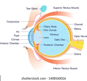
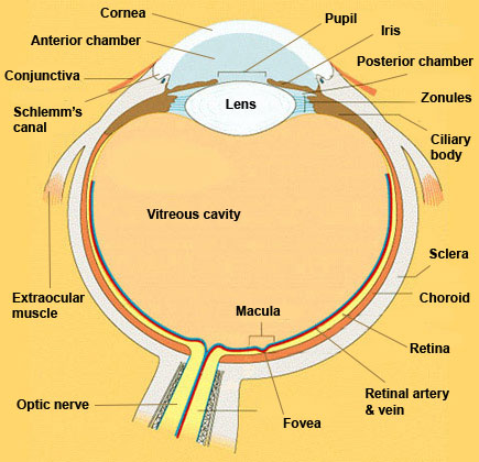
The bulbar conjunctiva is found on the eyeball over the anterior sclera. Anatomy of the eye includes lacrimal gland cornea conjunctiva uvea iris choroid ciliary body lens blood supply retina vitreous optic nerve. The conjunctiva is a mucous membrane that serves to attach.
Tenons capsule binds it to the underlying sclera. Conjunctiva palpebral conjunctiva marginal tarsal orbital bulbar conjunctiva scleral limbal. Conjunctiva thin transparent mucous membrane lining the posterior aspect.
The conjunctiva has an average thickness of 33 microns. Anatomy of conjunctiva 1. The conjunctiva here is comparatively thicker and loosely attached in order to allow free movement of the globe.
The eyelids lid a portion of the conjunctiva. For ophthalmologists optometrists medical dental and optometry students eye anatomy forms the basis for eye pathology in diseases. The palpebral conjunctiva lines the eyelids.
Conjunctiva of the fornix. Conjunctiva is continuous anteriorly with the epithelium of the cornea. The conjunctiva is a tissue that lines the inside of the eyelids and covers the sclera the white of the eye.
It is composed of unkeratinized stratified squamous epithelium with goblet cells and stratified columnar epithelium. It is fold lining the cul de sac formed by conjunctiva covering the posterior surface of the lids to the conjunctiva covering the anterior surface of the globe. Anatomy of the human eye.

 Conjunctiva Definition And Detailed Illustration
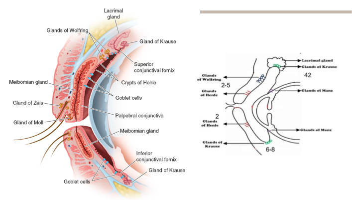
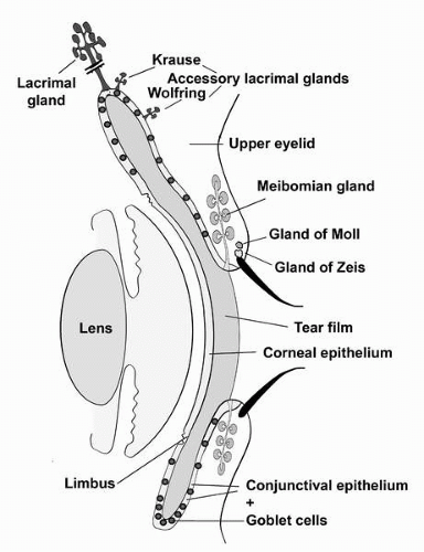
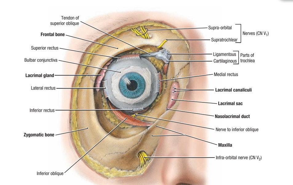 Easy Notes On Lacrimal Apparatus Learn In Just 3 Minutes
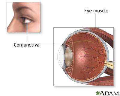 Eye Muscle Repair Series Normal Anatomy Medlineplus
 Conjunctiva Anatomy Pi Uptodate
 Conjunctival Anatomy Differential Diagnosis Of

 Anatomy Of The Conjunctiva Eye Anatomy Medical Pictures
 Review Your Eye Anatomy In Order To Understand Eye Disease
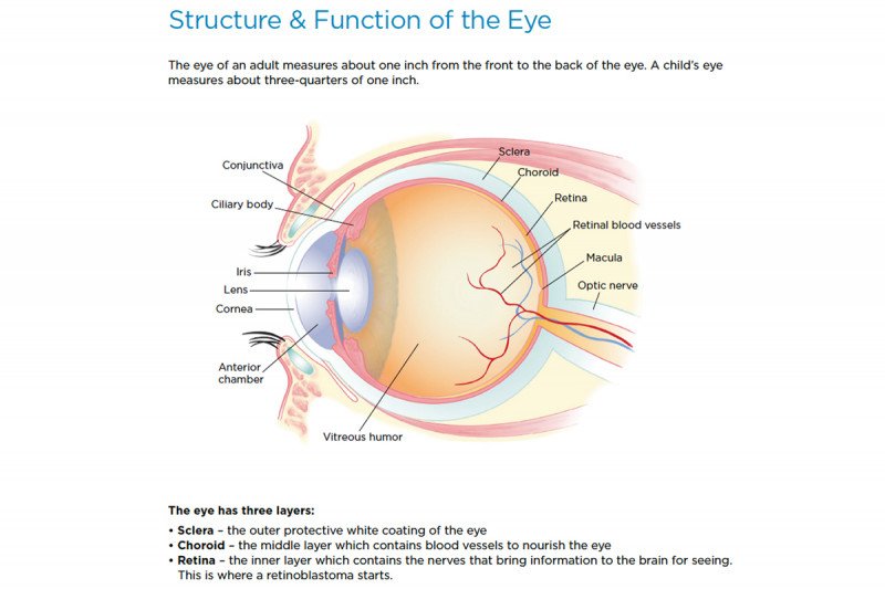 Retinoblastoma Anatomy Of The Eye Memorial Sloan
Conjunctiva Wikipedia
 Eye Injuries Real First Aid
Conjunctival Scleral Anatomy American Academy Of Ophthalmology
 Anatomy Of The Eyelid Eliminating Trachoma
 Eye Structure And Function In Dogs Dog Owners Merck
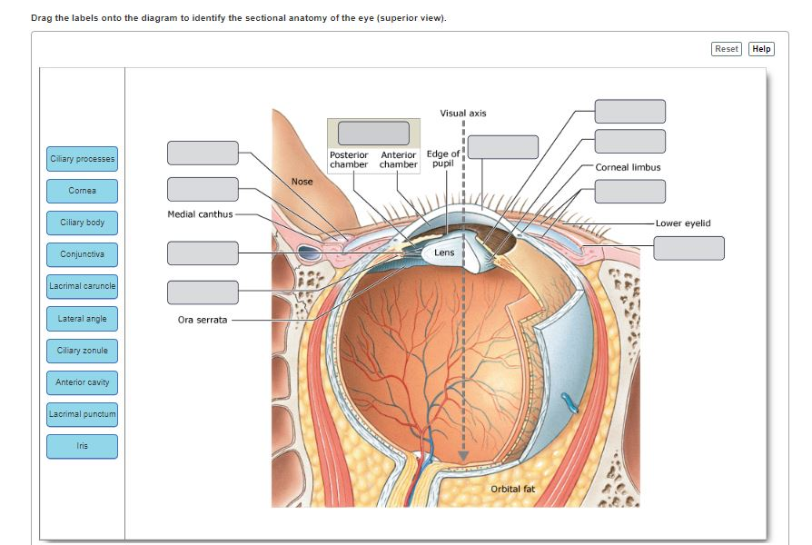 Solved Drag The Labels Onto The Diagram To Identify The S
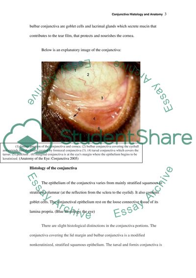 Conjunctiva Histology And Anatomy Essay Example Topics And
 Orbits And Eyes Anatomical Illustrations
 Conjunctiva Anatomy Britannica
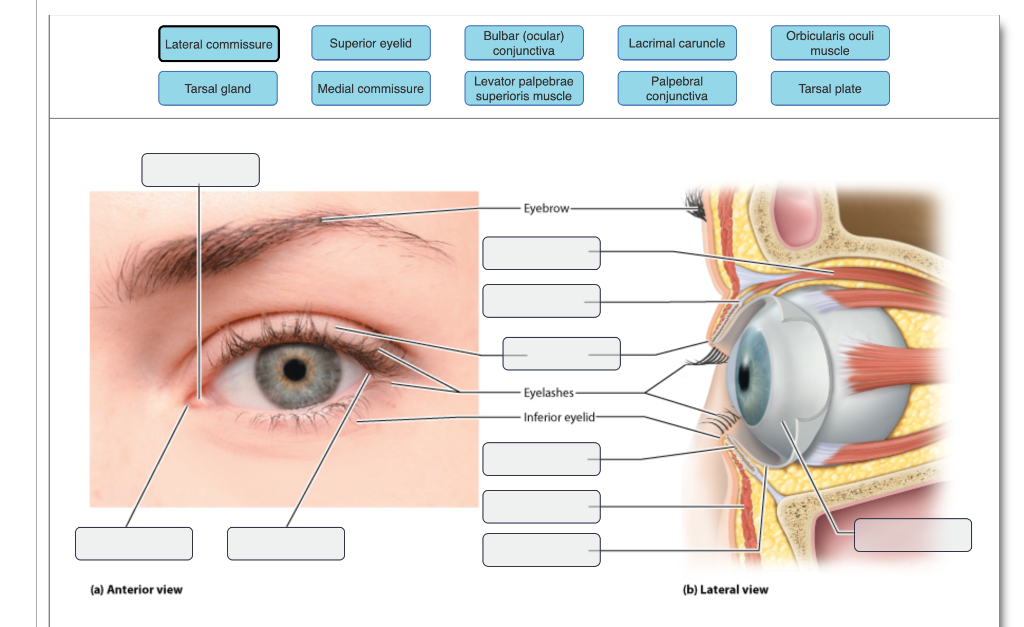 Solved Bulbar Ocular Conjunctiva Orbicularis Oculi Musc

 Royalty Free Conjunctiva Stock Images Photos Vectors
:max_bytes(150000):strip_icc()/GettyImages-695204442-b9320f82932c49bcac765167b95f4af6.jpg) Structure And Function Of The Human Eye
Structure And Function Of The Human Eye
 The Anatomy And Cell Biology Of The Human Cornea Limbus
Belum ada Komentar untuk "Anatomy Of Conjunctiva"
Posting Komentar