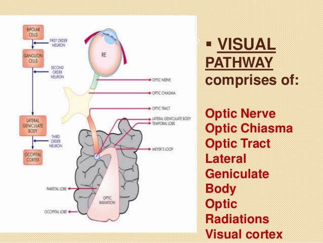
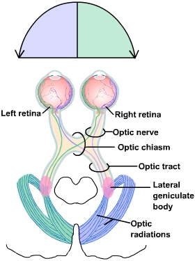
Visual Pathway Anatomy
Gross anatomy below the visual pathway is. The visual pathway describes the anatomical pathway by which electrical signals generated by the retina are sent to the brain fig.
 Visual Pathway An Overview Sciencedirect Topics
Click on a label to display the definition.
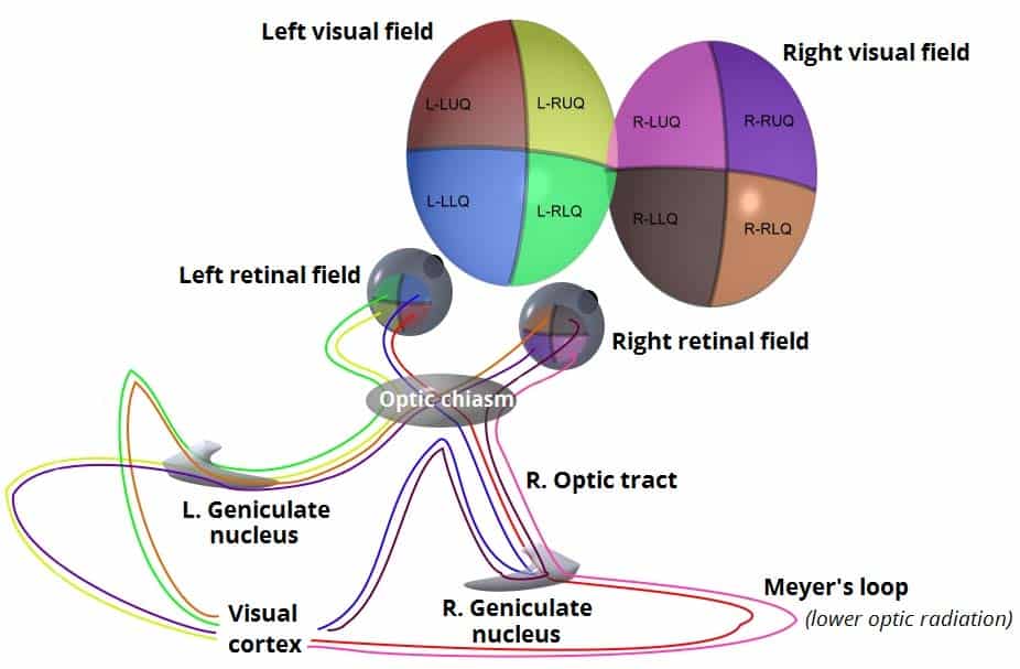
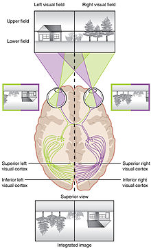
Visual pathway anatomy. Sivateja challa sssihms anatomy of visual pathway 2. Discover the worlds. The visual pathway shows a precise retinotopical organization at all levels that gives the anatomical background for symptoms when some part of optic pathway is damaged.
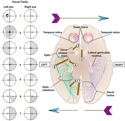
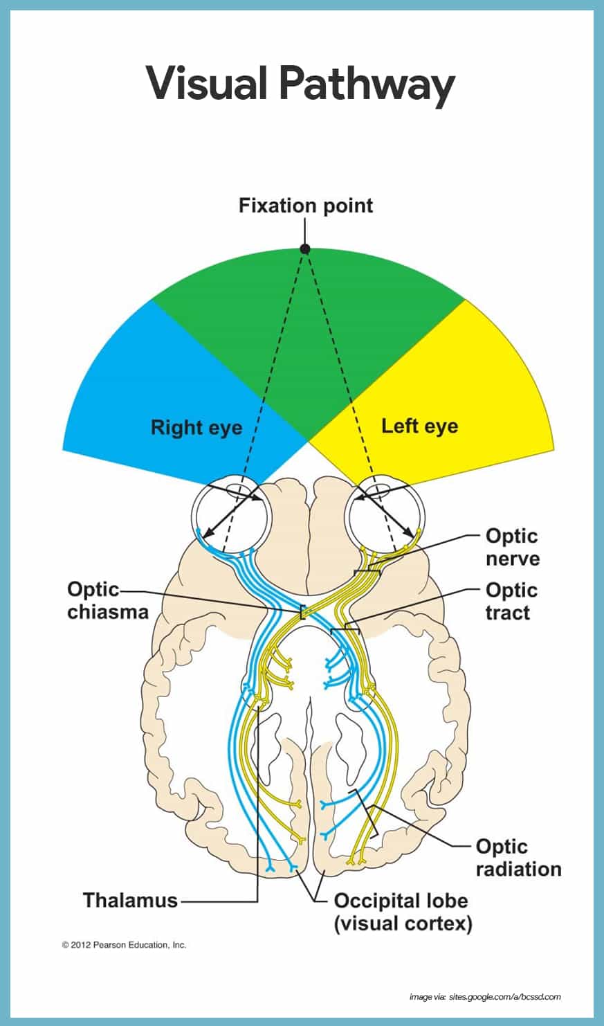
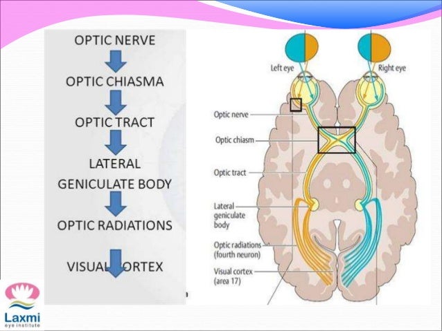
It begins at the retina and terminates at the primary visual cortex with several intercortical tracts. Intracranial the visual pathway within the middle cranial fossa the optic nerves from each eye unite to form the optic chiasm. Choose from 500 different sets of visual pathways anatomy flashcards on quizlet.
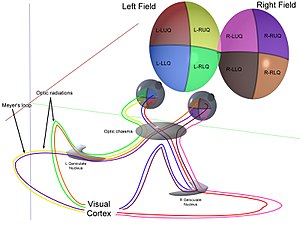
Visual feld retinal quadrent head eye are maintained in a fixed posture one eye is closed area seen by the open eye constitute the visual field of that eye visual field of the two eye overlap to a great extent on either side there is a small area which is seen only by eye of that side. The visual system transmits visual information from the retina within the eyes to the primary visual cortex of the occipital lobe as well as the pretectal nuclei and superior colliculi of the midbrain. Anatomy of visual pathway 1.
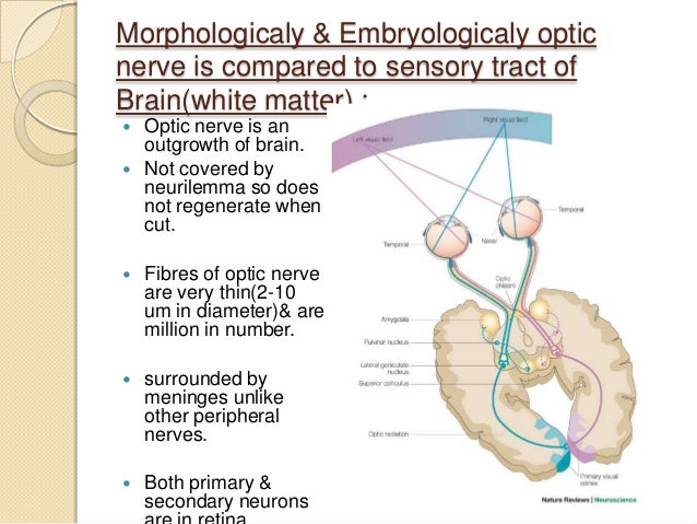
Tap on the image or pinch out and pinch in to resize the image. The visual pathway refers to the anatomical structures responsible for the conversion of light energy into electrical action potentials that can be interpreted by the brain. The nerve fibers of the retina representing the axons of the ganglion cells collect together at the optic disc before passing out of the eye through the orbital bones and into the brain via the optic nerve the second cranial nerve.
Learn visual pathways anatomy with free interactive flashcards. At the chiasm fibres from the nasal medial half of each retina cross over to the contralateral optic tract while fibres from the temporal lateral halves remain ipsilateral.
 Patterns Of Shared Variation In Visual Pathway Anatomy A
 Ecr 2019 C 3361 Visual Pathway Lesions From Visual
 Visual System Wikipedia

 Anatomy Of Visual Pathway And Its Lesions
 Visual Processing Cortical Pathways Section 2 Chapter 15

 Neural Pathways What Are They How Types Dysfunction

 Jaypeedigital Ebook Reader
 Visual Fields And Lesions Of The Visual Pathways Cn Ii
 Eye Opener Physiology Visual Pathway
 Visual System Radiology Reference Article Radiopaedia Org
 Visual Processing Cortical Pathways Section 2 Chapter 15
 Visual Fields And Lesions Of The Visual Pathways Cn Ii
 The Optic Nerve Visual Pathway Chiasm Tract
 Anatomy Of The Human Visual Pathway Springerlink
 Special Senses Anatomy And Physiology Nurseslabs
Anatomy Of Visual Pathway And Its Lesions
 Vision Introduction To Psychology
Visual Pathways
 Visual System Wikipedia

 Retinohypothalamic Tract Visual Acuity Alpf Medical Research
 Human Vision And Function Part 1 How The Eye Works 1 4
 Visual Pathway
 Patterns Of Individual Variation In Visual Pathway Structure
 Epos
Epos
 Visual Cortex Areas
 Visual Processing Cortical Pathways Section 2 Chapter 15
Belum ada Komentar untuk "Visual Pathway Anatomy"
Posting Komentar