Normal Heart Anatomy
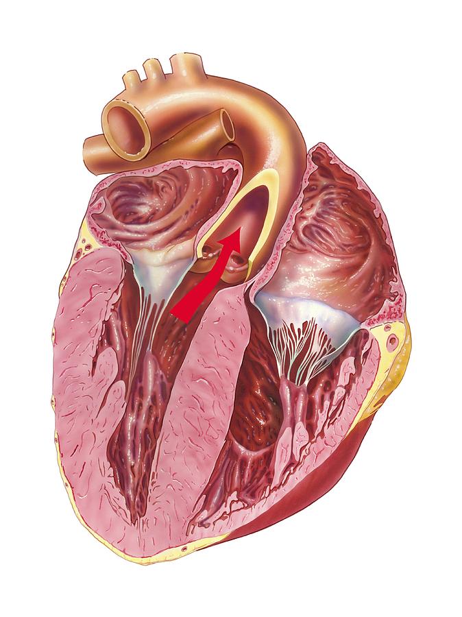
The left ventricle pumps blood into the aorta sending oxygenated blood to the rest of the body. Normal heart anatomy the main function of the heart is to deliver oxygen rich blood to every cell in the body.
 4 Savealifex Pals Normal Heart Anatomy Physiology
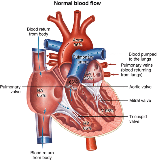
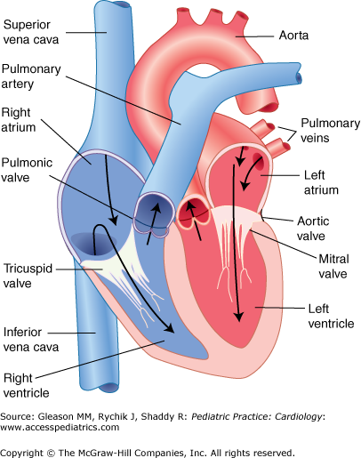
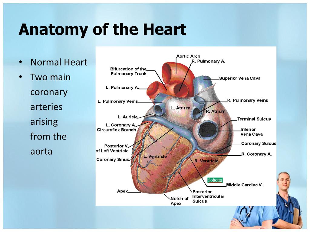
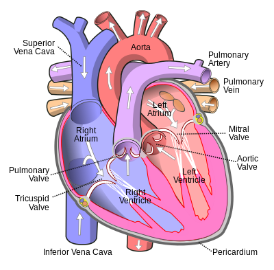
The heart the heart itself is made up of 4 chambers 2 atria and 2 ventricles.

Normal heart anatomy. At rest a normal heart beats around 50 to 99 times a minute. The atria are the two upper chambers and the ventricles are the two lower chambers. The heart is a hollow muscle comprised of four chambers surrounded by thick walls of tissue septum.
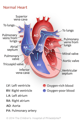
An average human has around 5 liters 8 pints of blood which is constantly pumped throughout the body. The atria are the receiving chambers for blood returning from the body and the lungs. De oxygenated blood returns to the right side of the heart via the venous circulation.
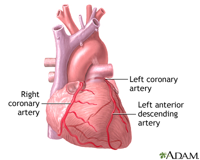
The arteries are the passageways through which the blood is delivered and the veins are the passageways through which the blood is collected and returned to the heart. It is pumped into the right ventricle and then to the lungs where carbon dioxide is released and oxygen is absorbed. The atria are smaller than the ventricles and have thinner less muscular walls than the ventricles.
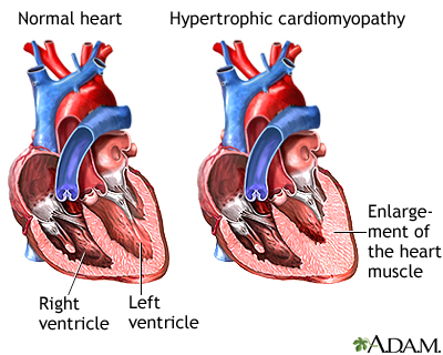
The heart is made up of the two atria which receive blood and two ventricles which are the actual pumps of the heart. Exercise emotions fever and some medications can cause your heart to beat faster sometimes to well over 100 beats per minute. The right side of the heart has less myocardium in its walls than the left side because the left side has to pump blood through the entire body while the right side only has to pump to the lungs.
The heart contains 4 chambers. The heart is a hollow muscle comprised of four chambers surrounded by thick walls of tissue septum. Its primary job is to pump blood throughout the body.
The inside of a normal heart is divided into 4 areas. The left and right halves of the heart work together to pump blood throughout the body. Blood in need of oxygen flows in from the body and enters the right atrium.
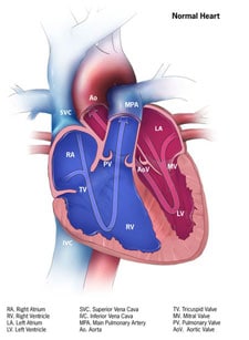
Normal heart anatomy and blood flow the heart has four separate chambers. The atria are the two upper chambers and the ventricles are the two lower chambers. The right atrium left atrium right ventricle and left ventricle.
The heart is a hollow muscular organ about the size of a fist. Chambers of the heart. Normal heart anatomy and physiology understanding normal cardiac anatomy and physiology is an important component of performing acls.
The heart blood and blood vessels combined are referred to as the circulatory system. The upper two chambers are called the right and left atria ra and la.
 Heart Anatomy And Types Of Heart Disease Vector Illustration
 Critical Congenital Heart Disease Genetics Home Reference
 What Is Atrial Fibrillation Afib
Pulmonic Stenosis In Dogs And Cats Veterinarypartner Com
Anatomy Tutorial Cardiac Valve Nomenclature Atlas Of
 Mayo Clinic Radio Structural Heart Disease Mayo Clinic
 Normal Cardiac Anatomy And Clinical Evaluation Springerlink
 How The Healthy Heart Works American Heart Association
 Chapter 1 Normal Cardiac Physiology And The Cardiac Exam
Details About 1 1 Human Heart Anatomy Model Anatomical Medical Circulation System Internal Us
 In Depth Reports Heart Bypass Surgery Series
 Cardiac Muscle Definition Function Structure Britannica
 Congenital Heart Defects How The Heart Works Cdc
 Anatomy Of The Heart And Lungs And Thoracic Surgery Ppt
 Heart Failure Lima Memorial Health System
 4 Pals Normal Heart Anatomy Physiology
 How The Normal Heart Works Children S Hospital Of Philadelphia
The Normal Heart Congenital Heart Disease Cove Point



Belum ada Komentar untuk "Normal Heart Anatomy"
Posting Komentar