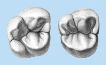
Molar Anatomy
Dental anatomy is a field of anatomy dedicated to the study of tooth structure. These teeth erupt at around age 18 but are often surgically removed.

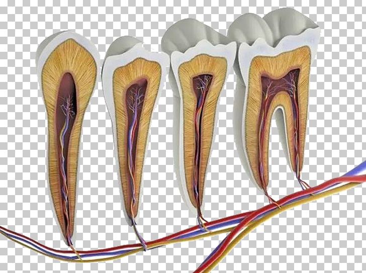
Flat teeth in the rear of the mouth best at grinding food.
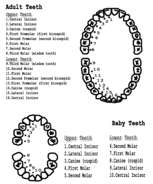
Molar anatomy. Wisdom teeth or third molars 4 total. Carabelli documented the particular anatomy of maxillary molars as early as 1844. Molars 8 total.
There are 12 permanent molars. Dental anatomy is a field of anatomy dedicated to the study of human tooth structures. The development appearance and classification of teeth fall within its purview.
Usually there are 20 primary te. Dental anatomy is also a taxonomical science. 300 dental anatomy facts part 5 molars nbde part 1 duration.
They are the sixth seventh and eighth teeth from the mid line. Numerous subsequent publications discussed the complexity of maxillary molar anatomy. Most often the mesiobuccal root and the occurrence of a second mesiobuccal mb2 canal have been in the main focus.
Pass the dental boards 80723 views. Twelve molars are usually present in an adult human in groups of three. The anatomy of maxillary molars is very complex and the root canal treatment of this particular group of teeth represents a major challenge for dentists.
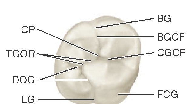
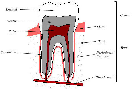
It makes up approximately two thirds of the tooth. Dental anatomy is also a taxonomic science as it is concerned with the naming of teeth and their structures. Anatomical characteristics of permanent primary molars learn with flashcards games and more for free.
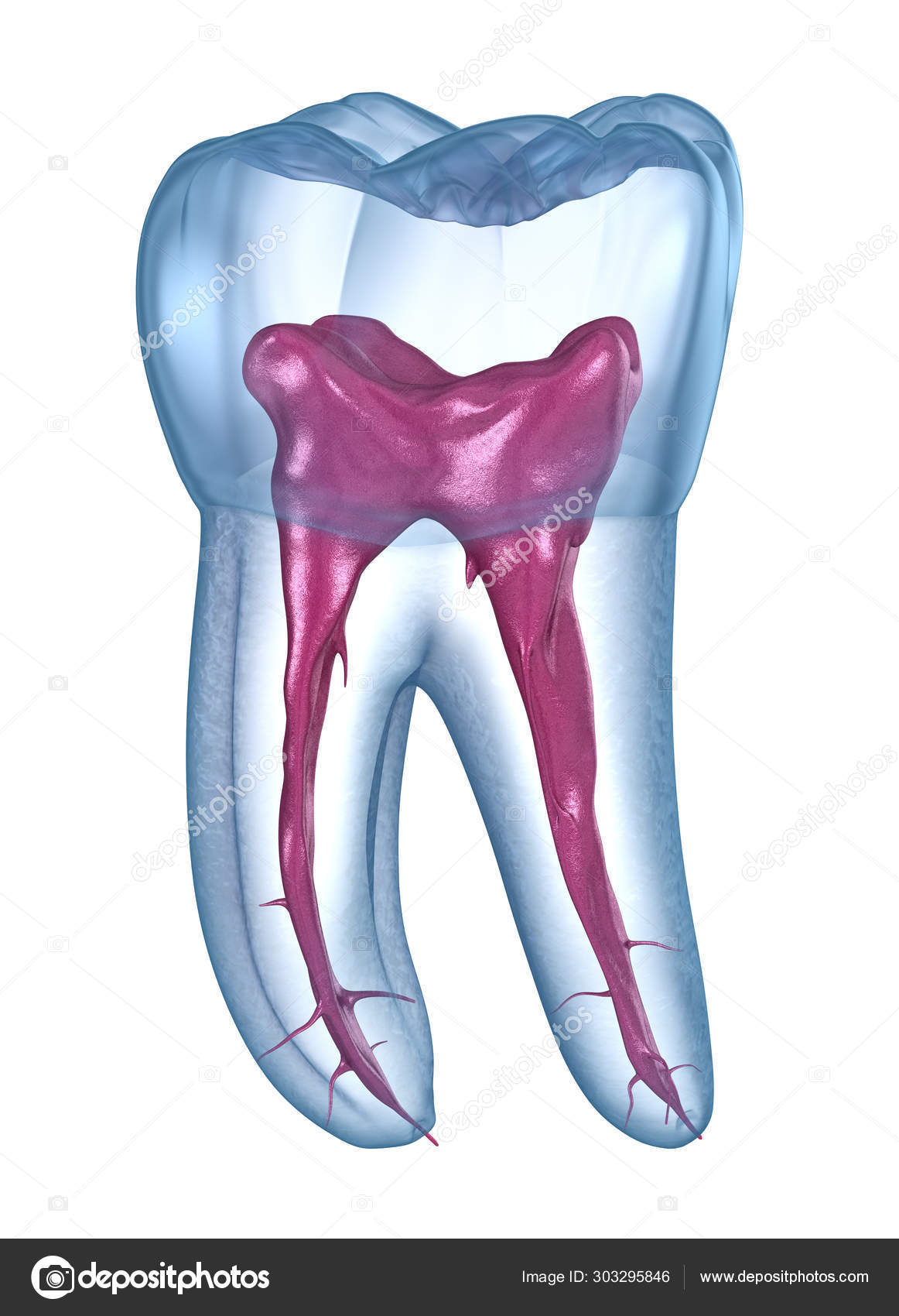
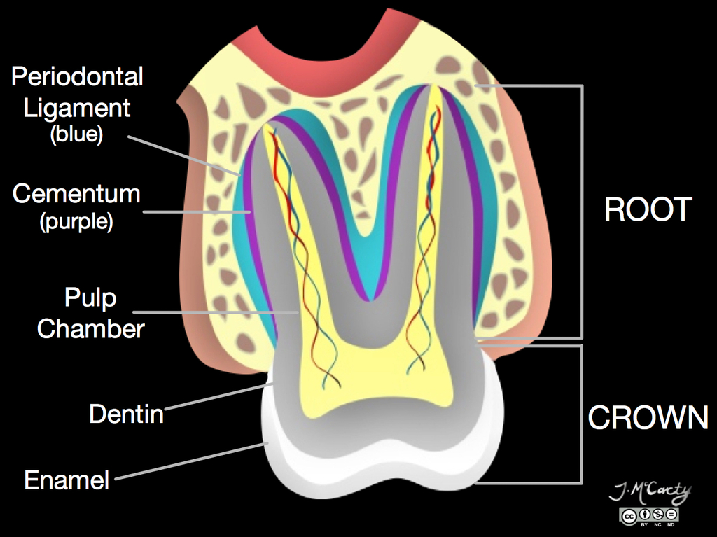
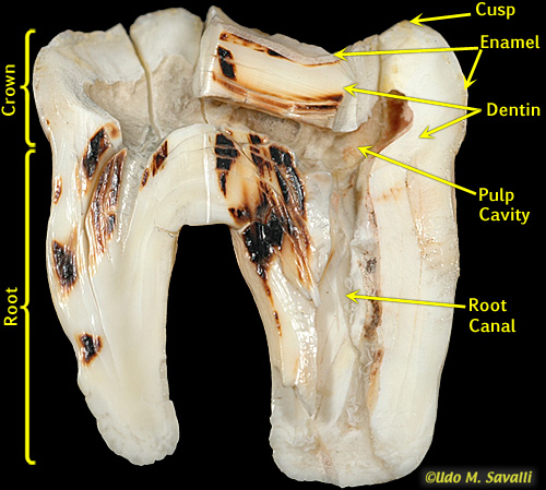
The root canal is a passageway that contains pulp. Its made up of several parts. The development appearance and classification of teeth fall within its field of study though dental occlusion or contact between teeth does not.
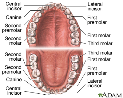
It is concerned with the naming of teeth and the structures of which they are made this information serving a practical purpose in dental treatment. Six maxillary and six mandibular. It is found in most mammals that use their posterior teeth to grind food.
A molar tooth is located in the posterior back section of the mouth. Also called cement this bone like material covers the tooths root. These teeth are typically larger than the premolars and have a larger surface area to in order to chew.
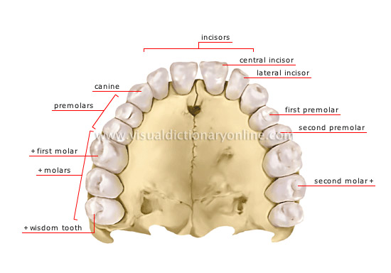
The six permanent molars in each arch are the first sec ond and third molars on either side of the arch. The root is the part of the tooth that extends into the bone and holds the tooth in place. Tooth formation begins before birth and the teeths eventual morphology is dictated during this time.
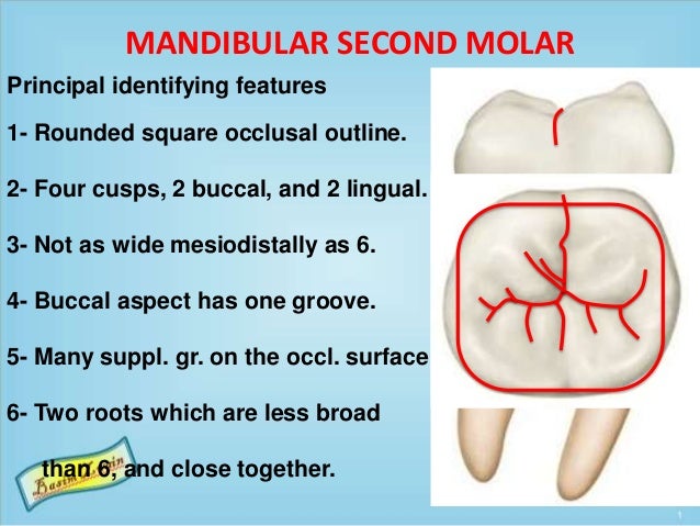 Mandibular Molars
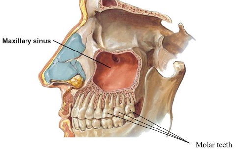 Sinus Lifts And Dental Implants Dr Nima Massoomi Dmd Med Md

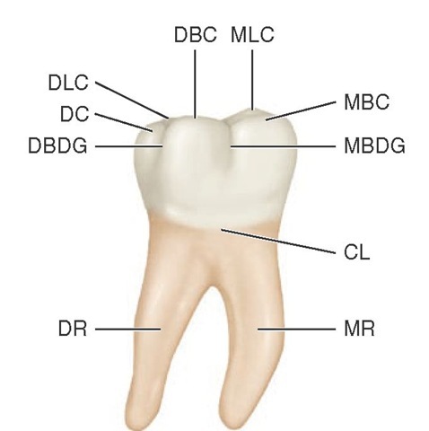
 Maxillary Molars Blackboard Quiz Dentistry 1512 With
 Science Source Structure Of A Molar Artwork
 Step By Step Protocol Restoring The First Upper Molar
 15 Molars Pocket Dentistry
 Dental Anatomy Wikipedia
 Maxillary Molar Root Canal Morphology And Anatomy Key

 Molar Anatomy Shared By Dr Gregory Bowen San Antonio
 Human Tooth Anatomy Stock Illustration Illustration Of
 General Anatomical Features Of Maxillary Third Molars Part
 Dental Anatomy Medlineplus Medical Encyclopedia Image
 Dental Anatomy Maxillary Molar Ppt English Flashcards
 Maxillary Molar Anatomy Illustration Radiology Case
 Dental Anatomy Permanent Molars
 Anatomical Teaching Models Plastic Human Dental Models
 External And Internal Root Canal Anatomy Of The First And
 Mandibular First Molar Wikipedia
Dental Root Anatomy First Maxillary Molar Tooth Medically
 Bio370 Mammal Teeth
 Human Tooth Anatomy Molar Pulp Png Clipart Anatomic
 Stock Illustration
Belum ada Komentar untuk "Molar Anatomy"
Posting Komentar