Femoral Nerve Anatomy
Its principal function is to carry instructions to the muscles that straighten the leg such as the quadriceps found in the anterior thigh. Nerves are complex structures that branch out like a tree.
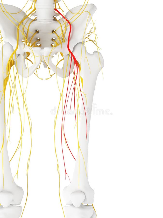 The Femoral Nerve Stock Illustration Illustration Of
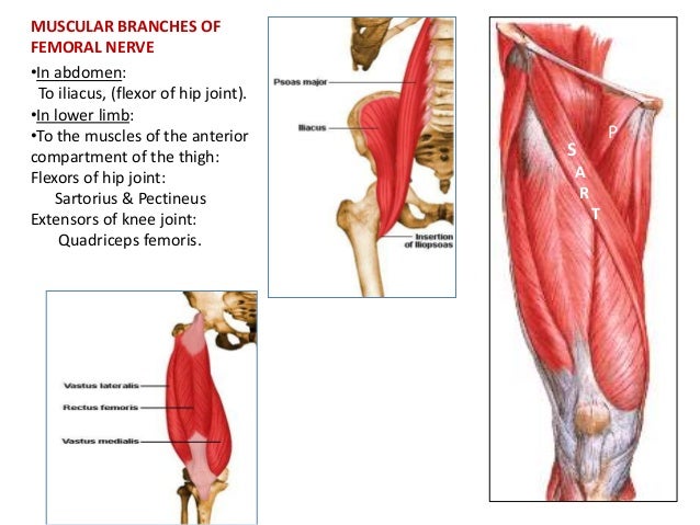
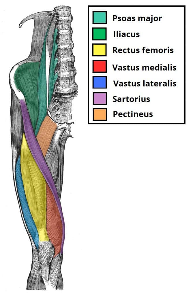
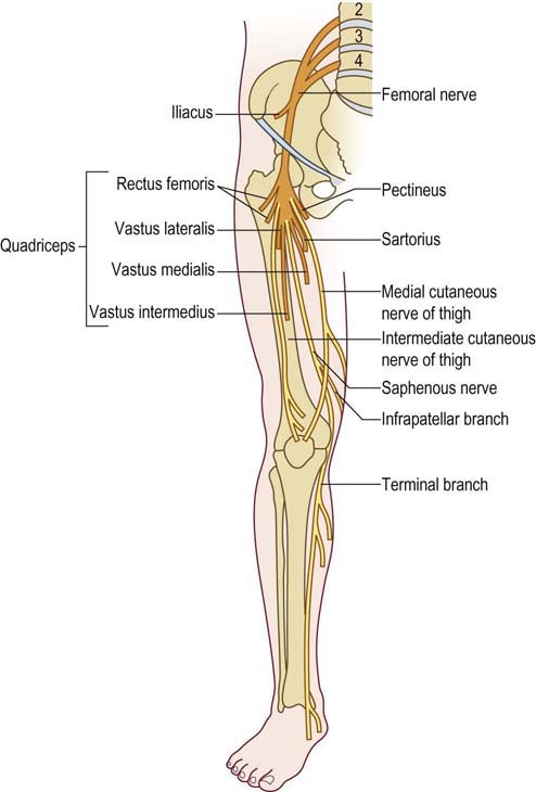
The first motor branch innervates iliacus.

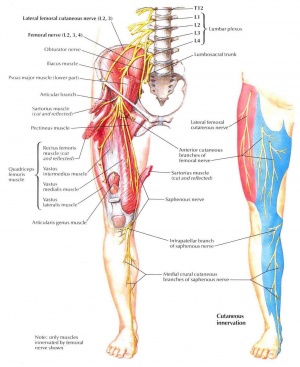
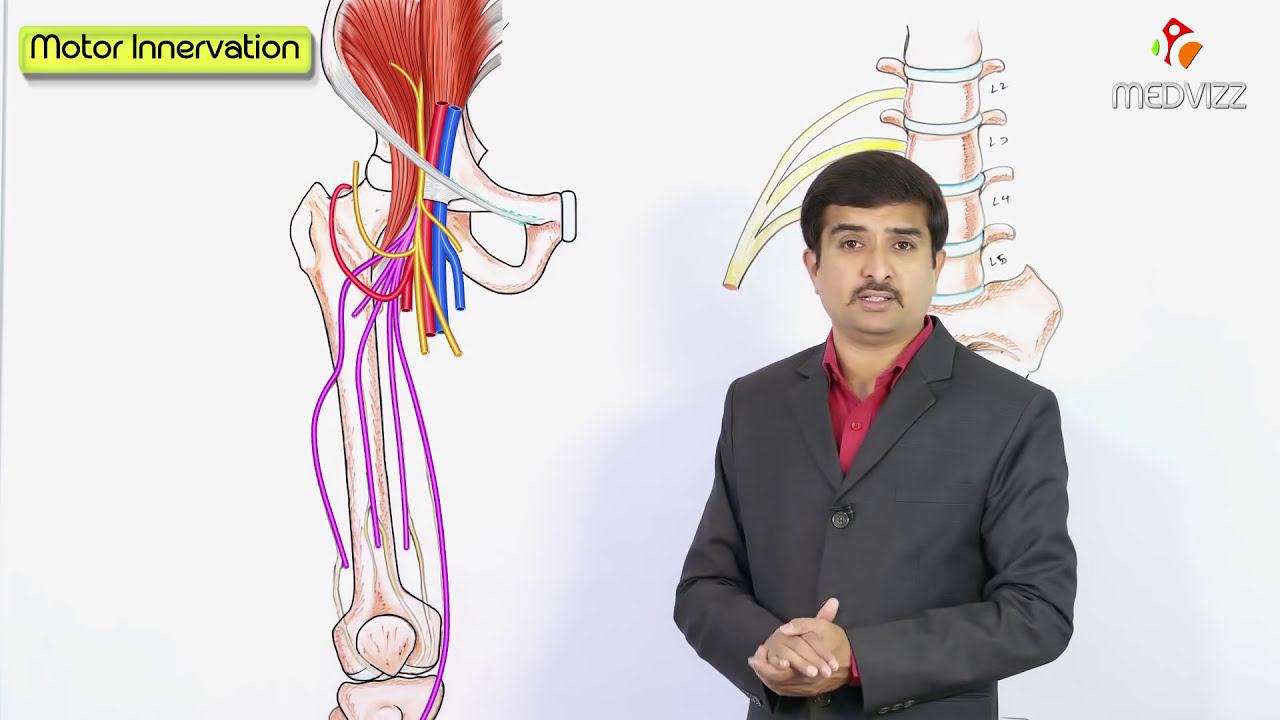
Femoral nerve anatomy. It may seem counter intuitive that the femoral nerve the nerve of the anterior thigh. It is the largest branch of the lumbar plexus and arises from the dorsal divisions of the ventral rami of the second third and fourth lumbar nerves l2 l3 and l4. The femoral nerve innervates the muscles which allow for the flexion of the hip and extension of the knee.
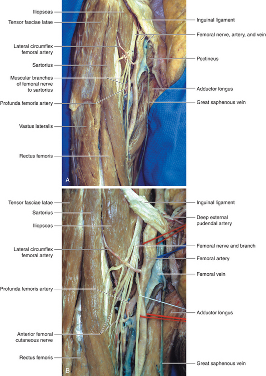
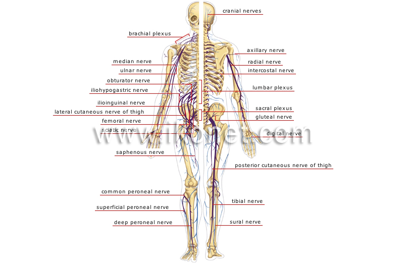
The femoral nerve is one of the major nerves that innervates the legs. The superficial branch of the femoral nerve first gives rise to the anterior. Femoral nerve is the major nerve supplying the anterior compartment of the thigh.
Fibres arise separately in lumbar plexus passes anterior to femoral nerve may terminate as saphenous nerve cutaneous branch. We may also use selective stimulation of the compound femoral nerve to control distal muscles and functions such as leg extension at the knee joint. The femoral nerve helps in hip muscle movement and straightening of the legs and this forms the motor function.
The femoral nerve helps move your hips and knees anatomy. Femoral nerve dysfunction can occur when the nerve is compressed entrapped. The femoral nerve anatomy is conducive to selective stimulation with multicontact nerve cuff electrodes.
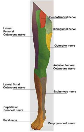
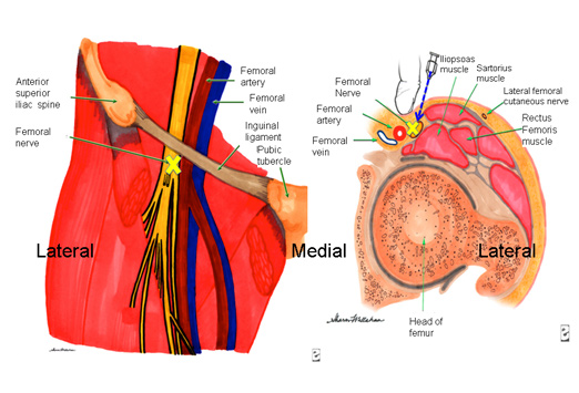
It provides sensory innervation to the skin of the anterior thigh. The femoral nerve supplies the muscles of the anterior thigh. This nerve has a triangular transverse cross section and passes through the pelvic region in the grove between.
The femoral nerve handles several important functions. The femoral nerve is the major nerve that serves the tissues of the thigh and leg including the muscles and skin. Femoral nerve splits into two or three separate slips within the psoas muscle but unites to descend as a single bundle 2.
Piereces the sartorius and fasica lata medial to the knee and provides cutaneous innervation to the skin anteriorly over the patella. While the much larger sciatic nerve also passes through the thigh on its way to the lower leg and foot only the femoral nerve innervates the tissues of the thigh.
 Femoral Nerve Anatomy Orthobullets
 Femoral Nerve Physiopedia

 Femoral Nerve Neupsy Key
 Femoral Nerve
 The Femoral Nerve Course Motor Sensory Teachmeanatomy
 Femoral Nerve Anatomy Everything You Need To Know Dr Nabil Ebraheim
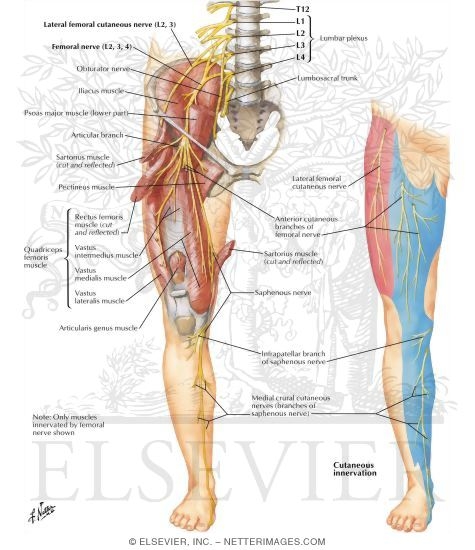 Femoral Nerve And Lateral Femoral Cutaneous Nerves
 Instant Anatomy Diagram
 Femoral Neuropathy Neupsy Key
 Femoral Nerve Anatomy Orthobullets
 Femoral Nerve Block Sciencedirect

 Femoral Nerve Transfers For Restoring Tibial Nerve Function
 Femoral Artery Wikipedia
 Femoral Nerve Anatomy Lecture Usmle Step 1 Gross Anatomy Usmle Videos By Dr G Bhanu Prakash
 Untitled Document

 Femoral Nerve Block

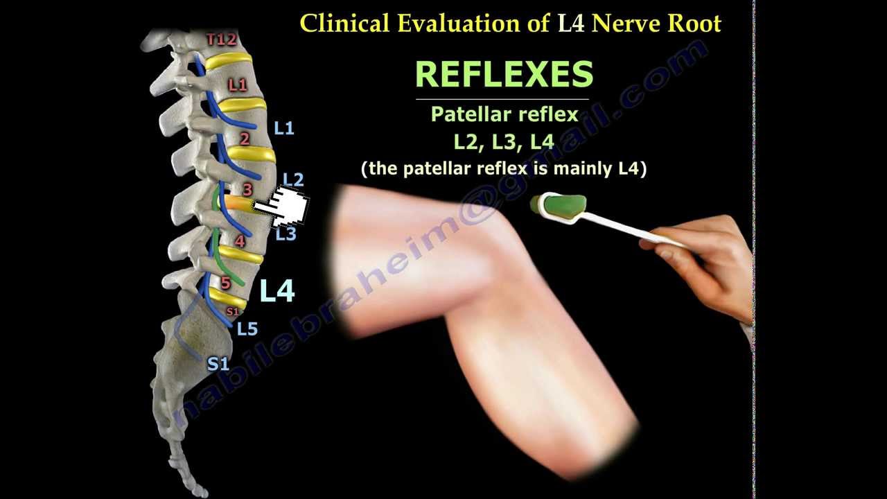
 Femoral Nerve L2 L4 Emg Review
![]() Femoral Nerve Anatomy And Clinical Notes Kenhub
Femoral Nerve Anatomy And Clinical Notes Kenhub
 Femoral Nerve Anatomy Pictures And Information
 Anterior Cutaneous Branches Of The Femoral Nerve Wikipedia
Belum ada Komentar untuk "Femoral Nerve Anatomy"
Posting Komentar