Smv Anatomy
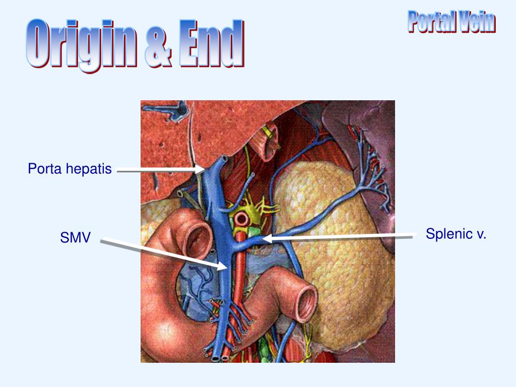
At its termination behind the neck of the pancreas the smv combines with the splenic vein to form the hepatic portal vein. Intestinal malrotation is a congenital anatomical anomaly that results from an abnormal rotation of the gut as it returns to the abdominal cavity during embryogenesis.
 The Pancreas Radiology Key
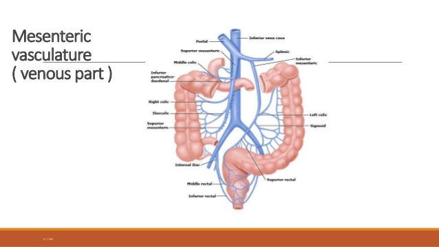
In human anatomy the inferior mesenteric vein imv is a blood vessel that drains blood from the large intestine.
Smv anatomy. It follows a path similar to that of the superior mesenteric artery. The superior mesenteric vein smv accompanies the superior mesenteric artery sma and drains the midgut to the portal venous system. Although some individuals live their entire life with malrotated bowel withou.
This vein is located in the abdominal cavity next to the superior mesenteric artery. Where it ends near the neck of the pancreas. For a general discussion refer to intestinal ischemia.
The inferior mesenteric vein usually enters the splenic adjacent to the confluence but it may also enter the smv either at or just caudal to the confluence. It usually terminates when reaching the splenic vein which goes on to form the portal vein with the superior mesenteric vein smv. Gross anatomy origin single vessel arising anteriorly from the abdom.
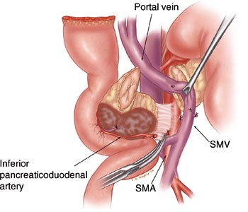
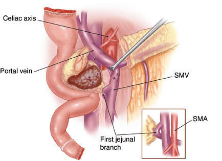
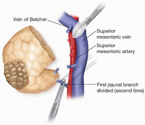
The splenic vein and superior mesenteric vein smv join to form the main portal vein see chapter 2. The anatomy of the smv as it pertains to the performance of pd and describe our surgical approach to tumors that involve either of the 2 first order branches of the smv. Acute superior mesenteric vein thrombosis is one of the less common causes of intestinal ischemia.
Often despite thrombosis of the smv small bowel necrosis does not occur presumably due to persistent arterial supply and some venous drainage via collaterals. Gross anatomy origin and course mesenteric venous arcades which accompany the arteries unite to form the j. The superior mesenteric artery sma is one of the three non paired major visceral arteries in the abdominal cavity arising from the abdominal aorta and supplying the midgut.
The superior mesenteric vein also known as smv transports blood from the small intestine and the cecum. The superior mesenteric vein smv is a blood vessel that drains blood from the small intestine jejunum and ileum. In human anatomy the superior mesenteric artery sma arises from the anterior surface of the abdominal aorta just inferior to the origin of the celiac trunk and supplies the intestine from the lower part of the duodenum through two thirds of the transverse colon as well as the pancreas.
 Illustration Of Superior Mesenteric Vein Smv Resection
 Mapping Of The Superior Mesenteric Vessels For Artery First
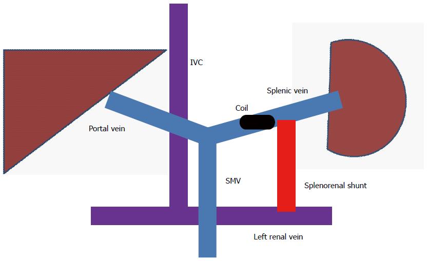 Novel Therapy For Non Cirrhotic Hyperammonemia Due To A
 Medical Management Of Portal Hypertension Complications
 Mapping Of The Superior Mesenteric Vessels For Artery First
 Normal Venous Anatomy Arrows Indicate The Directional Flow
 Pancreatic Carcinoma Springerlink
 Pancreatic Carcinoma Springerlink
 Smv X Ray Anatomy Diagram Quizlet
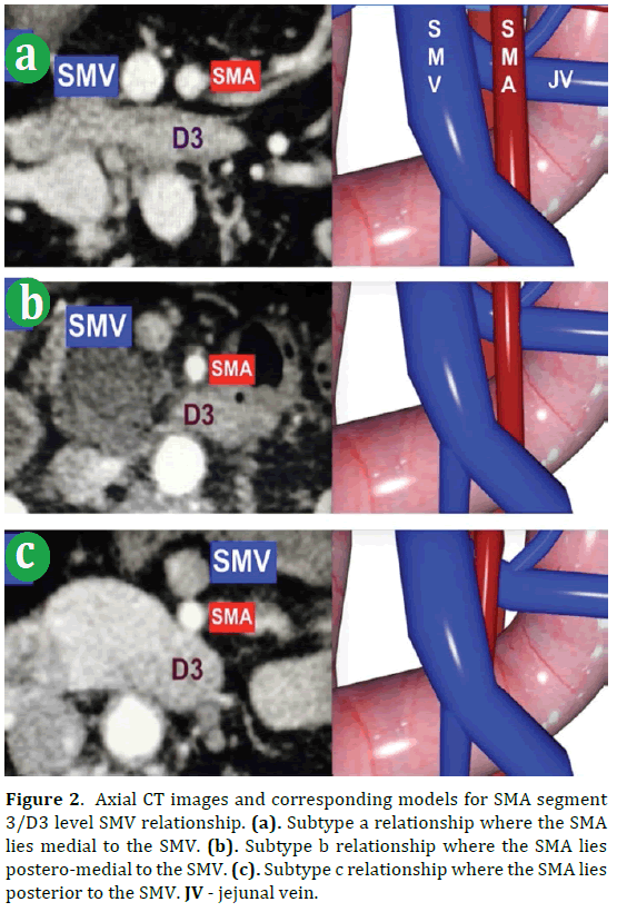
 Figure 1 From Anatomy Of The Superior Mesenteric Vein With
 Pin On Smv Portal Vein Thrombosis

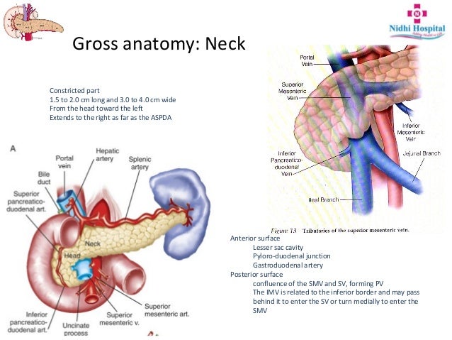 Resectional Anatomy Of Pancreas
 Normal Venous Anatomy Arrows Indicate The Directional Flow
 Pancreas Basicmedical Key
 Pin On X Ray
 Surgical Anatomy Of The Superior Mesenteric Vessels Related
 The Role Of Neoadjuvant Therapy In Surgical Treatment Of
 Smv Sinuses Anatomy Diagram Quizlet
 View Large Accessmedicine Mcgraw Hill Medical
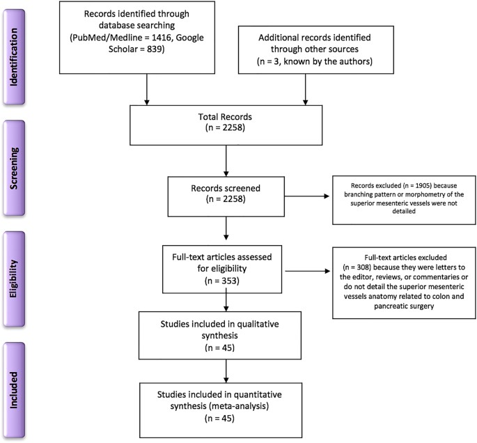 Surgical Anatomy Of The Superior Mesenteric Vessels Related
 Portal Vein Anatomy And Conditions Docsity
 Acute Smv Thrombosis
 X Ray Anatomy Smv Cranium Schuller Diagram Quizlet
Where There Is Blood There Is A Way Unusual Collateral
 Anatomy Of The Superior Mesenteric Vein Smv Portal Vein
Ppt Portal Vein Powerpoint Presentation Free Download
 Pancreaticobiliary Surgery General Considerations
Belum ada Komentar untuk "Smv Anatomy"
Posting Komentar