Anal Canal Anatomy
Structure of rectum serous muscular ouer longitudinal inner circular submucous mucous 4. Levator anus muscles components 3.
Anatomy Of The Rectum And Anal Canal Anat10110 Ucd Studocu
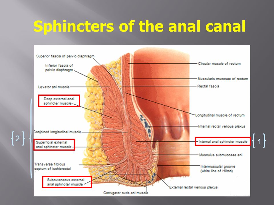
Describe the anal sphincters internal and external and provide a comparison between.
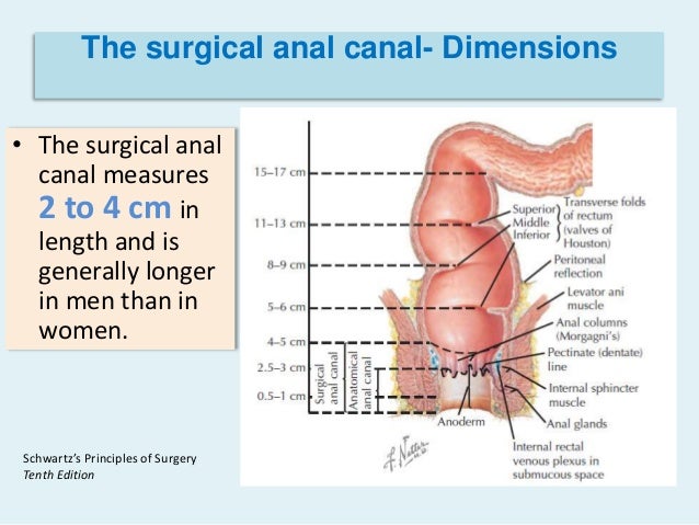
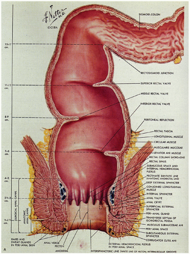
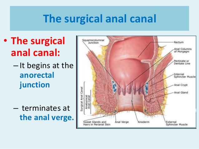
Anal canal anatomy. Define and describe the internal features of the anal canal. Anal canal anatomy it is about 1 15 inches long. This part differs from the part above the pectinate line in several respects including innervation venous and lymphatic drainage and possibly lining epithelium.
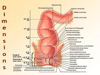
Triangles pelvic outlet 2. The anal canal is between 25 and 5 in length and is guarded by two muscles that control the release of waste from the rectum. Anatomically the anal canal extends from the level of the upper aspect of the pelvic diaphragm to the anus.
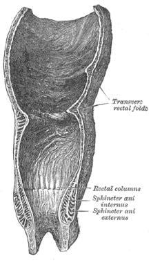
It is encircled by a muscle called sphincter which controls intestinal movements. The upper region has 5 to 10 rectal columns each column containing a small artery and vein. The anal canal is surrounded by internal and external anal sphincters which play a crucial role in the maintenance of faecal continence.
It is surrounded by a muscular sphincter system which tightly closes the lumen. The anal canal is the most terminal part of the lower gi tractlarge intestine which lies between the anal verge anal orifice anus in the perineum below and the rectum above. The anal canal is the short terminal portion of the rectum through which wastes from the large intestine are excreted from the body.
The anal canal connects with the rectum at the point where it passes through a muscular pelvic diaphragm. Anatomy of anal canal 1. The internal anal sphincter is permanently contracted through the sympathetic tonus and relaxes under parasympathetic influence.
The upper two thirds of the anal canal is supplied with blood mainly from the superior rectal artery a branch of the inferior mesenteric artery and drains into the superior rectal veins. The anatomy of the anal canal is also key to understanding the spread of neoplastic disease based on its origin in the upper or lower anal canal. It is formed from a thickening of the involuntary circular smooth muscle in the bowel wall.
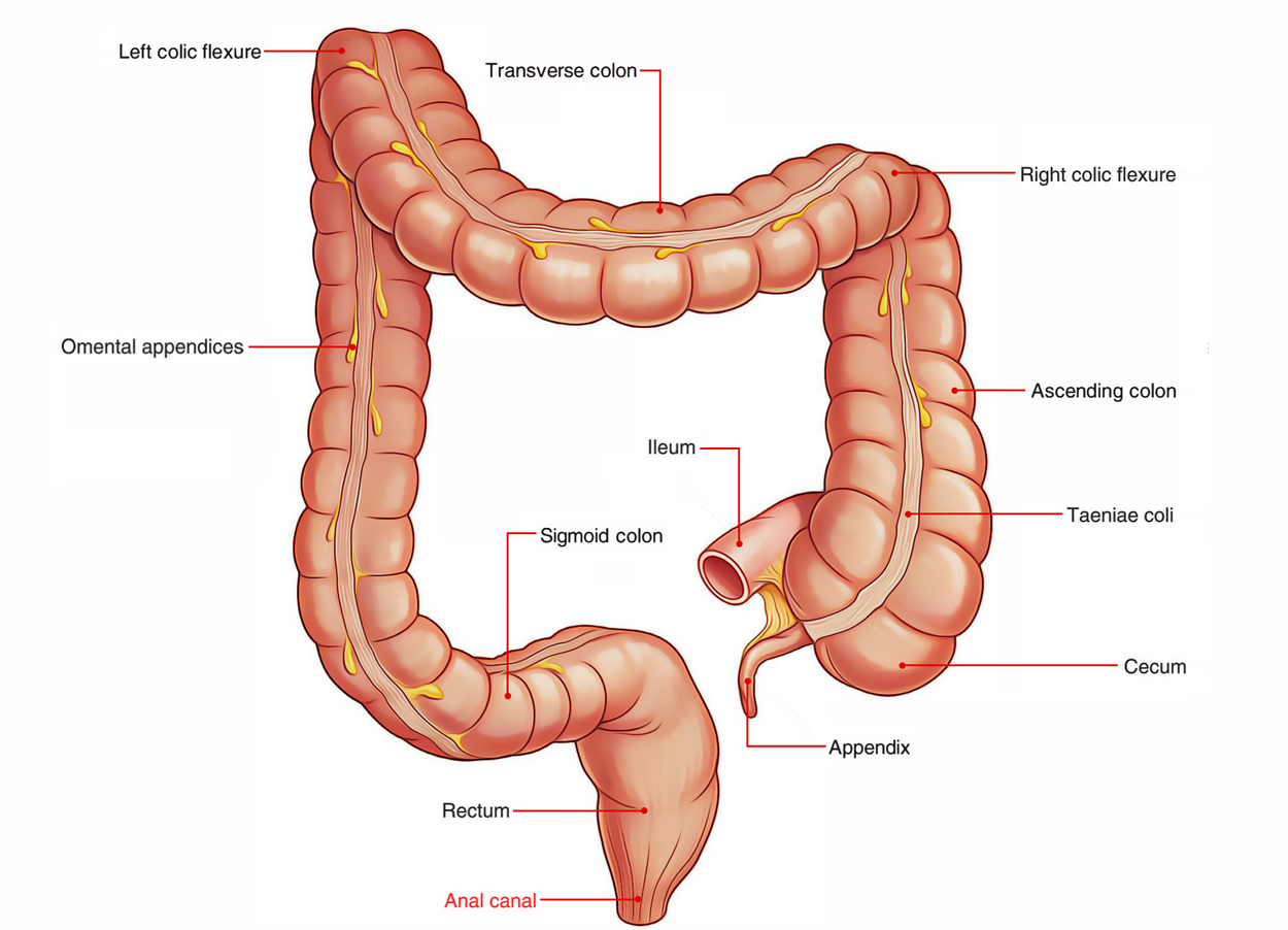
It has a narrower diameter than that of the rectum to which it is joined. The ring at the terminal portion of the anal canal is called the anus. Pectinate line anal columns valves and sinuses.
The external anal sphincter surrounds the anal canal like a clamp. The anal canal is an important part of the continence organ. Internal anal sphincter surrounds the upper 23 of the anal canal.
In surgical usage however the anal canal is frequently limited to that part of the intestine below the pectinate line.
Common Anorectal Problems Glowm
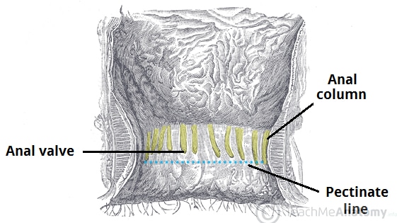 The Anal Canal Structure Arterial Supply Teachmeanatomy
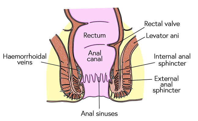 The Structures Involved In Maintaining Continence And
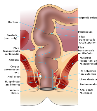 Anal Canal Anatomy Gross Anatomy Tissue Nerves And
 Anal Canal Radiology Reference Article Radiopaedia Org
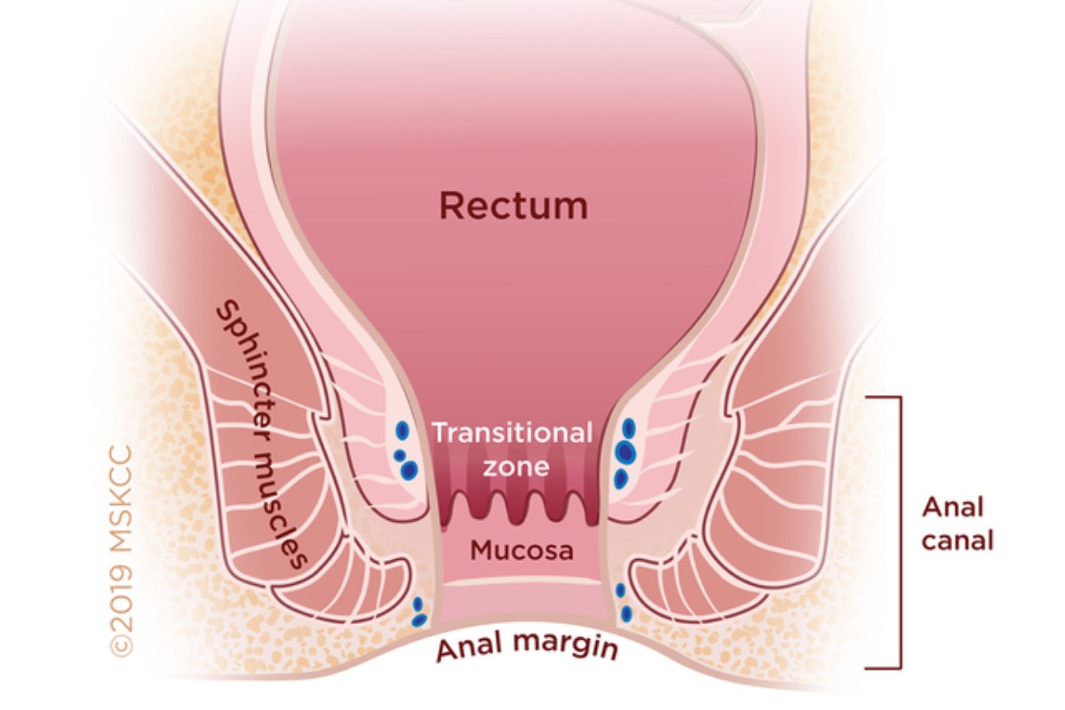 Anal Cancer Memorial Sloan Kettering Cancer Center
 Rectum Anatomy And Rectum Function Differentiate Anus Vs Rectum
 Anal Canal Anatomy Gross Anatomy Tissue Nerves And
 Anatomy Labeling Of Anal Canal Purposegames
 Easy Notes On Anal Canal Learn In Just 4 Minutes
 Viscera Rectum And Anal Canal Ranzcrpart1 Wiki Fandom
 Rectum Anal Canal Ppt Video Online Download
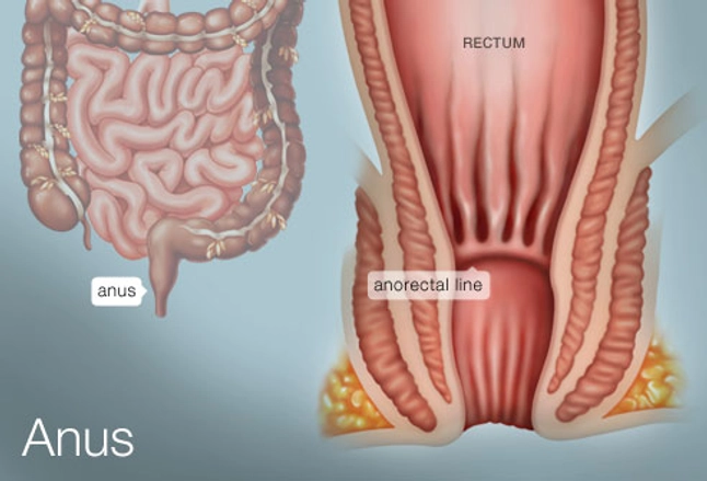 The Anus Human Anatomy Picture Definition Conditions
 Anal Canal An Overview Sciencedirect Topics
 1 Anatomy Of The Anal Canal Reprinted With The Permission
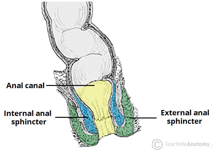 The Anal Canal Structure Arterial Supply Teachmeanatomy
 Git Anatomy The Rectum And The Anal Canal
 Figure 1 From Functional Ultrasound Of The Anal Canal The
 Anatomy Of The Anus Anal Cancer Information
 Why Does Anal Sphincter Muscle Damage Cause Faecal
 3 Venous Drainage Of The Rectum And Anal Canal Note The
 Surgical Anatomy Of The Anal Canal The Cleator Clinic
 Carcinoma Of The Anal Canal Nejm



Belum ada Komentar untuk "Anal Canal Anatomy"
Posting Komentar