Tm Anatomy
It also serves as the lateral wall of the tympanic cavity separating it from the external auditory canal. Review the anatomy of the tympanic membrane image courtesy of eprom1 try to identify the marked anatomical structures and areas belowhold the cursor over a letter to display the answerwhen you feel ready proceed to the self quiz.
The tympanic membrane is a thin membrane that separates the external ear from the middle ear.

Tm anatomy. Dysfunction of the tmj can cause severe pain and lifestyle limitation. Since the tmj is connected to the mandible the right and left joints must function together and therefore are not independent of each other. The tympanic membrane is shaped like a flat cone pointing into the middle and inner ear.
The temporomandibular joints are the two joints connecting the jawbone to the skull. This joint is unique in that it is a bilateral joint that functions as one unit. It is from these bones that its name is derived.
Tmj anatomy the temporomandibular joint tmj or jaw joint is a bi arthroidal hinge joint that allows the complex movements necessary for eating swallowing talking and yawning. Created by a team of doctors and medical students each topic combines anatomical knowledge with high yield clinical pearls seamlessly bridging the gap between scholarly learning and improved patient care. The temporomandibular joint tmj is an atypical synovial joint located between the condylar process of the mandible and the mandibular fossa and articular eminence of the temporal bone.
It is a bilateral synovial articulation between the temporal bone of the skull above and the mandible below. It is divided into a superior discotemporal space and infer. Each topic combines anatomical knowledge with high yield clinical pearls seamlessly bridging the gap between scholarly learning and improved patient care.
Tympanic membrane also called eardrum thin layer of tissue in the human ear that receives sound vibrations from the outer air and transmits them to the auditory ossicles which are tiny bones in the tympanic middle ear cavity. It acts to transmit sound waves from air in the external auditory canal eac to the ossicles of the middle ear. The tympanic membrane is a vital component of the human ear and is more commonly known as the eardrum.
It is a thin circular layer of tissue that marks the point between the middle ear and the. Teach me anatomy is a comprehensive easy to read anatomy reference. Lettering scheme taken from jerger j clinical experience with impedance audiometry.
Containing over 1000 vibrant full colour images teachmeanatomy is a comprehensive anatomy encyclopaedia presented in a visually appealing easy to read format.
 Do Thai Massage Therapists Need To Know Anatomy
 Pin By Jinjutha Aomm On Anatomy Of Ears Anatomy
 Anatomy Atlases A Digital Library Of Human Anatomy
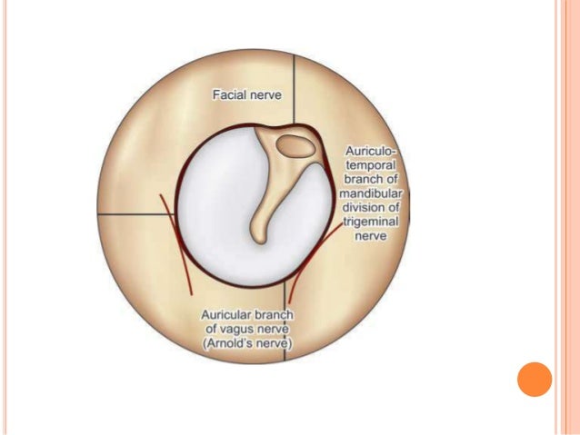 Anatomy Of External Ear And Middle Ear
 Sterling Human Body Understanding Anatomy Flexibound Book
 Gross Anatomy The Big Picture Second Edition Smartbook Ebook By K Bo Foreman Rakuten Kobo
 Tm Anatomy Of Tecta Tecta C1509g Gly And
 Artstation Anatomy Study Trilina Mai
 9780135205051 Anatomy Physiology Plus Mastering A P With
 Leptaxinus Indusarium Sp Nov Anatomy Imaged Using
 Some Basic Science For The Party Of Science Tm Anatomy
 Chapter 20 Anatomy At Minnesota Southeast Technical
 Arms Arms Anatomy Arms Print Anatomy Med School Doctor For Doctor Medical Office Decor Watercolor Arms Shoulder Anatomy
 Tympanic Membrane Tm Pars Flaccida Superior To Middle
 Tm Joint Pain Find Out What The Difference Is Between Tmd
 T M Joint Of Facial Bone Close And Open Mouth Position Anatomy And Physiology Part 70
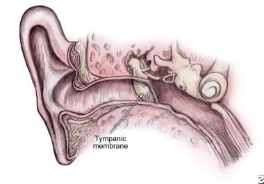 What Is The Anatomy Of The Tympanic Membrane
 Silverstein Tm Instructor S Manual And Test Fi Le T A
 Anatomy And Physiology Of The Ear Ppt Video Online Download
Plos One Cranial Anatomy Of The Gorgonopsian Cynariops


Belum ada Komentar untuk "Tm Anatomy"
Posting Komentar