Soleal Vein Anatomy
The soleus muscle and surrounding structures from grays anatomy. Detailed anatomical knowledge is required for early diagnosis using noninvasive ultrasound techniques.
 Pdf Pathophysiology Of Venous Thromboembolism With Respect
These are large veins with thin walls and are found in skeletal muscles.
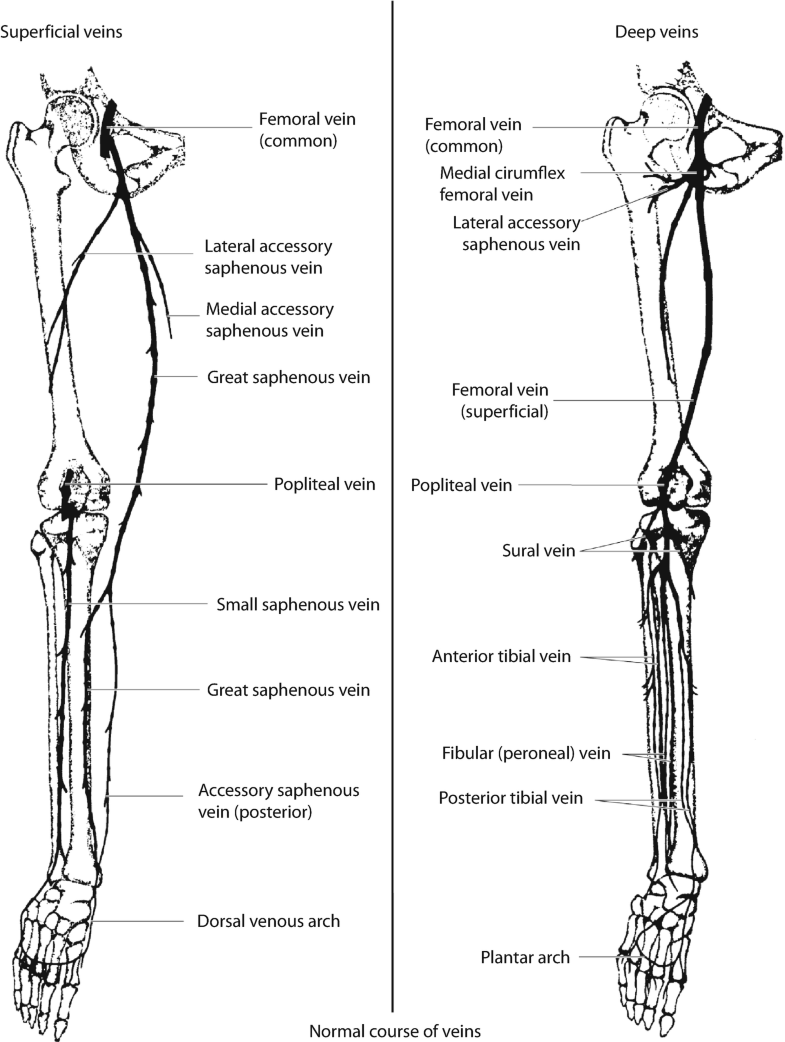
Soleal vein anatomy. Most of the gastrocnemius muscle has been removed. There are many pics related to parts of female anatomy out there. The soleal vein is the most important among the crural veins as the first site of thrombi formation.
We curate gallery of parts of female anatomy. This is a view of the back of the right leg. Once identified these patients medical records were.
Soleus veins have been implicated as the site for deep vein thrombosis dvt. 12 photos of the parts of female anatomy parts of female anatomy posted on anatomy diagram. The soleal sinusoids or soleal sinus veins are part of the deep system of the veins of the leg.
In the present work we describe the anatomy of the veins that emerge from the ventral surface of the soleus muscle. Concomitant venous thrombosis in other venous segments wereexcludedonehundredfifty eightpatientswereiden tified who had thrombus localized to the gastrocnemius andor soleal veins and had a repeat duplex ultrasound examination within 30 days of diagnosis performed in our laboratory. In contrast to the involvement of hydrostatic pressure and the structure of venous valves the anatomic characteristics of connections in the crural vein play a key role in the etiology of thrombus propagation.
Parts of female anatomy parts of female anatomy. These are large veins with thin walls and are found in skeletal muscles. Find out more other soleal vein anatomy.
This is a view of the back of the right leg. Hope you make use of it. Most of the gastrocnemius muscle has been removed.
 Isolated Distal Deep Vein Thrombosis What We Know And What
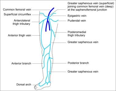 Varicose Vein Surgery Practice Essentials Anatomy
 Venous At Nova Southeastern University Studyblue
Medical Exhibits Demonstrative Aids Illustrations And Models
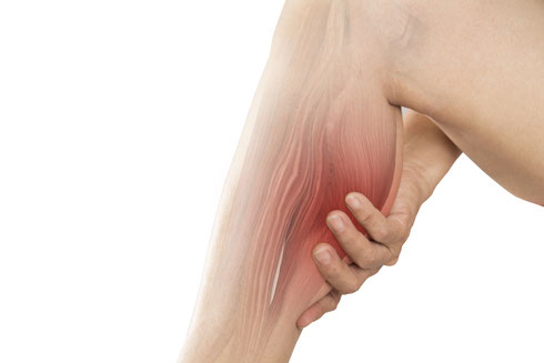 Best Venous Ultrasound For Dvt Truffles Vein Specialists
 Pdf Relationship Between Specific Distributions Of Isolated
 Popliteal Vein Radiology Reference Article Radiopaedia Org
 Anatomical Features Of The Gastrocnemius Muscle Soleus
 Doppler Ultrasound In Deep Vein Thrombosis
 Leg Dvt Normal Ultrasoundpaedia
 In Type A The Medial Gastrocnemius Veins Mgvs And The
 Venous Anatomy Physiology And Pathophysiology Plastic
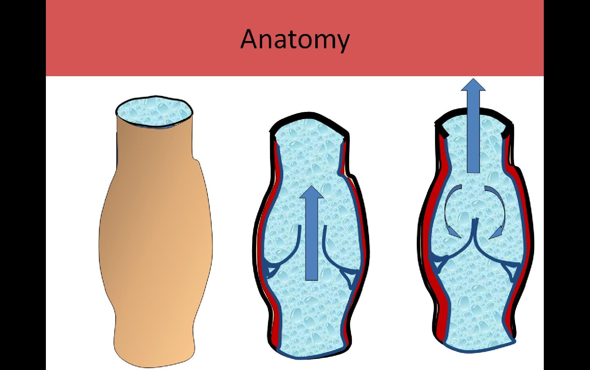 Ultrasound Registry Review Extremity Venous
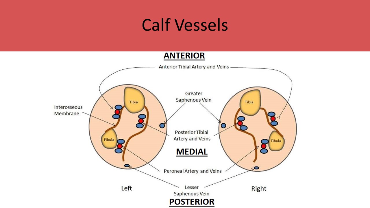 Ultrasound Registry Review Extremity Venous
010 Posterior Compartment Of Leg Anatomy Flashcards
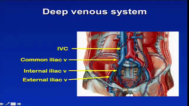 Lower Extremity Venous Imaging Anatomy And Hemodynamics
 Hot Tips Locating The Calf Vein With Ultrasound
Venous Hemodynamics What Happens When Flow Is Wrong
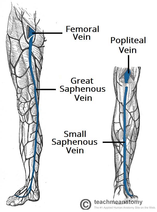 Venous Drainage Of The Lower Limb Teachmeanatomy
 Pdf Relationship Between Specific Distributions Of Isolated
 Anatomical Features Of The Gastrocnemius Muscle Soleus
 The Concordance Rates Of Leg Deep Vein Thrombi To Soleal
 Deep Calf Vein Superficial Vein Imaging Flashcards Quizlet
 The Level Of The Small Saphenous Vein Ssv Insertion Can Be
Best Venous Ultrasound For Dvt Truffles Vein Specialists
 The Hemodynamics And Diagnosis Of Venous Disease Sciencedirect
 Pdf Pathophysiology Of Venous Thromboembolism With Respect
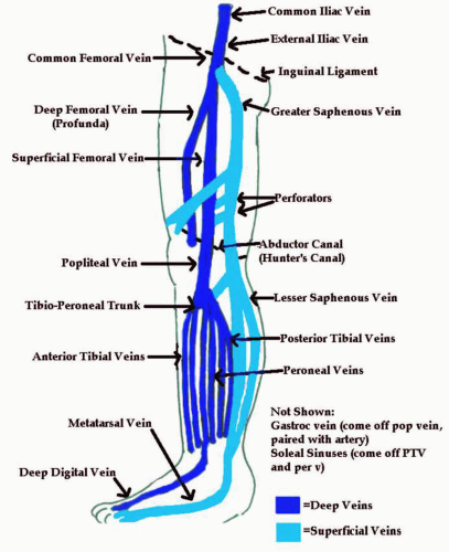 Peripheral Venous Systems Thoracic Key
 Leg Vein Anatomy Veinspecialistsofarizona Com

Belum ada Komentar untuk "Soleal Vein Anatomy"
Posting Komentar