Pelvis Hip Anatomy
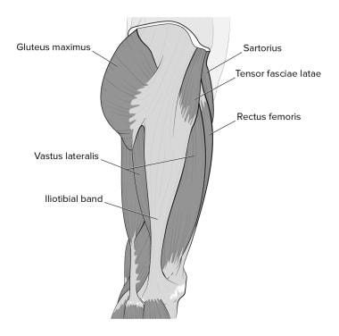
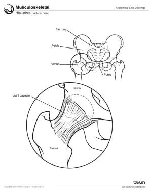
The hip joint is a ball and socket type joint. Hip muscles that both support the joint and enable.
 Yoga For Hip Stability Understanding Hypermobility
Knee shoulder shoulder arthrogram ankle elbow wrist hip contact.
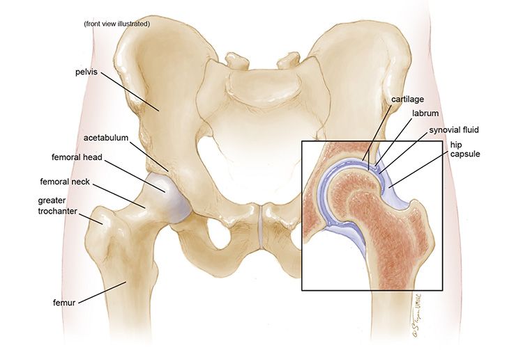
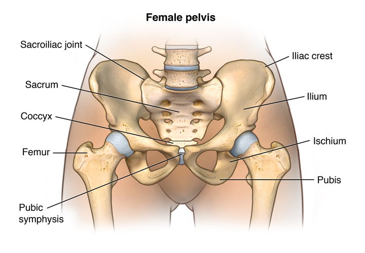
Pelvis hip anatomy. The muscles of the thigh and lower back work together to keep the hip stable. The pelvis is the lower portion of the trunk located between the abdomen and the lower limbs. Ligaments of the pelvis and hip.
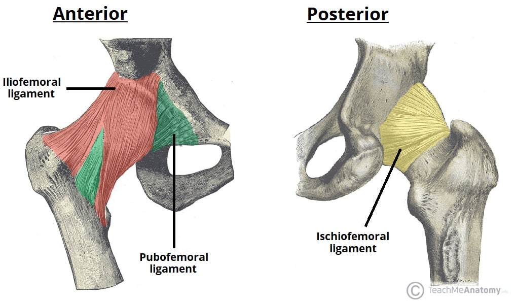
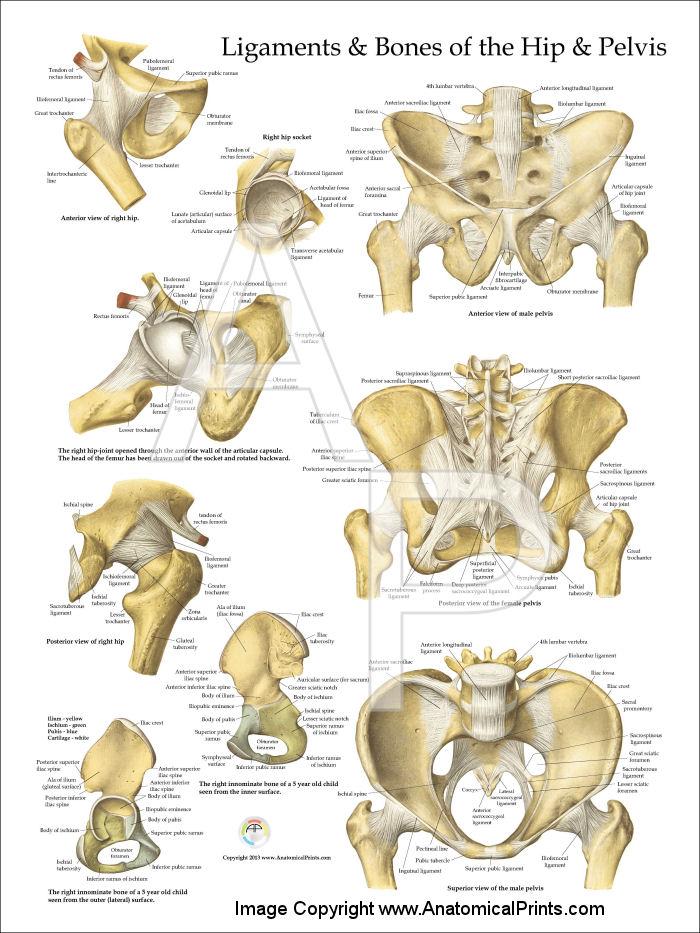
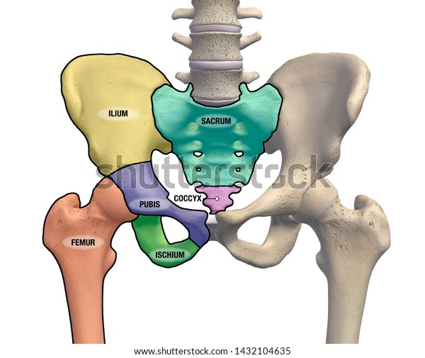
Also a couple of ligaments in the pelvis participate in forming the pelvis cavity. Copyright c 2005 2019 alex freitas md. The hip is a major ball and socket joint connecting the long bones.
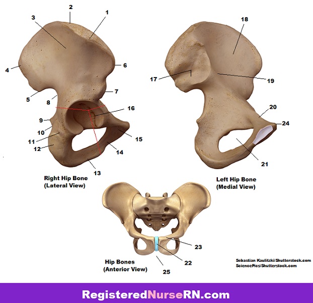
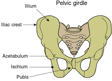
The gap enclosed by the bony pelvis called the pelvic cavity is the section of the body underneath the abdomen and mainly consists of the reproductive organs sex organs and the rectum while the pelvic floor at the base of the cavity assists in supporting the organs of the abdomen. The pelvic girdle hip girdle is formed by a single bone the hip bone or coxal bone coxal hip which serves as the attachment point for each lower limb. Muscles of the hip.
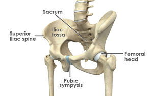
Anatomy of the pelvis the sacrum. Use the mouse to scroll or the arrows. Anatomy of the hip the hip joint.
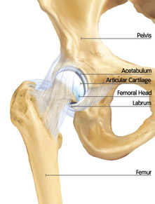
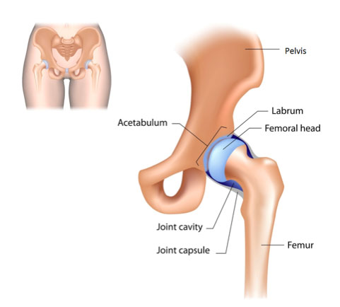
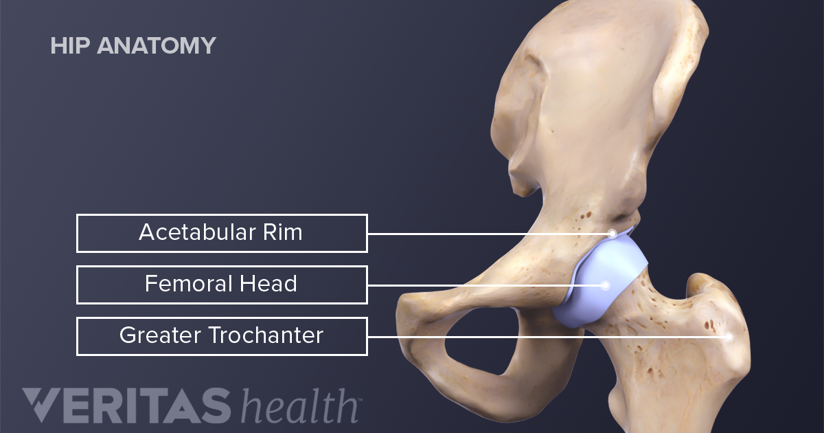
Anatomy of the femur. It is clear that the anatomy of pelvis is complex and consists of the several bones that are connected with mutual joints. The stability of the hip is increased by the strong ligaments that encircle the hip.
The sacrum consists of five fused vertebrae. Hip articular cartilage that decreases friction between the bones and allows for a smooth gliding. Anteroposterior compressions lateral compressions vertical shears combined fractures.
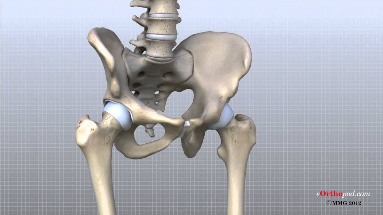
The hip bones join to the upper part of the skeleton through attachment at the sacrum. Each hip bone in turn is firmly joined to the axial skeleton via its attachment to the sacrum of the vertebral column. Also known as the acetabulofemoral joint the hip joint is comprised of these basic components.
Each hip bone is made of three smaller. Together they form the part of the pelvis called the pelvic girdle. Anatomy of the hip.
Hip bones including the femur and pelvic bones. The pelviss frame is made up of the bones of the pelvis which connect the axial skeleton to the femurs and therefore acts in weight bearing of the upper body. The femur or thighbone is the longest and strongest bone in.
 Hip Bone Wikipedia
 Hip Canadian Orthopaedic Foundation Canadian Orthopaedic
 Hip Anatomy Pictures Function Problems Treatment
Hip Dislocation Orthoinfo Aaos
 Pelvis Hip Anatomy
 Hip Surgery Illustrations Pelvis Hip Anatomy Medical
![]() Hip And Thigh Bones Joints Muscles Kenhub
Hip And Thigh Bones Joints Muscles Kenhub
 The Hip Joint Articulations Movements Teachmeanatomy
 Anatomy Of The Hip Mu Health Care
 Hip Pelvic And Spinal Anatomy
 Pelvis Hip Anatomy
 Musculoskeletal Pelvic Anatomy Sciencedirect
 Pelvis Hip Anatomy
 Pelvis Anatomy Quiz
 Anatomy Of The Male And Female Pelvis Comprehensive
 Issues Around The Hip From Tendonitis To Bursitis Beacon
 The Dancer S Hip Anatomy And How The Hip Functions
 Canine Hip Anatomical Model Lfa 9060
 Hip Joint Anatomy Overview Gross Anatomy
 Hip Anatomy
 Hip Anatomy Diagram From Bones To Joints Science Trends
 Pelvis Hip Anatomy
 Hip Anatomy
 The Pelvic Girdle Of Human Hip Bone Anatomy Vector
 Pelvis Hip Anatomy
 Pelvis And Hip Anatomy Poster
 Hip Anatomy Animated Tutorial
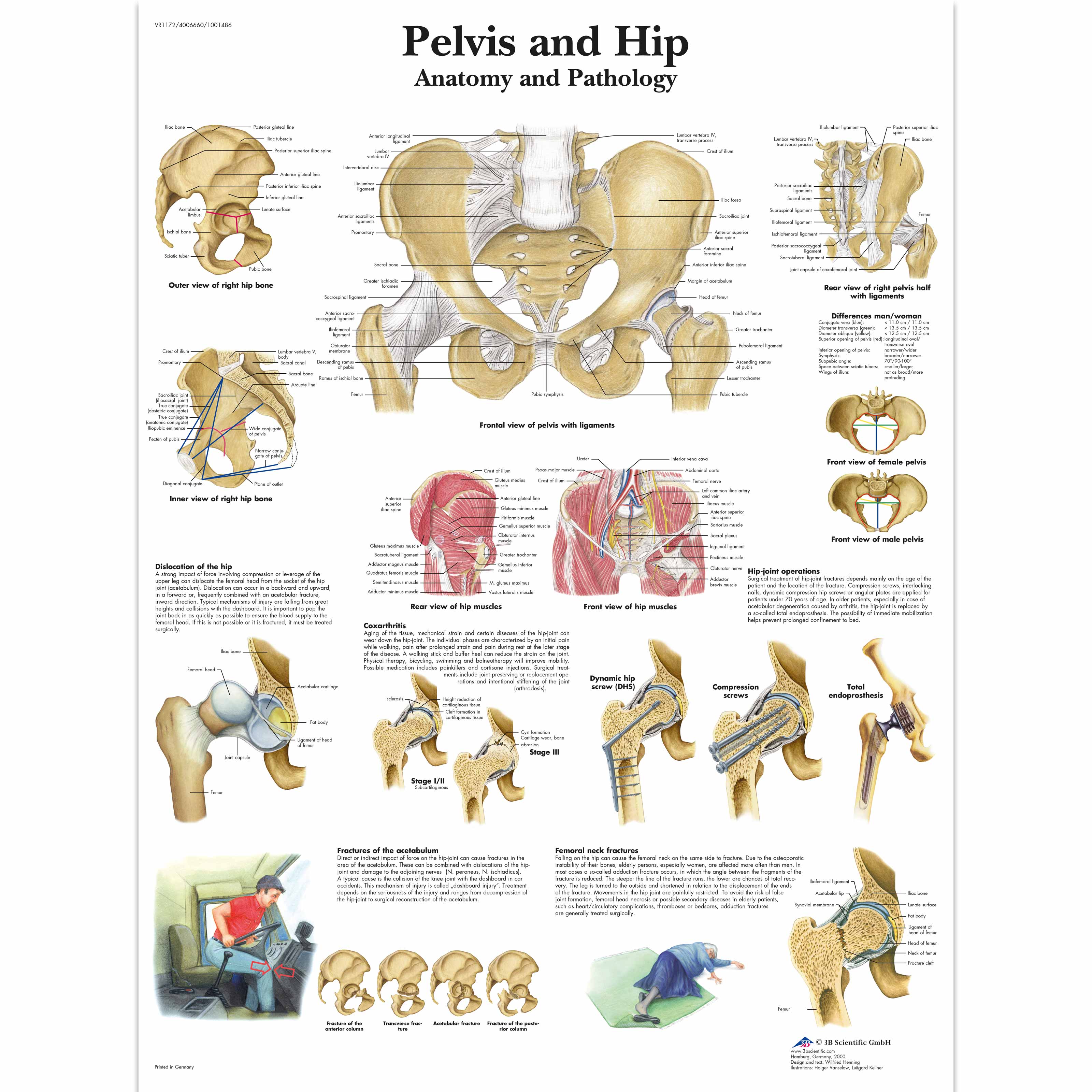 Pelvis And Hip Chart Anatomy And Pathology
Hip Bursitis Orthoinfo Aaos
 Hip Joint Anatomy Overview Gross Anatomy
 3d Rendering Pelvis Hip Anatomy Front Royalty Free Stock Image
Belum ada Komentar untuk "Pelvis Hip Anatomy"
Posting Komentar