Canine Spinal Anatomy
The spinal cord is divided into spinal cord segments. There is a partial fusion with ossification of the right sacroiliac joint.
Comparative Anatomy Of The Horse Ox And Dog The Vertebral
The bone between the hip and knee is the femur.

Canine spinal anatomy. Anatomy of intervertebral disc disease ivdd intervertebral discs are situated between the vertebrae. Each segment gives rise to paired spinal nerves click left image. A dogs spine is located along the top or dorsal side of a dogs body.
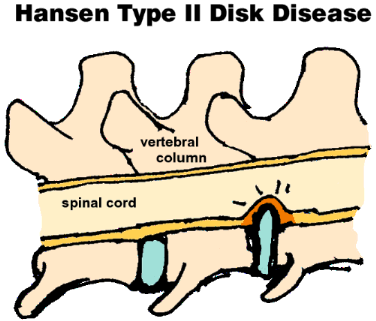
In dogs and cats the disc sits just beneath or ventral to the spinal canal. The 13 th right rib is a rudimentary rib. Spinal cord anatomy close.
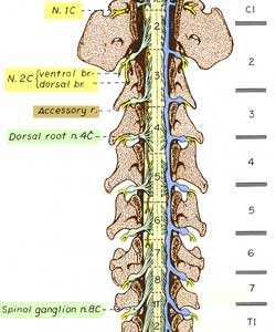
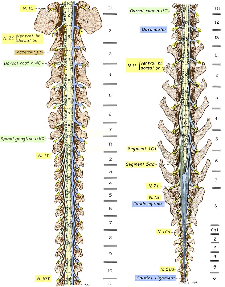
Vertebrae develop segmentally from somitic sclerotomes whereas muscles develop from somitic myotomes. A spinal ganglion is present distally on each dorsal root. Dorsal and ventral spinal roots arise as a series of rootlets.
Note from authors about anatomical variations. A dogs spine is designed to support weight and protect the spinal cord. Canine spine anatomy significance.
Dog hind leg anatomy the hind leg can be confusing to some owners but it has some of the same features as a human. Dorsally the roof of all three cavities is formed by the spinal column and associated muscles. Spinal cord segments are labeled and locations of vertebral bodies separated by intervertebral discs are shown to the right.
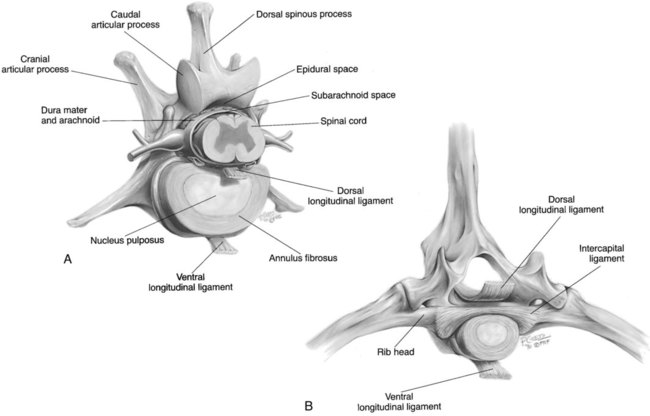
The canine spinal cord has 8 cervical 13 thoracic 7 lumbar 3 sacral and 5 caudal segments. The discs are comprised of a fibrous outer ring called the annulus fibrosis and an inner gel called the nucleus pulposes. The pelvic cavity is defined by the borders of the bony pelvis and communicates with the abdominal cavity.
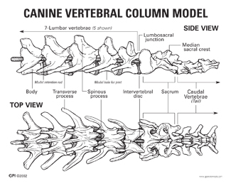
Normal dog have usually 7 lumbar vertebrae and 3 sacral vertebrae. The first caudal vertebra cd1 is partially fused with the last. Cranial and caudal halves of a canine vertebral column are illustrated after a laminectomy to expose the spinal cord.
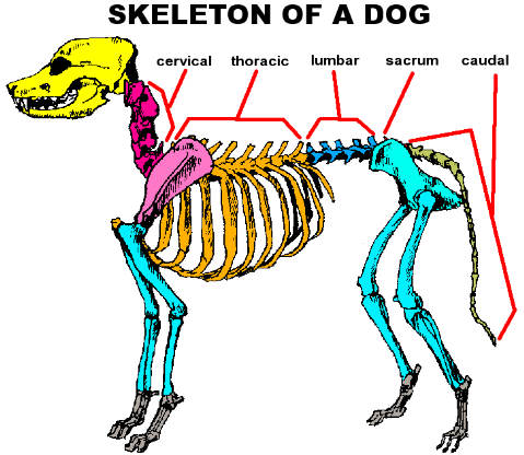
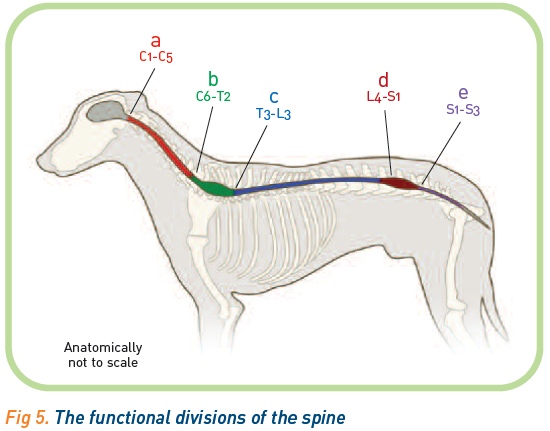
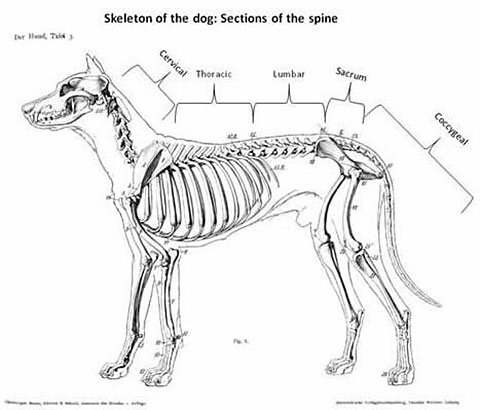
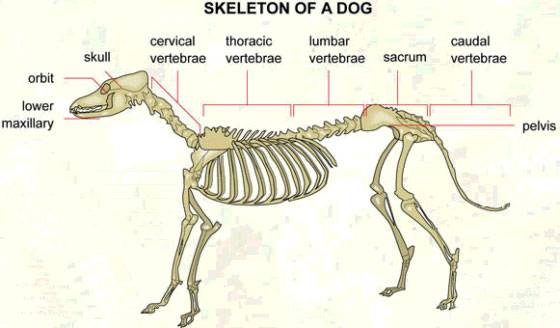
Below the knee is the tibia and fibula. The canine spinal column is separated into four main regions.
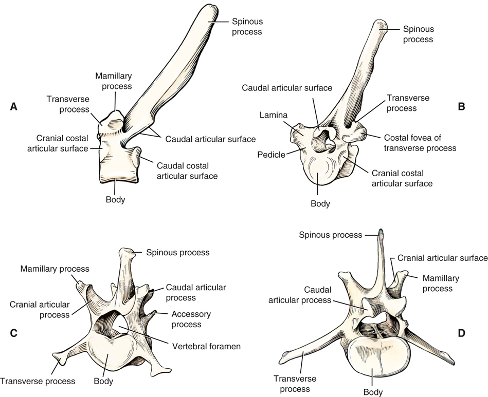
 The Canine And Feline Vertebrae Veterian Key
 Canine Spine Anatomy Preview 3d Veterinary Anatomy Ivala
 Comparative Anatomy Of The Horse Ox And Dog The Vertebral
 Lumbar Spine Of The Dog On Ct
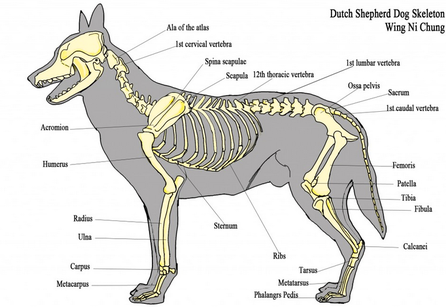 Pelvis Anatomy The Institute Of Canine Biology
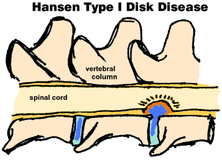
 Intervertebral Disk Disease Mar Vista Animal Medical Center
 Biomimetics Free Full Text The Spine A Strong Stable
 Intervertebral Disk Disease Mar Vista Animal Medical Center
 Comparative Anatomy Of The Horse Ox And Dog The Vertebral

 Cervical Spinal Nerve 8 Wikipedia
 Canine Vertebrae Model 9080 For Sale Anatomy Now
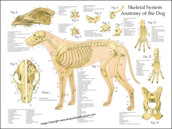 Dog Skeletal Skull Anatomy Poster 18 X 24 Veterinary Chart
 Canine Anatomy Veterian Key
 Ligaments Of The Canine Vertebral Column 1 Supraspinous
 Intervertebral Disk Disease Mar Vista Animal Medical Center
 Spinal Nerves Neuroanatomyofthedog
 The Dog S Body Systems A Double Sided Uv Protected Laminated Dog Anatomy Chart A Learning And Teaching Chart For Veterinary Science
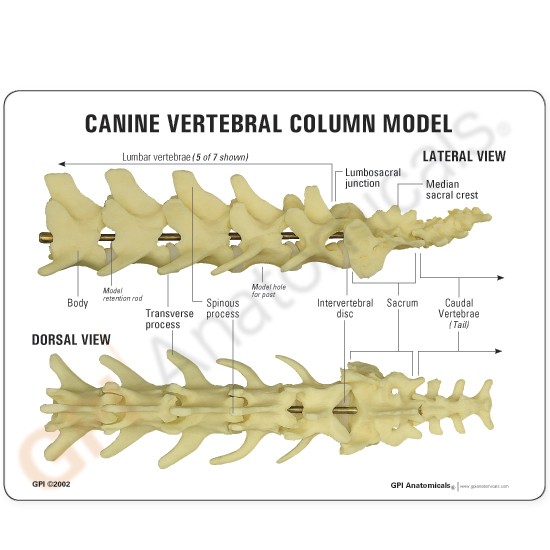 Canine Spine Anatomical Model Lfa 9080

 French Bulldog Hemivertebrae Ufaw
 Intervertebral Disc Disease A Major Pain In The Neck Or Back
 Canine Whole Spine Skeletal Anatomy Resource Wikivet English
 Labeled Atlas Of Anatomy Illustrations Of The Dog
 Vna Veterinary Neuronal Adjustment Long Beach Animal
 Spinal Column Anatomy Physiology Wikivet English
 Parts Of The Nervous System In Dogs Dog Owners Merck
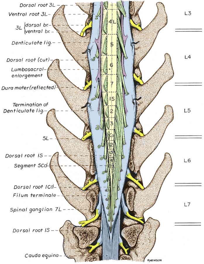
 Anatomy Lumbar Dog Spinal Cord Nerve Anatomy Spinal
 Lumbosacral Syndrome In Dogs Vca Animal Hospital
Belum ada Komentar untuk "Canine Spinal Anatomy"
Posting Komentar