The Anatomy Of The Hip Joint
There are two other protrusions near the top of the femur known as the greater and lesser trochanters. Hip problems occur when any one of these components starts to degenerate or is in some way compromised or irritated.
 Hip Joint Treatment Orange County Hip Surgeon Fountain
It allows us to walk run and jump.
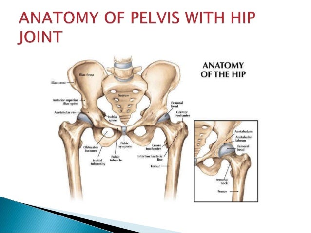
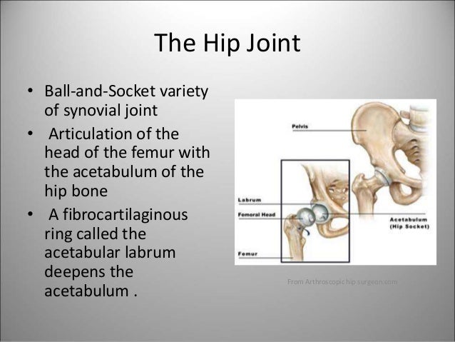
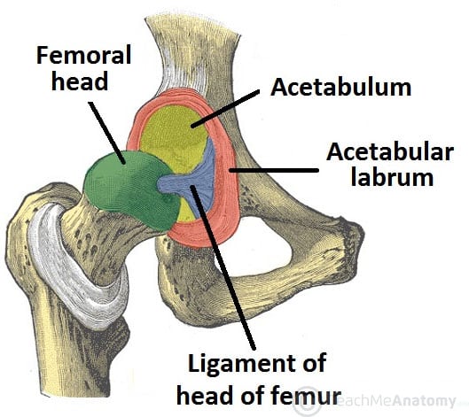
The anatomy of the hip joint. Femoral head a ball shaped piece of bone located at the top of your thigh bone or femur acetabulum a socket in your pelvis into which the femoral head fits. It is the largest bone in the body. It is the largest ball and socket joint in your body.
The round head of the femur rests in a cavity the acetabulum that allows free rotation of the limb. At the top of the femur is a rounded protrusion which articulates with the pelvis. A healthy hip can support your weight and allow you to move without pain.
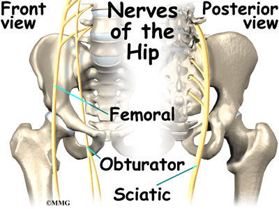
Hip in anatomy the joint between the thighbone femur and the pelvis. So the anterior portion of the hip joint is innervated by the femoral nerve 2. The hip joint consists of two main parts.
This portion is referred to as the head of the femur or femoral head. The hip joint is a ball and socket synovial joint formed by an articulation between the pelvic acetabulum and the head of the femur. The femoral nerve innervates the flexors of the hip joint which pass anterior to the hip joint.
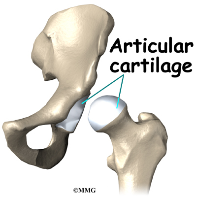
The hip joint is made up of two bones. Bones of the hip joint. And synovial membrane and fluid which encapsulates the hip joint and lubricates it respectively.
The hip joint is the articulation of the pelvis with the femur which connects the axial skeleton with the lower extremity. Hip ligaments and tendons tough fibrous tissues that bind bones to bones and muscles to bones. Weight bearing stresses on the hip during walking can be 5 times a persons body weight.
It bears our bodys weight and the force of the strong muscles of the hip and leg. Hip anatomy function and common problems. The hip joint is a ball and socket joint.
The hip joint is one of the most important joints in the human body. The pelvis and the femur the thighbone. It forms a connection from the lower limb to the pelvic girdle and thus is designed for stability and weight bearing rather than a large range of movement.
The hip joint is one of the largest joints in the body and is a major weight bearing joint. Yet the hip joint is also one of our most flexible joints and allows a greater range of motion than all other joints in the body except for the shoulder. The adult os coxae or hip bone is formed by the fusion of the ilium the ischium and the pubis which occurs by the end of the teenage years.
Amphibians and reptiles have relatively weak pelvic girdles and the femur extends horizontally. It is the largest ball and socket joint in your body. Also the area adjacent to this joint.
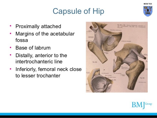
Bands of tissue called ligaments connect the ball to the socket stabilizing the hip and forming the joint capsule.
 Ligaments Tendons And Muscles Of The Hip Joint Naples
Hip Joint Treatment Sydney Hip Ligaments Treatment Sydney
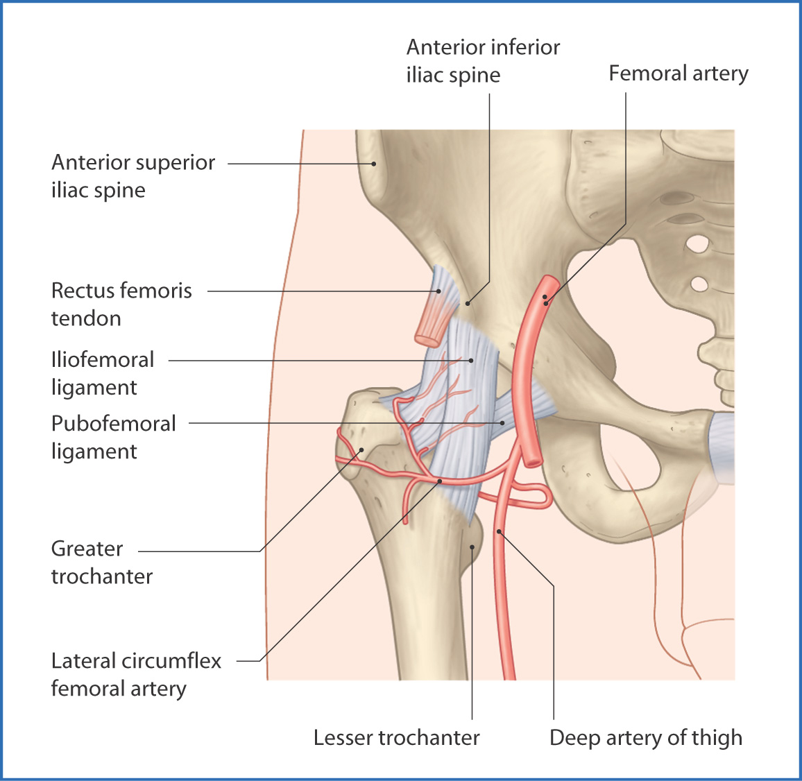 Hip Anatomy Recon Orthobullets
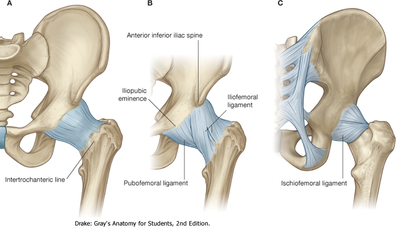 Hip Joint Anatomy Movement Muscle Involvement How To
 Laminated Anatomy And Injuries Of The Hip Poster Hip Joint Anatomical Chart 18 X 27
 Radiographic Views For Hip Joint
 Anatomy 101 Learn To Balance Mobility Stability In Your
 Anatomy And Injuries Of The Hip Anatomical Chart
 Anatomy Of The Hip Joint Left Dissected Joint Right
 Hip Joint Labral Tears Corona Physio Downtown Edmonton
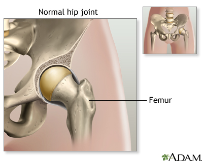 Hip Joint Replacement Series Normal Anatomy Medlineplus
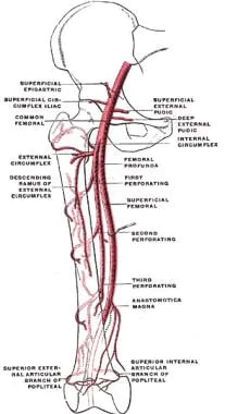 Hip Joint Anatomy Overview Gross Anatomy
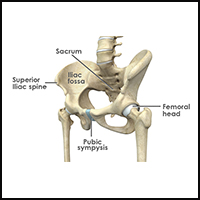 Hip Surgery Anterior Hip Replacement Revision
 Hip Joint Radiology Reference Article Radiopaedia Org
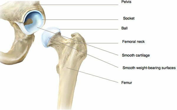 Hip Joint Anatomy Hip Bones Ligaments Muscles
 Yoga For Hip Stability Understanding Hypermobility
 The Gross Antomy Of The Hip Hoint And Applied Anatomy
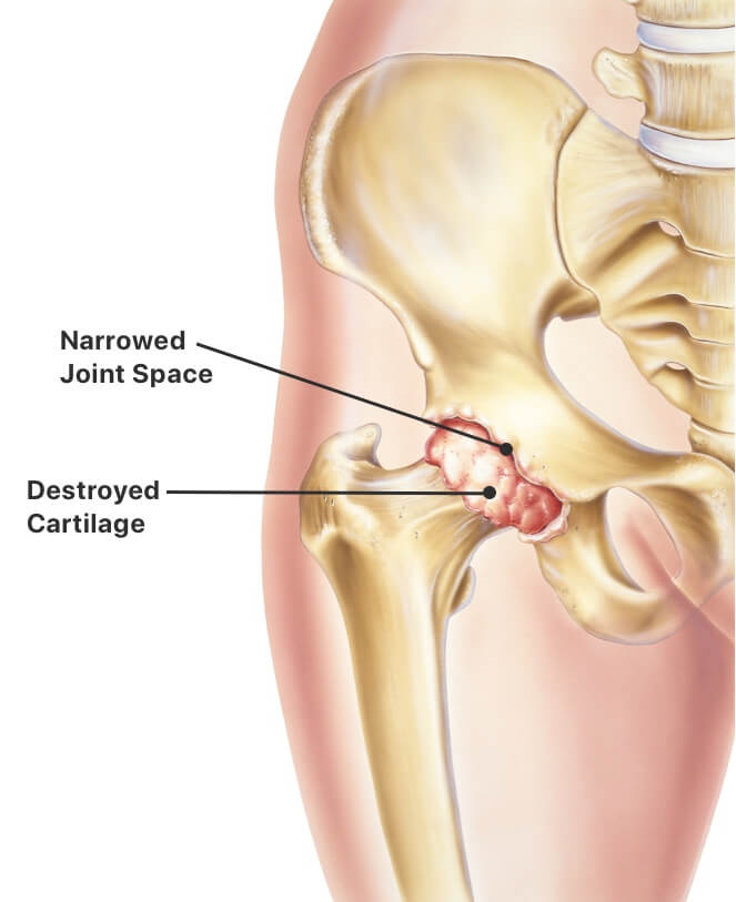 Hip Replacement Procedure Types Recovery Time And Risks
 The Hip Joint Articulations Movements Teachmeanatomy
 Hip Anatomy Pictures Function Problems Treatment





Belum ada Komentar untuk "The Anatomy Of The Hip Joint"
Posting Komentar