Mri Anatomy Knee
Click on a link to get t1 coronal view t2 fatsat axial view t2 fatsat coronal view t2 fatsat sagittal view. This webpage presents the anatomical structures found on knee mri.
 How To Read Knee Mri Of Normal Knee Anatomy Of The Knee Colorado Knee Specialist
After all an entire year of fellowship training is dedicated to musculoskeletal imaging.

Mri anatomy knee. Use the mouse scroll wheel to move the images up and down alternatively use the tiny arrows on both side of the image to move the images. Anatomy of the knee mri atlas of the human body using cross sectional imaging. Use the mouse to scroll.
This mri knee sagittal cross sectional anatomy tool is absolutely free to use. This atlas of cross sectional anatomy of the knee is based on imagery by magnetic resonance mri. By now you probably know that the anatomy is deceptively complex combinations of injuries can be challenging and of course the referring clinicians expectations are as high as the range of meniscus injuries is wide.
Knee seems like it should be pretty easy right. This tool is at the same time useful for the training and teaching of the anatomy. Robert laprade discusses how to read an mri of a normal knee.
Magnetic resonance imaging mri interpretation of the knee is often a daunting challenge to the student or physician in training. Colorado knee specialist dr. The iliotibial band bursa is situated between the tibia and distal iliotibial band immedi ately proximal to its insertion on gerdys tubercle.
The fcl biceps femoris bursa is found lateral to the distal fcl and insinuates anterior and antero medial in relation to this ligament. Each anatomical structure is labelled interactively. Through the use of magnetic resonance imaging clinicians can diagnose ligament and meniscal injuries along with identifying cartilage defects bone fractures and bruises.
Atlas of knee mri anatomy. Normal mri anatomy of the knee 639. Anatomy of the knee can be complicated and hard to understand.
 I Love Physical Therapy Atlas Of Knee Mri Anatomy
Stanford Msk Mri Atlas C 2019

 Mri Knee Anatomy Knee Sagittal Anatomy Free Cross

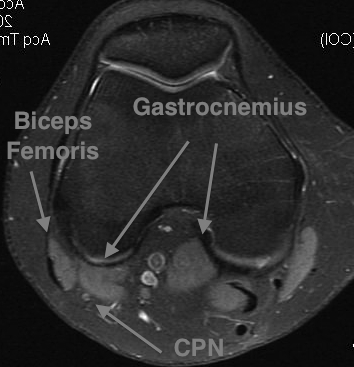
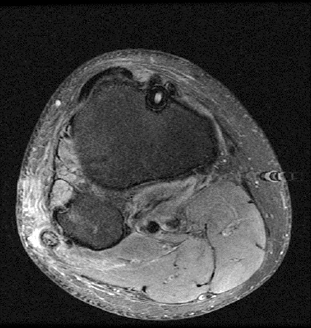
 Knee Mri Sequences
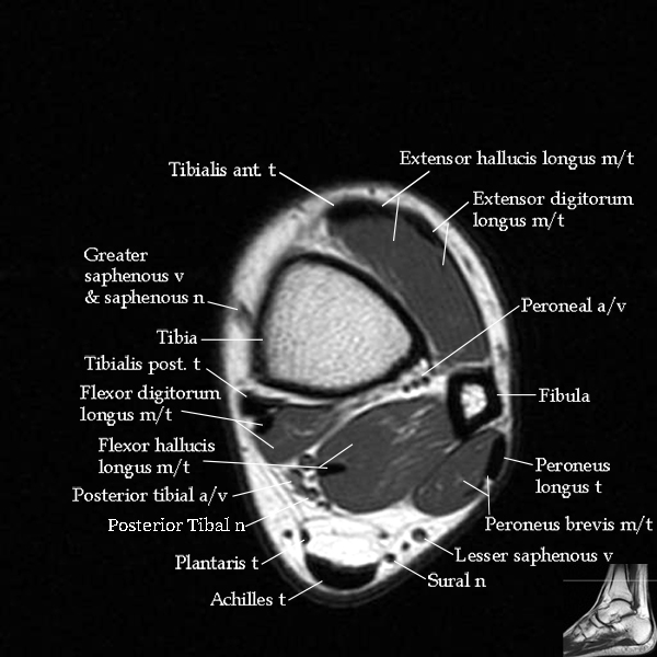 Mri Ankle Anatomy
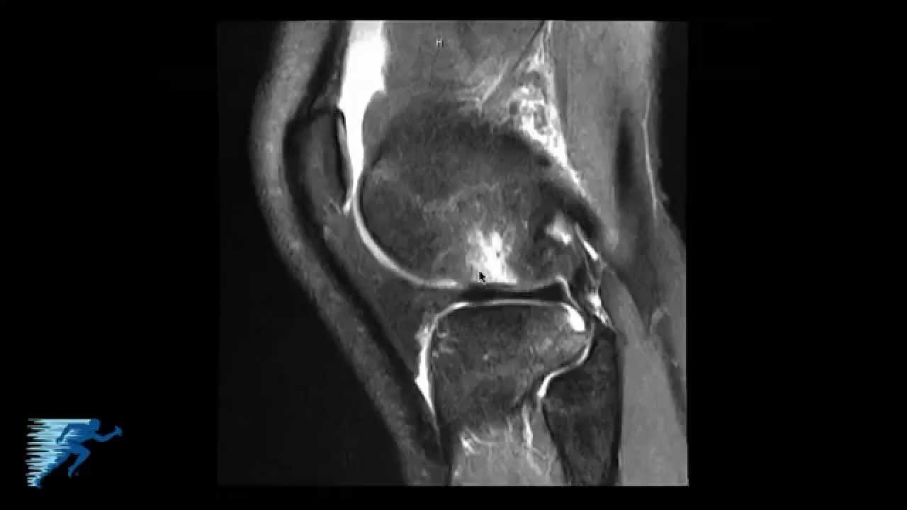
 Knee 29 Mov
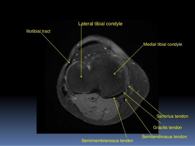
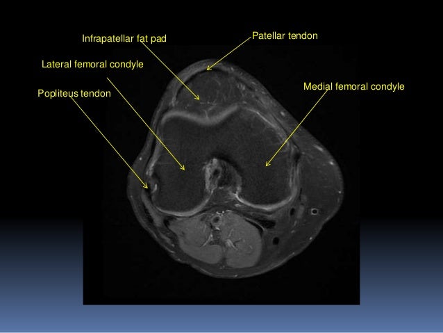 Mri Knee Joint Anatomy
 Multi Ligament Knee Injuries Sterling Ridge Orthopaedics


 Multi Ligament Knee Injuries Sterling Ridge Orthopaedics
 Mri Knee Joint Anatomy

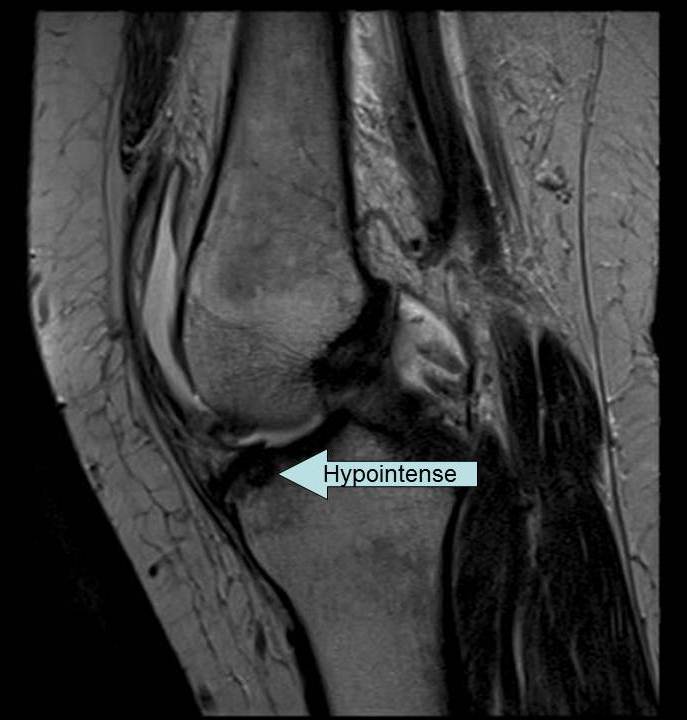
 Magnetic Resonance Imaging Of Cruciate Ligament Injuries Of
 How To Read Knee Mri Of Acl Tear Anterior Cruciate Ligament Injury Colorado Knee Surgeon
 Mrt Der Knie T2 Gewichtete Fatsat Axialen Schnitte
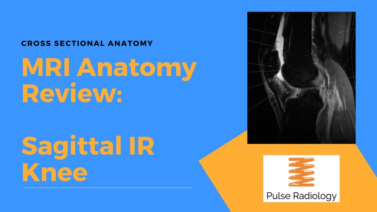 Mri Knee Sagittal Ir Anatomy In Under 60 Seconds
 The Knee Mri Atlas Of Anatomy In Medical Imagery


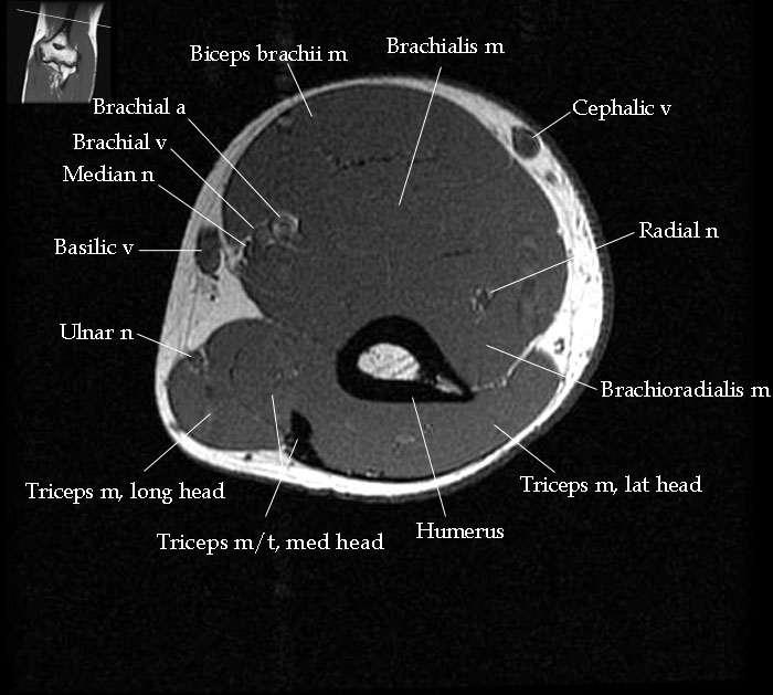 Mri Elbow Anatomy
 A Radiologist S Exploration Of The Stanford Ml Group S Mrnet
 Radiologic Evaluation Of The Knee Fundamentals Of
 Figure 14 From Normal Mr Imaging Anatomy Of The Knee

 James Y Song Msiv Gillia
 The Knee Mri Atlas Of Anatomy In Medical Imagery
Belum ada Komentar untuk "Mri Anatomy Knee"
Posting Komentar