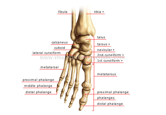
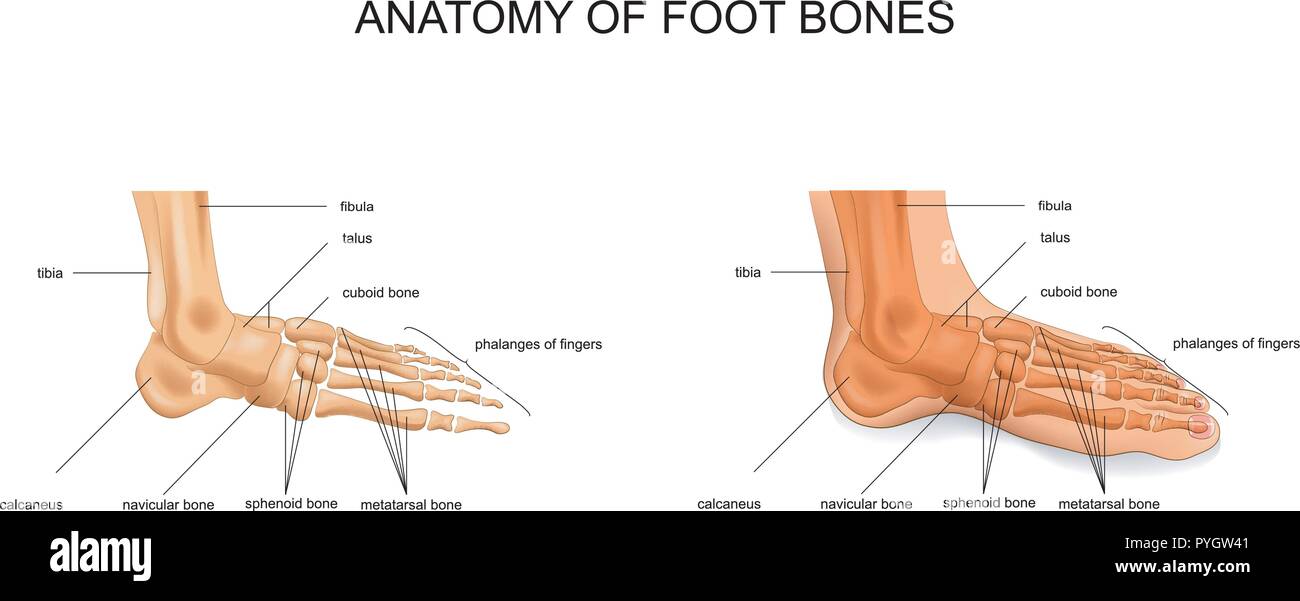
Anatomy Of The Foot Bones
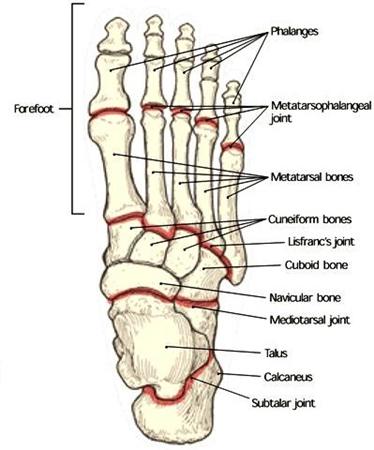
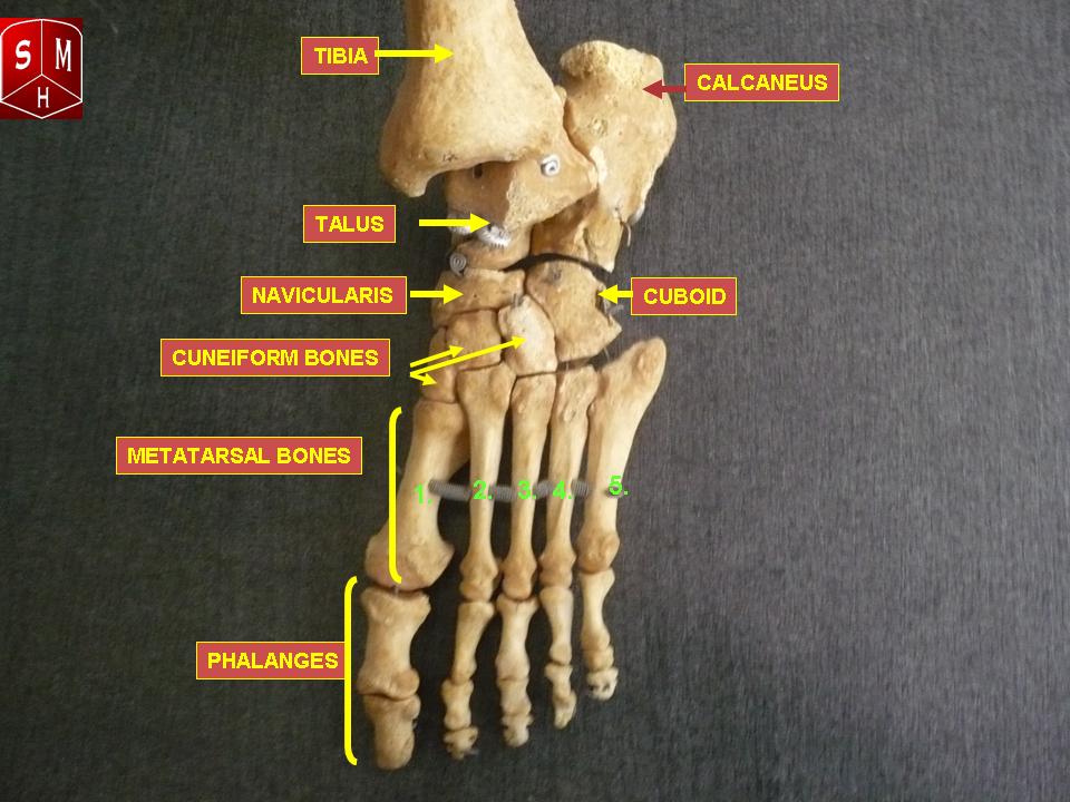
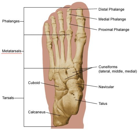
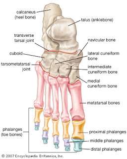
The hindfoot midfoot and the forefoot. The calcaneus heel bone is the largest bone in the foot.
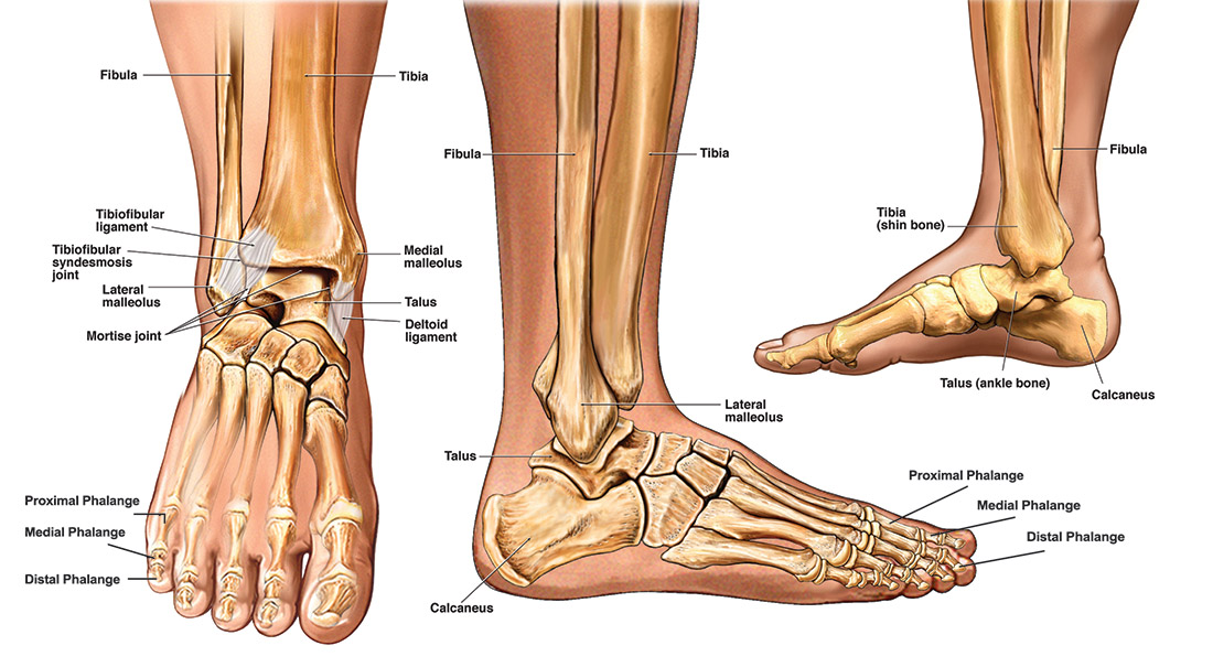
 Ankle Foot Anatomy
The phalanges which are the bones in your toes.

Anatomy of the foot bones. The calcaneus which is the bone in your heel. Foot anatomy bones the complex structure of human feet originates from grasping feet cells like and hand like that can be seen in primates today. Tarsals five irregularly shaped bones of the midfoot that form the foots arch.
Sign up for your free kenhub account today and join over 1234952 successful anatomy students. This unlabeled quiz of the bones of the foot will test your knowledge on how to label the structures of these bones. Foot bone quiz for anatomy and physiology.
Calcaneus the largest bone of the foot which lies beneath the talus to form the heel bone. The bone in the foot frequently associated with an accessory is the navicular bone. The cuneiform bones the navicularis and the cuboid all of which function to give your foot.
Parts of foot bones. The parts of the foot bones. The foot is a firm platform that support the weight of the body.
The hindfoot consists of bone from the leg and the ankle joint. The different bones on each section of the foot. The other bones of the foot that create the ankle and connecting bones include.
The hindfoot is the posterior part of the foot. The accessory navicular bone forms when the tuberosity of the navicular develops from a secondary center of. Want to learn more about it.
The foot is located after the long shin bones and it starts from the back of your ankle to your toes. Anatomically the foot is divided into 3 sections. Talus the bone on top of the foot that forms a joint with the two bones of the lower leg.
Hind means posterior so it basically the backward part of the foot. The metatarsals which run through the flat part of your foot. Our ancestors were tree dwellers and used to hang with all four limbs on branches.
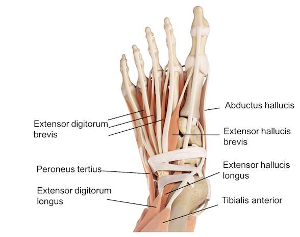
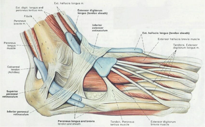
Muscles tendons and ligaments run along the surfaces of the feet allowing the complex movements needed for motion and balance. The bones of the feet are. Our engaging videos interactive quizzes in depth articles and hd atlas are here to get you top results faster.
You will be required to label the cuboid navicular calcaneus lateral cuneiform medial cuneiform medial cuneiform talus metatarsals and distalmiddleproximal phalanges. Bones of the foot. This enables them to evolve complex extraordinary hand and feet which they use for gripping grasping and rotating.
Bones of the foot as seen from the medial arch side. It is made up of many bones including the tarsal bones the metatarsal bones and the phalanges described in more detail below. The talus which is the.
 Foot Anatomy Bones Ligaments Muscles Tendons Arches
 Foot Anatomy Spokane Valley Wa Foot Doctor
 Foot Ankle Anatomy Dr Sanford Bone And Joint Specialists
 Understanding The Anatomy Of The Bones Of The Foot
 Bones Of The Leg And Foot Interactive Anatomy Guide
 Foot Anatomy And Biomechanics Foot Ankle Orthobullets
 Foot Anatomy Bones Ligaments Muscles Tendons Arches
 Ankle Foot Anatomy
 Ankle Foot Atlas Of Anatomy
 Bones Of Foot Human Anatomy The Diagram Shows The Placement
 Bones Of The Foot Illustrations Foot Anatomy Illustrations
 Anatomy Of The Foot Bones Buy This Stock Vector And
 Foot Bones Foot Pain Anatomy Info
 Foot Bones Anatomy
 Anatomy Moment 52 Foot Bones Corpo Kinetic Pilates Rehab
 Human Anatomy Skeleton Foot Bones By Da Vinci Letterhead
 Foot Bones Photos 40 827 Foot Stock Image Results
 Foot Anatomy
 The Bones In The Foot Inferior View Picture Illustrated
 Vector Illustration Of Anatomy Of The Foot Bones Stock
 Toe Wikipedia
 Foot Bones Anatomy Conditions And More
Belum ada Komentar untuk "Anatomy Of The Foot Bones"
Posting Komentar