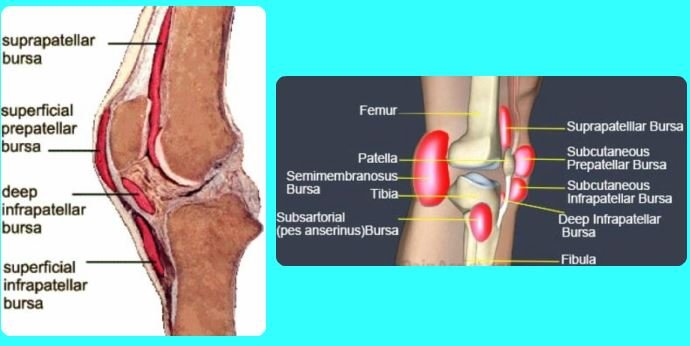
Anatomy Of The Knee Bursa
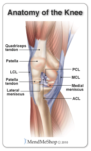
The knee is the largest joint in your body and one of the most easily injured. A mild sprain is simply a stretched ligament which usually heals just fine on its own.
 Knee Wikipedia
A moderate sprain involves some tearing of the ligament.
Anatomy of the knee bursa. It is a pivotal hinge joint in the leg that allows for a variety of movements ie. Knee bursitis causes pain and can limit your mobility. The prepatellar bursa is one of the larger bursae of the knee and is located on the front of the patella hence pre patellar just under the skin.
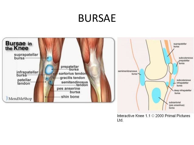
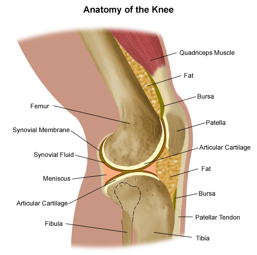
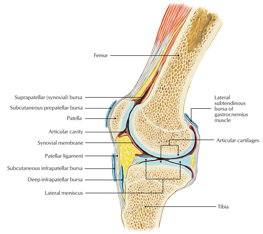
The knee bursae are the fluid filled sacs and synovial pockets that surround and sometimes communicate with the knee joint cavity. The knee contains three important groups of bursae. This allows everything to move smoothly preventing inflammation.
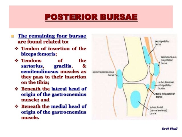
Its most common causes are overuse and injury. There are four bursae anterior to the knee joint. It is filled with synovial fluid or lubricant made by the membrane.
They represent the weak point of the joint but also provide enlargements to the joint space. The bursae are thin walled and filled with synovial fluid. Knee sprains are graded on their severity.
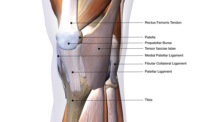
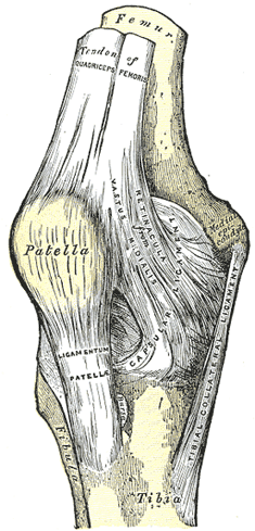
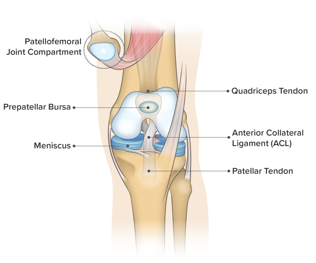
Anatomy of the knee bursae a bursa is a small sac made of fibrous tissue that has an inner lining of synovial type membrane. It protects the patella. They sit between two surfaces usually muscle and bone to reduce friction a bit like ball bearings.
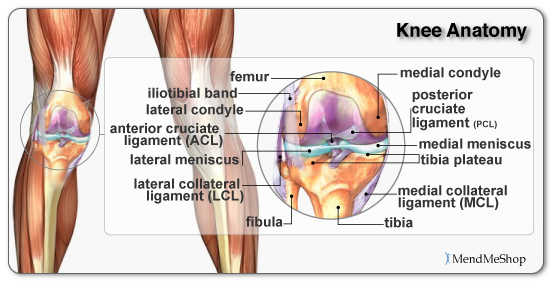
Bursa are found all over the body and there are approximately fourteen around the knee. Flexion extension medial rotation and lateral rotation and it connects the tibia and the fibula with the thigh bone femur. Between the femur and quadriceps femoris it is attached to the articularis genu muscle and communicates with the synovial cavity.
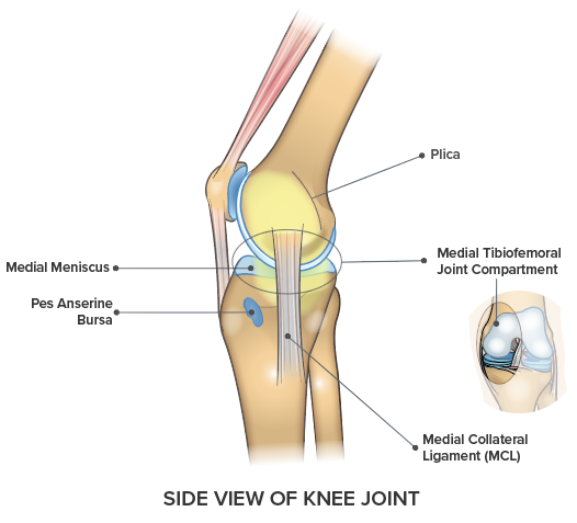
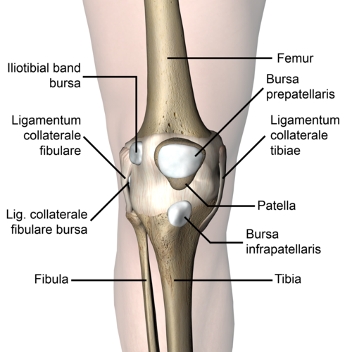
Bursitis of the knee bursa also known as pes anserine bursitis or goosefoot bursitis causes individuals especially runners to restrain motion. There are bursa located underneath the tendons and ligaments on both the lateral and medial sides of the knee. Between the skin and patella.
Knee bursitis is inflammation of a small fluid filled sac bursa situated near your knee joint. Anatomy of the knee. Bursae one is a bursa are fluid filled sacs that help cushion the knee.
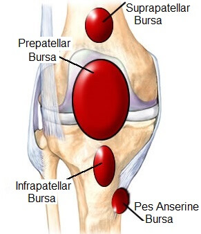
Treatment includes rest ice and specific exercises. Knee bursa are small fluid filled sacs which contain synovial fluid. The prepatellar bursae lie in front of the patella.
A severe sprain may include a complete rupture of a ligament.
 Knee Pain On The Inside Of Your Joint Causes Solutions
 Knee Joint Anatomy
 Bursitis Knee Knee Bursitis Symptoms Treatment
 Knee Bursa Anatomy Function Injuries Knee Pain Explained
 Anatomy Knee Physician Assistant 2014 With Lockwood At
 The Knee Joint Human Anatomy
 Anatomy Of The Knee
 Articular Capsule Of The Knee Joint Wikipedia
Acupuncture For Knee Pain Sample Acupuncture Continuing

 Prepatellar Bursitis Physiopedia
 A Medical Illustration Of A Knee With An Inflamed Prepatellar
 Knee Bursae Locations Anatomy Study Com
 Anatomy Of The Knee Joint
 Knee Joint Anatomy Motion Knee Pain Explained
 Understanding The Anatomy Of The Knee Bodyheal
 Prepatellar Bursitis
 Amazon Com Semtomn Canvas Wall Art Print Patella Knee Joint
 Anatomy Of The Knee Joint Download Scientific Diagram

 Bursitis Ankle Bursa Care And Prevention
Baker S Cysts And Other Knee Bursae Joint Replacement
 Prepatellar Bursitis
 Amazon Com Semtomn Shower Curtain Pain Patella Knee Joint
 Knee Pain On The Front Of Your Joint Learn Why Spring
 Knee Bursae Stock Illustration Illustration Of Kneecap
 Easy Notes On Knee Bursa Learn In Just 4 Minutes
Belum ada Komentar untuk "Anatomy Of The Knee Bursa"
Posting Komentar