Lower Leg Anatomy Bones
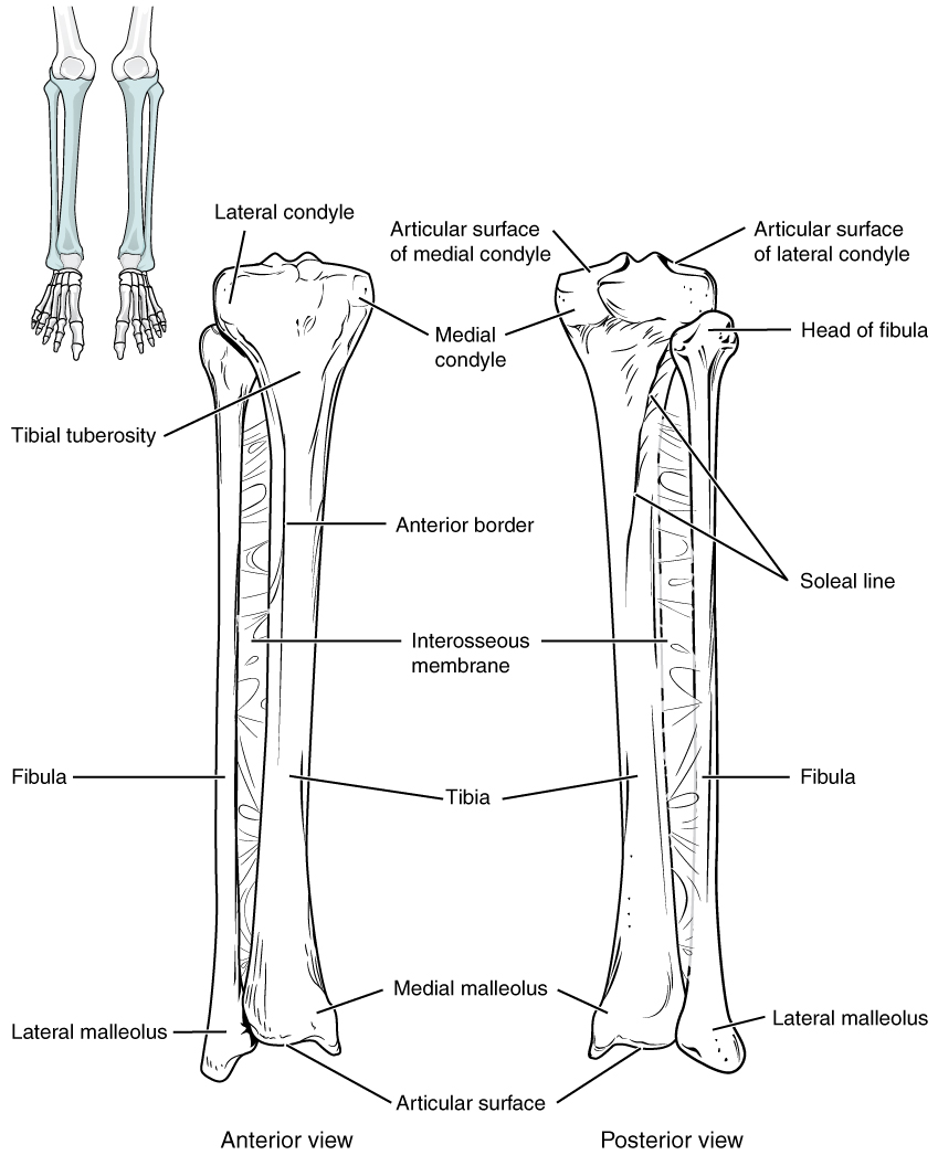
The tibia is the main weight bearing bone of the lower leg and the second longest bone of the body after the femur. The tibia is the main weight bearing bone of the lower leg and the second longest bone of the body after the femur.
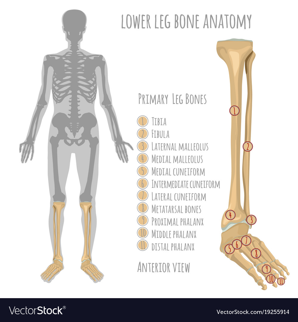
The tibia commonly known as the shin bone is the largest and most medial of the two.

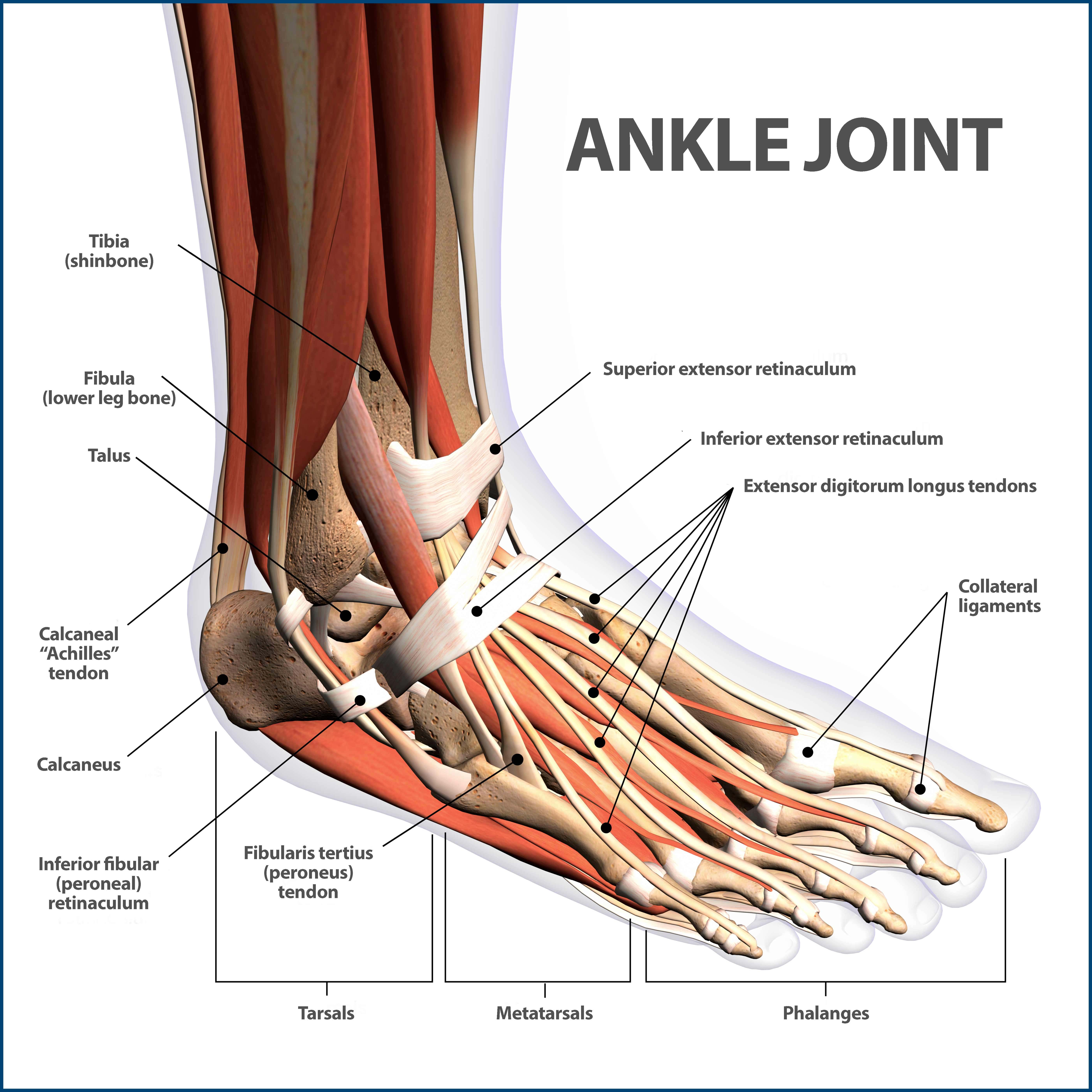
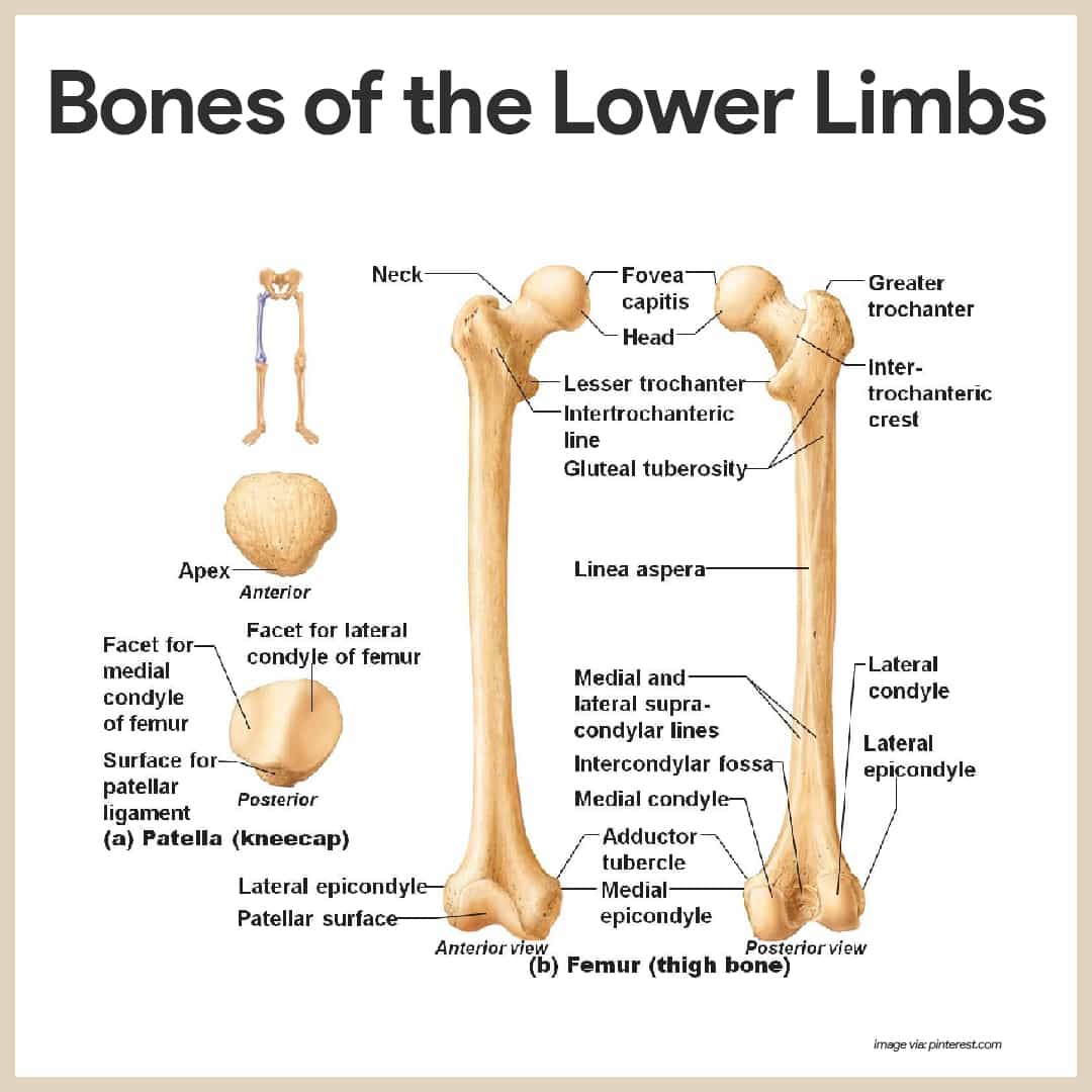
Lower leg anatomy bones. The tibia and fibula are two long bones that run parallel to each other forming the scaffold of the leg and providing attachment points for many muscles. The patella commonly known as the kneecap is at the center of the knee. At the distal end of the femur two rounded condyles meet the tibia and fibula bones of the lower leg to form the knee joint.
Several muscles attach to and act on the femur. The fibula is connected via ligaments to the two ends of the tibia. The knee is a strong but flexible hinge joint that uses muscles and ligaments to withstand the torques and strains of powerful leg movements.
The medial side of the tibia is located immediately under the skin allowing it to be easily palpated down the entire length of the medial leg. It mainly serves as an attachment point for the muscles of the lower leg. The lower leg contains two major long bones the tibia and the fibula which are both very strong skeletal structures.
Also called the shin bone the tibia is the longer of the two bones in the lower leg. The hip joint via its upper extremity and the knee joint via its lower extremity. It is also known as the calf bone as it sits slightly behind the tibia on the outside of the leg.
The tibia shin bone is the medial bone of the leg and is larger than the fibula with which it is paired. The tibia also called the shinbone is located near the midline of the leg and is the thicker and stronger of the two bones. It acts as the main weight bearing bone of the leg.
The fibula is located next to the tibia. The femur participates in two major joints in the lower leg. Next to the tibia is the fibula the thinner weaker bone of the lower leg.
Several muscles attach to and act on the femur. You can palpate its anterior border when you run your finger down the anterior aspect of your leg. It lies between the knee and the ankle while the upper leg lies between the hip and the knee.
:background_color(FFFFFF):format(jpeg)/images/library/11153/muscles-tibia-fibula_english__2_.jpg) Lower Extremity Anatomy Bones Muscles Nerves Vessels
Lower Extremity Anatomy Bones Muscles Nerves Vessels
 Infographic Diagram Of Human Skeleton Lower Limb Anatomy Bone
 Human Anatomy Bones Of Upper Limb Joints Of Lower Limb
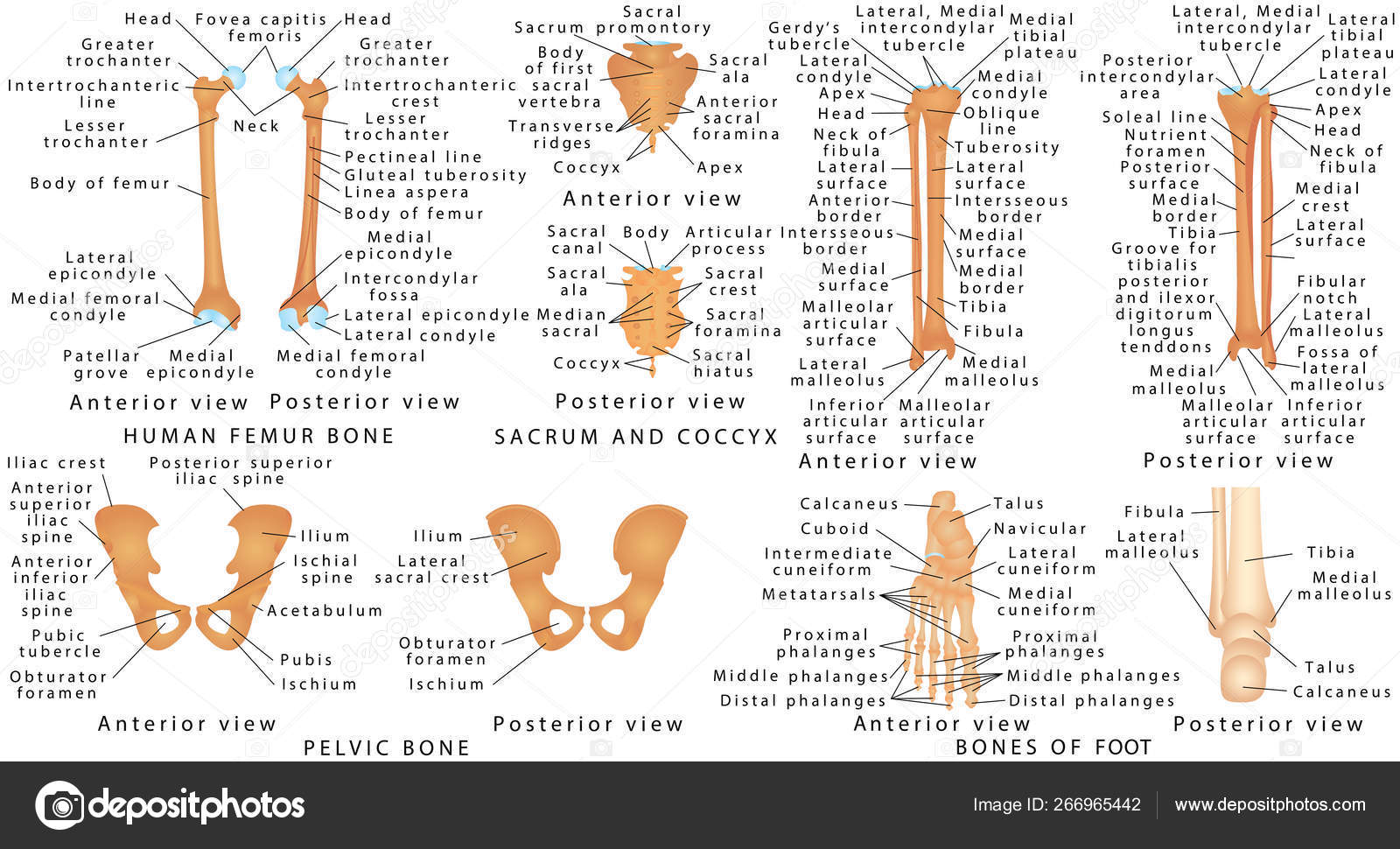 Bones Lower Limb Human Anatomy Human Pelvic Girdle Legs
 Lower Limb Bones Anatomy 1 With Clifford At Logan College
 Muscular Function And Anatomy Of The Lower Leg And Foot
 Bones Bony Landmarks Lower Extremity Anatomy And Cell
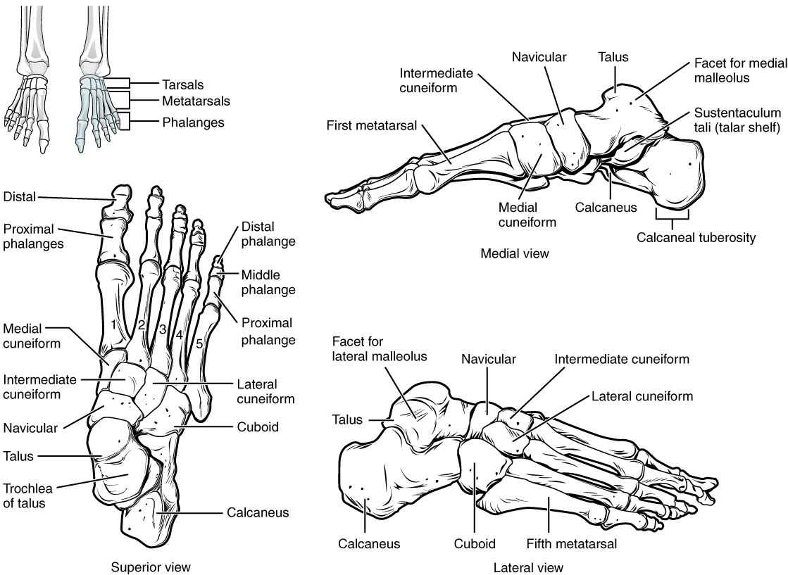 8 4 Bones Of The Lower Limb Anatomy And Physiology
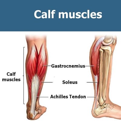 Anatomy Of The Lower Leg George Herald
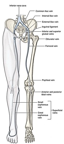 Easy Notes On Lower Limb Learn In Just 4 Minutes
 Amazon Com Medical Anatomical Unisex Adult Bone Socks
 8 4 Bones Of The Lower Limb Anatomy And Physiology
:background_color(FFFFFF):format(jpeg)/images/library/11078/bones-knee-tibia-fibula_english.jpg) Lower Extremity Anatomy Bones Muscles Nerves Vessels
Lower Extremity Anatomy Bones Muscles Nerves Vessels
 Bones Of The Lower Limb Anatomy And Physiology I
 Ankle Fractures Broken Ankle Florida Orthopaedic Institute
The Skeleton Of The Lower Extremity Human Anatomy
 Skeletal System Anatomy And Physiology Nurseslabs
 Macro Structures Of Animals Bi Peds Lower Leg Bones Leg
 Anatomy And Physiology Of Equine Tendons And Ligament


Belum ada Komentar untuk "Lower Leg Anatomy Bones"
Posting Komentar