Mandibula Anatomy
The mandible is the single midline bone of the lower jaw. The mandible lower jaw or jawbone is the largest strongest and lowest bone in the human face.
The mandible sits beneath the maxilla.
Mandibula anatomy. The lower set of teeth in the mouth is rooted in the lower jaw. The mandible is a u shaped bone. Intra and extracapsular condylar fractures are the most frequent mandibular fractures.
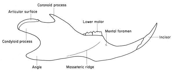
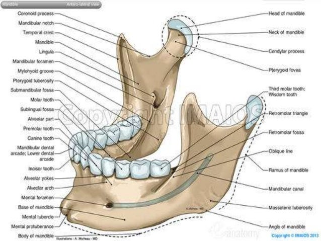
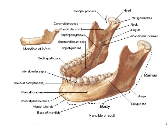
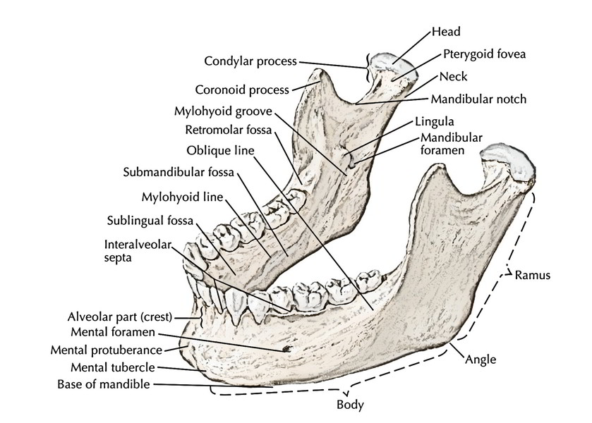
This is an article about the anatomy structures and clinical aspects of the mandible. Bony structures of the mandible. It consists of a curved horizontal portion the body and two perpendicular portions the rami which unite with the ends of the body nearly at right angles angle of the jaw.
Jaws function by moving in opposition to each other and are used for biting chewing and the handling of food. Learn all about the lower jaw now at kenhub. Other mandibular fracture areas include the body the angle the.
There is a lack. Four different muscles connect to the lower jaw to facilitate its movement. Alveolar bone resorption occurs when the teeth are lost.
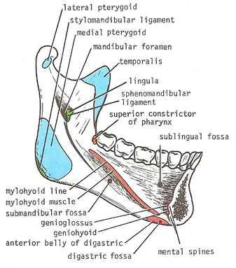
The mandible is composed of 2 hemimandibles joined at the midline by a vertical symphysis. The mandibles of a nauplius have two branches with a chewing or compressing lobe at the base. It articulates with both temporal bones at the mandibular fossa at the temporomandibular joints tmj.
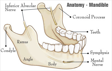
In this article we will discuss the anatomy its contents and clinical relevance of the mandibular foramen. It is the only mobile bone of the facial skeleton and since it houses the lower teeth its motion is essential for mastication. Inferior alveolar nerves and vessels to the lower teeth.
Mental nerve and vessels. The mandible l mandere to chew is the facial bone that forms the lower jaw and contains the lower teeth. Mandible anatomy mental foramen.
It forms the lower jaw and holds the lower teeth in place. Introduction to mandible bone anatomy. Movement of the lower jaw opens and closes the mouth and also allows for the chewing of food.
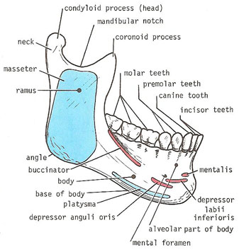
External lateral surface mentalis buccinator platysma depressor labii inferioris depressor anguli oris. It is the only movable bone of the skull discounting the ossicles of the middle ear. The anterior portion of the mandible called the body is horseshoe shaped and runs horizontally.
Jaw in jaw a movable lower jaw mandible and fixed upper jaw maxilla. It consists of right and left halves that fuse together early in life. It is formed by intramembranous ossification.
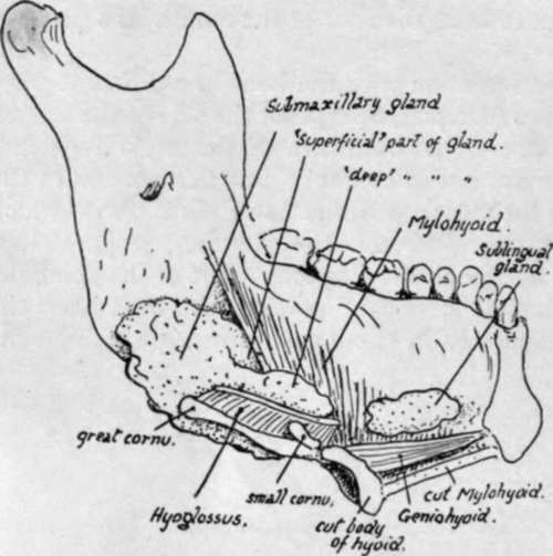
Internal medial surface genioglossus geniohyoid mylohyoid and digastric. Here the most common bony disturbances have been noted. They also may be used for swimming.
These muscles are the masseter the temporalis the medial pterygoid and the lateral pterygoid.
 Mandible Bone Anatomy Youtube
 Anatomy Of Oromandibular Cancer Headandneckcancerguide Org
 Surgical Anatomy Of Periodontium And Related Structures
 Mandible Radiology Reference Article Radiopaedia Org
The Skull Anatomy And Physiology Openstax
 Angle Of The Mandible Wikipedia
 Broken Jaw Mandibular Fracture Types Causes Symptoms
 Mandible
![]() The Mandible Anatomy Structures Fractures Kenhub
The Mandible Anatomy Structures Fractures Kenhub
 Mandible
 Mandible Anatomy Jaw Skull Infratemporal Fossa Png
 Mandible Authors Added Material Ao Surgery Reference
![]() The Mandible Anatomy Structures Fractures Kenhub
The Mandible Anatomy Structures Fractures Kenhub


 Anatomy Of Mandible Right Lateral View Diagram Quizlet
 Human Mandible Anatomy

 Mandible
Mandible Fractures American Academy Of Ophthalmology
 Mandible Wikipedia
Anatomy Of Maxilla And Mandible
 Easy Notes On Mandible Learn In Just 4 Minutes Earth S Lab
 Lower Jaw Or Mandible Part 2
 Lateral View Of The Skeletal Anatomy Of The Skull Mandible
 The Mandible Lower Jaw Human Anatomy
 Science Source Mandible Artwork
 Mandible Wikipedia
Belum ada Komentar untuk "Mandibula Anatomy"
Posting Komentar