Atrium Anatomy
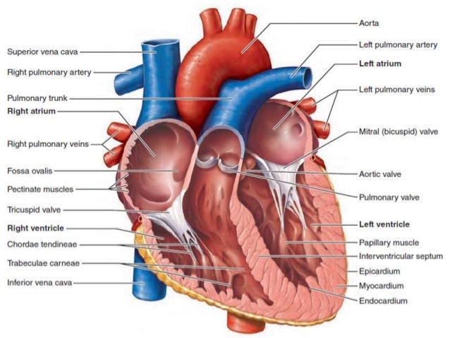
The right atrium is located in the upper portion of right side of heart consisting of the sinus venosus and the right atrial appendage. Components of the left atrium like the rigbt atrium the left atrium can be considered as having four components.
 Left Atrium Radiology Reference Article Radiopaedia Org
The atria receive blood while relaxed then contract to move blood to the ventricles.

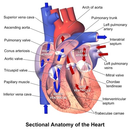
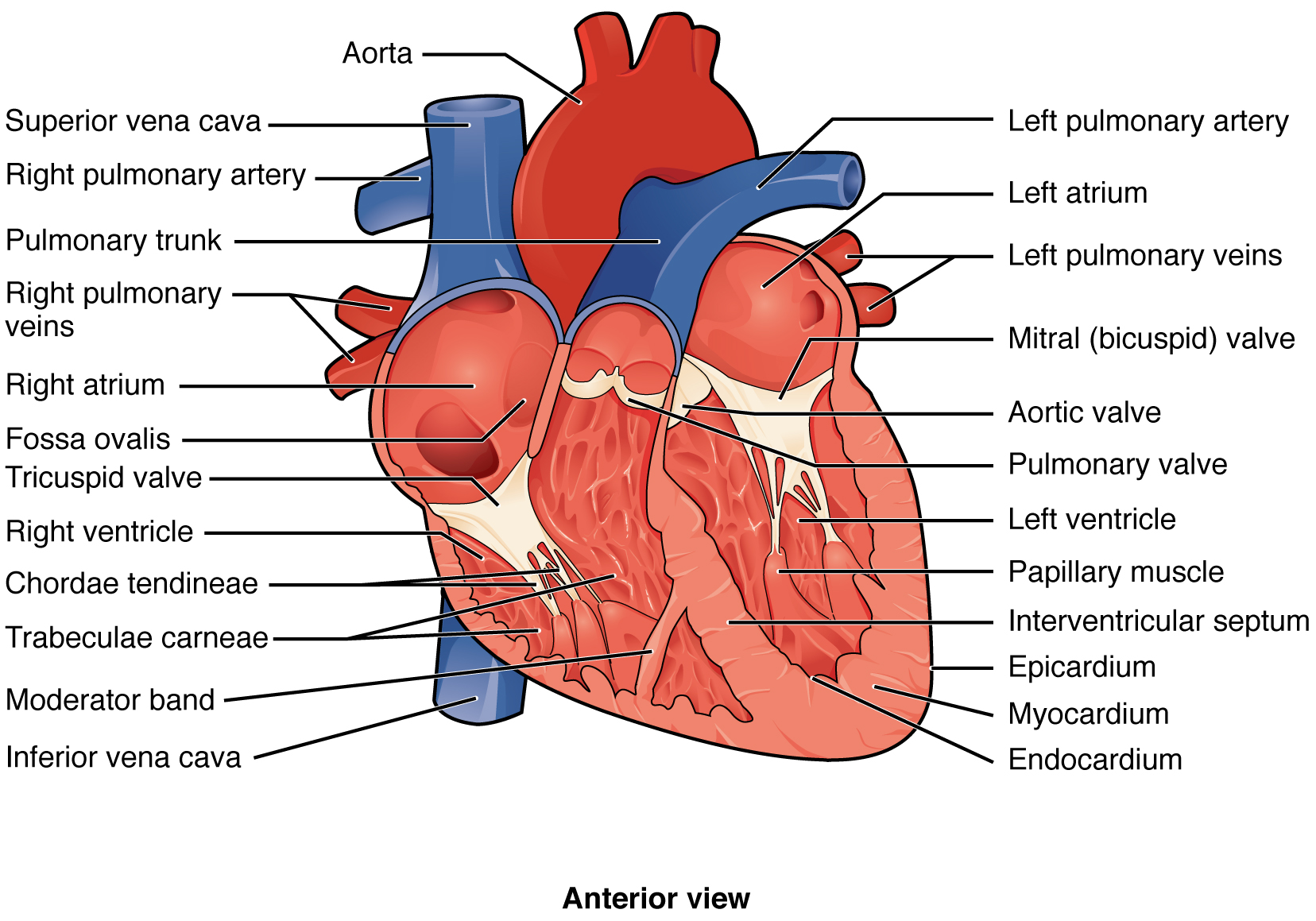
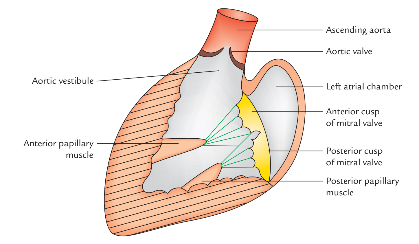
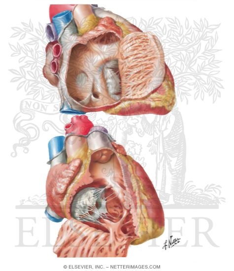
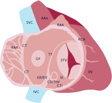
Atrium anatomy. The true extent is revealed by making sec tions through the heart fig. The atrium was formerly called the auricle. Blood enters the heart through the two atria and exits through the two ventricles.
The right atrium is the receiving chamber for oxygen poor blood deoxygenated returning from the systemic circuit. That term is still. Contains the sinoatrial node.
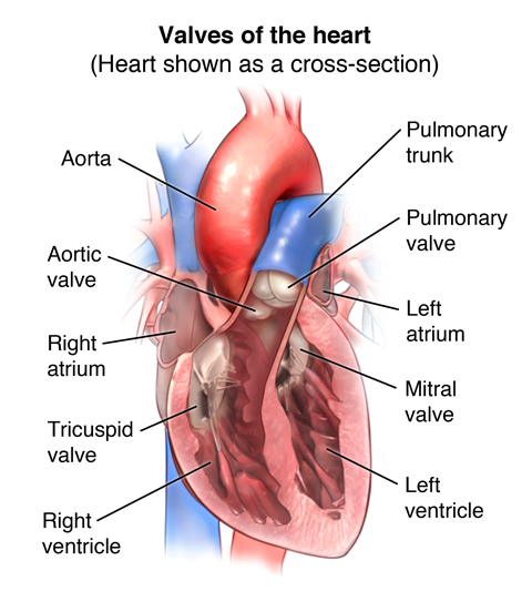
The superior vena cava inferior vena cava and coronary sinus figures 1 and 2. There fresh oxygen enters the blood stream. There are two atria in the human heart the left atrium receives blood from the pulmonary circulation and the right atrium receives blood from the venae cavae.
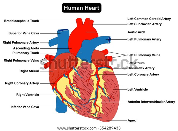
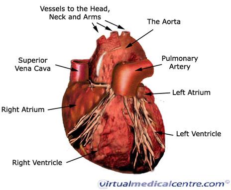
In this image you will find right atrium and left atrium anatomy brachiocephalic trunk superior vena cava right pulmonary artery ascending aorta pulmonary trunk right pulmonary veins right atrium right coronary artery left common carotid artery left subclavian artery aortic arch in it. Right atrium atlas of human cardiac anatomy. Septum appendage vestibule and venous component.
Deoxygenated blood enters the right atrium through the inferior and superior vena cava. On examination from the right atrial aspect the atrial septum at first sight appears extensive fig. All animals with a closed circulatory system have at least one atrium.
The atrium is the upper chamber through which blood enters the ventricles of the heart. The right side of the heart then pumps this deoxygenated blood into the pulmonary arteries around the lungs. The left atrium is located above left ventricle.
The right atrium receives oxygen poor blood from three veins. The right atrium forms the entire right border of the human heart. Humans have two atria.
Blood enters via the pulmonary veins and exits through the mitral valve.

 Heart Anatomy Diagram Quizlet
 What Is The Function Of The Auricle Of The Heart Quora
 Heart Anatomy Section Through Left Atrium And Ventricle W
 Pediagenosis
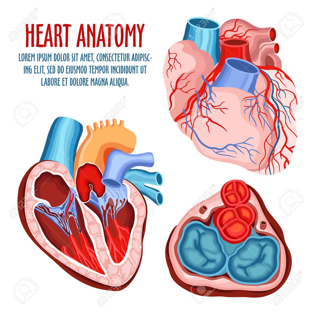
 Human Heart Muscle Structure Anatomy Infographic Stock

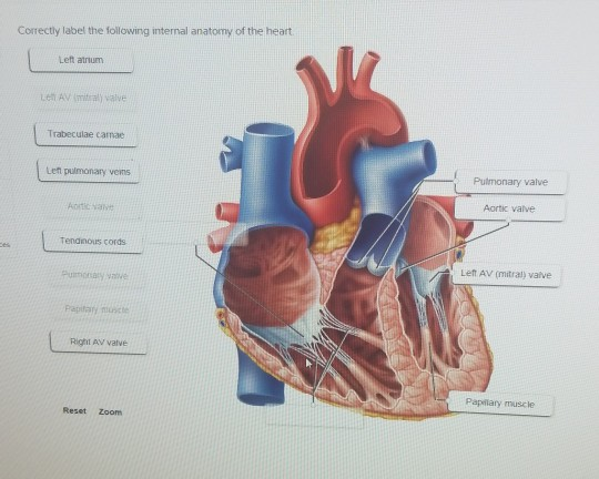 Solved Correctly Label The Following Internal Anatomy Of
 Anatomy Of Heart
:max_bytes(150000):strip_icc()/heart_inner_section-577d5c673df78cb62c939314.jpg) Atria Of The Heart Function
Atria Of The Heart Function
 Trabeculae Carneae Wikipedia
 Atrium Heart Wikipedia
 Right Atrium An Overview Sciencedirect Topics
 Cardiac Anatomy
 What Are The Differences Between The Ventricle And Atrium Of
 Pediagenosis
 Anatomy Of Heart Human Coronary Organ With Atrium And Aorta
![]() Heart Right And Left Atrium Anatomy And Function Kenhub
Heart Right And Left Atrium Anatomy And Function Kenhub
 Left Atrium Earth S Lab
 Right Atrium The Anatomy Of The Heart Visual Atlas Page
 Right Atrium And Right Ventricle
Gross Anatomy And Histology Of The Heart 02 09 2015
![]() Heart Right And Left Atrium Anatomy And Function Kenhub
Heart Right And Left Atrium Anatomy And Function Kenhub
 Chambers Of The Heart
 Cardiovascular System Heart Anatomy Healthengine Blog
 3 5 The Right Atrium 123sonography
 Science Source Heart Anatomy Illustration
Belum ada Komentar untuk "Atrium Anatomy"
Posting Komentar