Anatomy Of Brain In Ct Scan
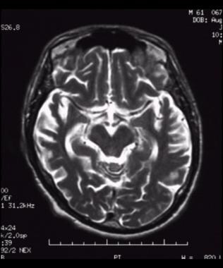
Amygdala on ct and mr images the amygdala is a large region of gray matter contiguous with the uncus of the medial temporal lobe and the most anterior portion of the hippocampus the pes hippocampi. Ct brain image orientation.
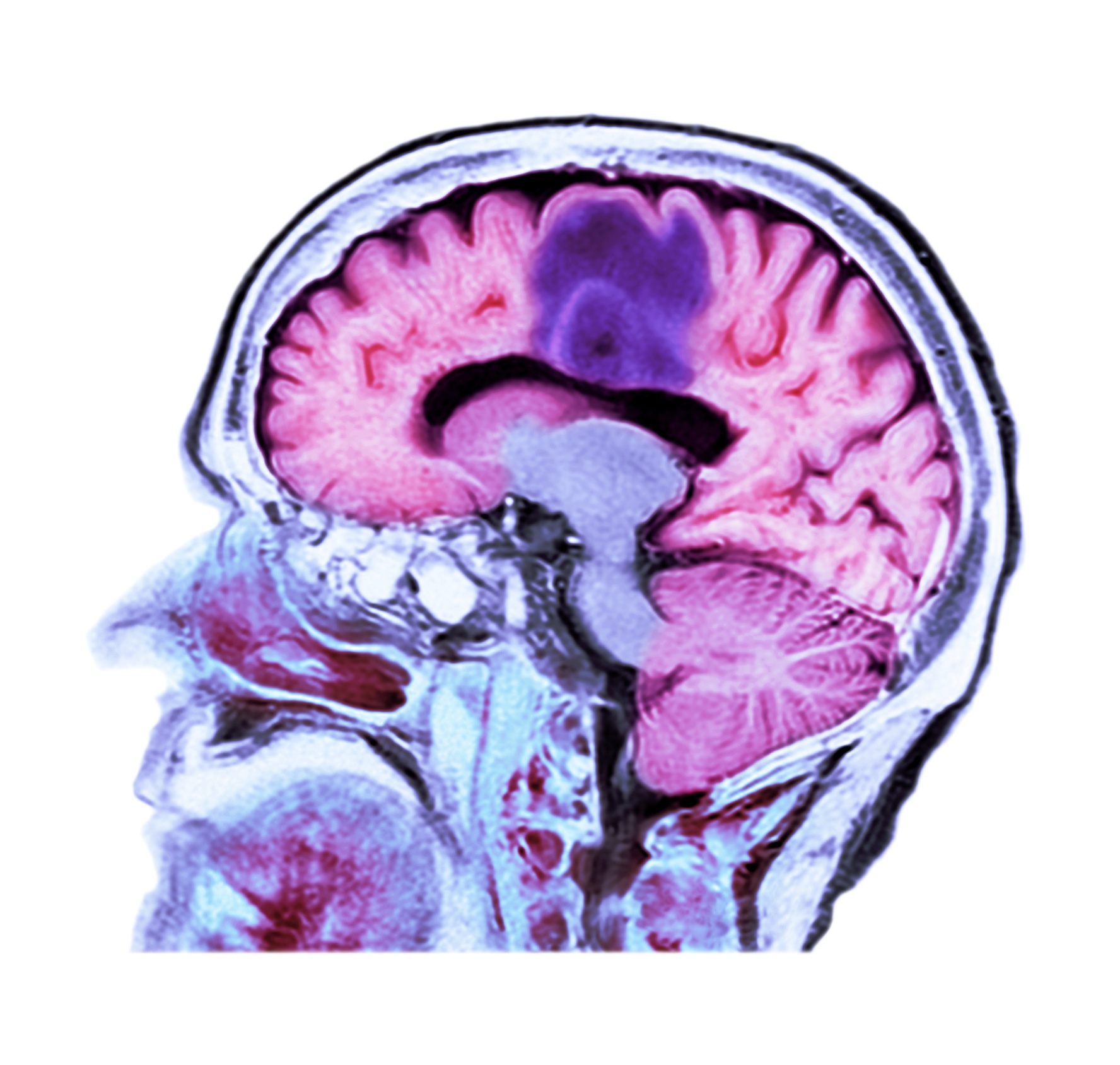
 Signs Of Stress In The Brain May Signal Future Heart Trouble
Brain bones of cranium sinuses of the face.
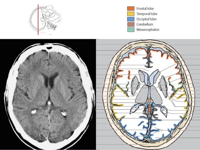
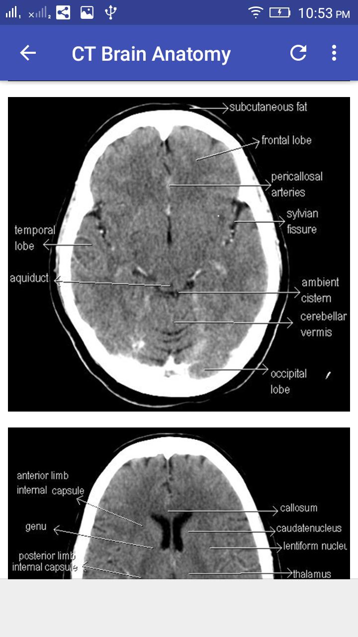
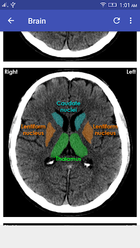
Anatomy of brain in ct scan. 6 frontal bone 27 occipital bone 32 optic nerve 43 frontal sinus 45 sigmoid sinus 46 internal carotid artery. Brain bones of cranium sinuses of the face. This means that the right side of the brain is on the left side of the viewer.
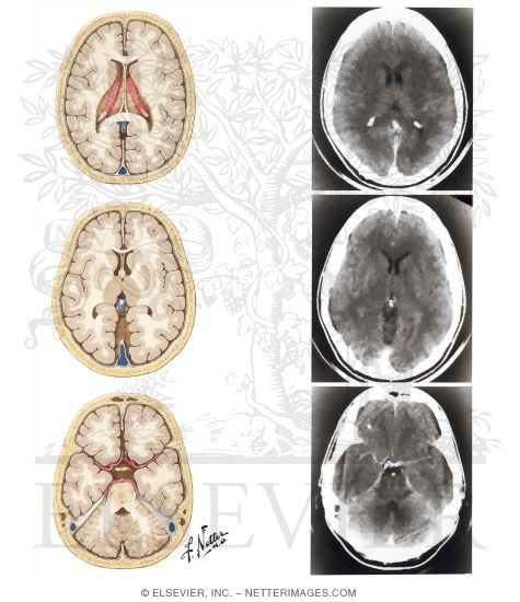
Third and fourth ventricles in midline. Focal abnormalities are not observed in the brain parenchyma. Brain and face ct.
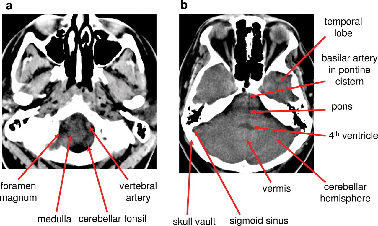
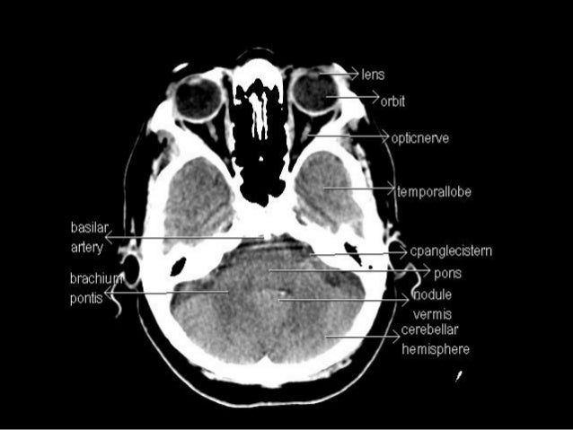
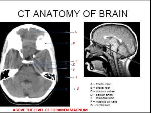
Brain ct scans can provide more detailed information about brain tissue and brain structures than standard x rays of the. Anatomy of the head on a cranial ct scan. Anatomy ct axial brain form no 19.
Anatomy of the head on a cranial ct scan. They lie on the ventricular surface of the hippocampus and become the fimbria of the fornix medially. Anatomy ct axial brain anatomy ct axial brain form no 1.
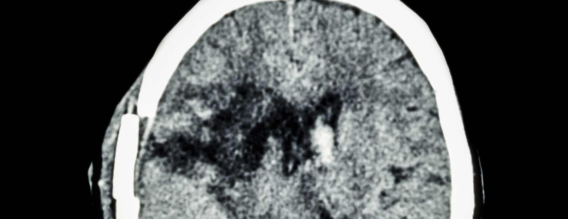
Ct scan provides a 3d display of the intracranial anatomy built up from a vertical series of transverse axial tomograms each tomogram represents a horizontal slice through the patients head. A ct of the brain is a noninvasive diagnostic imaging procedure that uses special x rays measurements to produce horizontal or axial images often called slices of the brain. The anterior part of the head is at the top of the image.
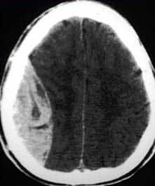
Learn ct scan learn the diagnosis of ct and methods of computed tomography. Fig 11 ct scan of a massive extradural haematoma extradural. Adequate gray matter white matter differentiation.
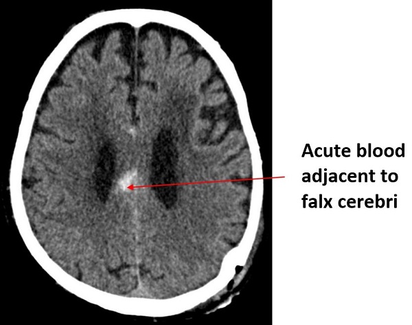
A bleed outside the dura mater which creates a lentiform lemon shaped bleed on ct. Ct images of the brain are conventionally viewed from below as if looking up into the top of the head. Brainstem and cerebellum without evidence of focal lesions.
Lateral ventricles of normal volume. Basal subarachnoid cisterns normal configuration. These are arterial and frequently related to blunt trauma.
 Head Ct
 Normal Anatomy Of The Brain On Ct And Mri With A Few Normal
 Ct Brain Hemorrhage Startradiology
 Medical Imaging Wikipedia
 Alzheimer Disease Imaging Practice Essentials Ct Scan Mri
 Brain Lobes Annotated Mri Radiology Case Radiopaedia Org
 Introduction To Brain Surface Anatomy
 Ct Brain For Android Apk Download
 Normal Brain Anatomy As Demonstrated By Computerized
 Brain Imaging
 Ct Anatomy
Basics Of Ct Head
 Brain And Face Ct Interactive Anatomy Atlas
 Brain And Face Ct Interactive Anatomy Atlas
 Ct Scans Interpretation Principles Basics Teachmeanatomy
 Computed Tomography Of The Head Wikipedia
 Radiology Basics Head Anatomy
 Texas A M Cvm Study Finds New Pathway For Potential

 Normal Ct Brain
 Brain Imaging
 Mri Anatomy Free Mri Axial Brain Anatomy
 The Ct Anatomy Tutor
 Normal Brain Ct Mri Imaging In Arabic Prof Dr Mamdouh Mahfouz
 Axial View Of A Head Computed Tomography Ct Scan Of Pineal
Belum ada Komentar untuk "Anatomy Of Brain In Ct Scan"
Posting Komentar