External Nasal Anatomy
Made up mainly of cartilage and bone and covered by mucous membranes. And two other portions called the latter and oblique sections.
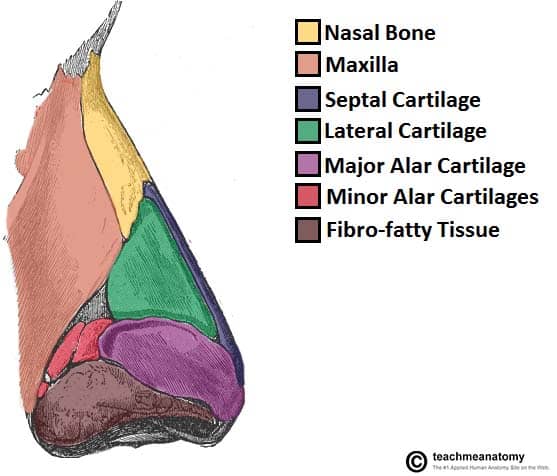
 The External Nasal Anatomy Bones Cartilage And Musculature
The external nose is said to have a pyramidal shape.

External nasal anatomy. These form the tip of the nose and nares they are called the major alar. Essentially nose anatomy can be divided into two parts. Two chambers divided by the septum.
Passages that are lined with mucous membranes and. External nasal anatomy can best be considered in structural thirds. This article reviews the relevant anatomy pathologic conditions and treatments for external nasal valve dysfunction including a detailed review of the nasal muscles and their contribution to external nasal valve patency.
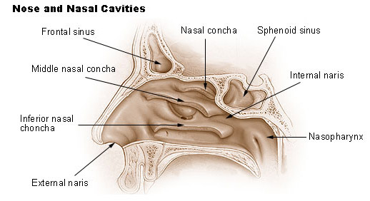
Found within the frontal bone each of these sinuses is triangular in shape. Theyre lined with a mucosal membrane and have small openings into the nasal cavity. The nose is made up of.
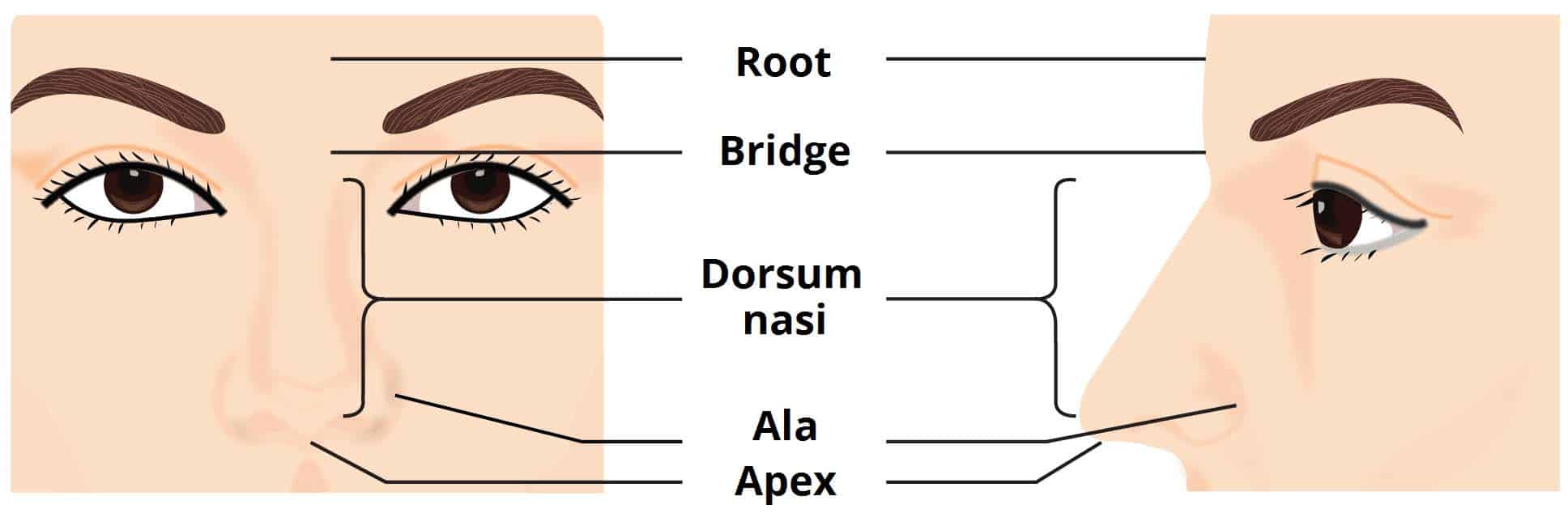
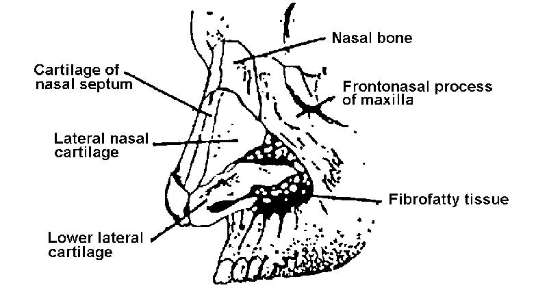
The external nasal nerve or external nasal branches are terminal branches of the anterior ethmoidal nerves from the ophthalmic division of the trigeminal nerve and provide sensory innervation to the skin of the lower half of the nose and of the septum mobile nasi. The nasal ridge nasal dorsum is the border between the root and the tip of the nose which in profile can be variously shaped. The middle third is composed of the stiff paired upper lateral cartilages fused.
The upper third includes the paired nasal bones. The external the part that protrudes from the face and is visually seen and the internal which comprises the nasal cavities that connect with the sinuses located under the forehead and cheekbones. Lets start with the external anatomy of the nose.
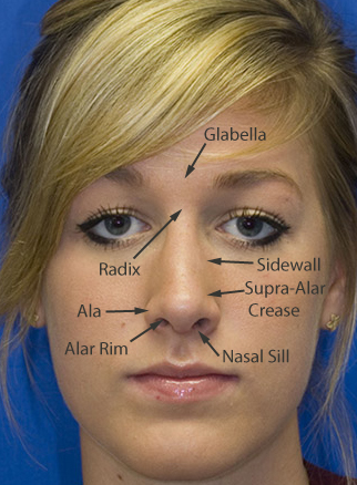
The external nose surface appearance. Bony component located superiorly and is comprised. The ala of the nose ala nasi wing of the nose is the lower lateral surface of the external nose shaped by the alar cartilage and covered in dense connective tissue.
A number of small muscles insert into the external nose contributing to facial expression. The surface of the human nose consists of a frontal portion comprised of the glabella nasion alar sidewalls and tip points. As such there is no single operation that can address all problems of the external valve.
Nasal septum this forms part of the dorsum it extends upward between the upper and lower lateral cartilages. This sinus is located in the body of the maxilla behind the cheek just above. A basal portion made up of the columella nostrils soft tissues and infra tip lobule.
Triangular shaped projection in the center of the face. The external nasal valve is a complex entity comprised of multiple structures and tissue types.
 Acfs Archives Of Craniofacial Surgery
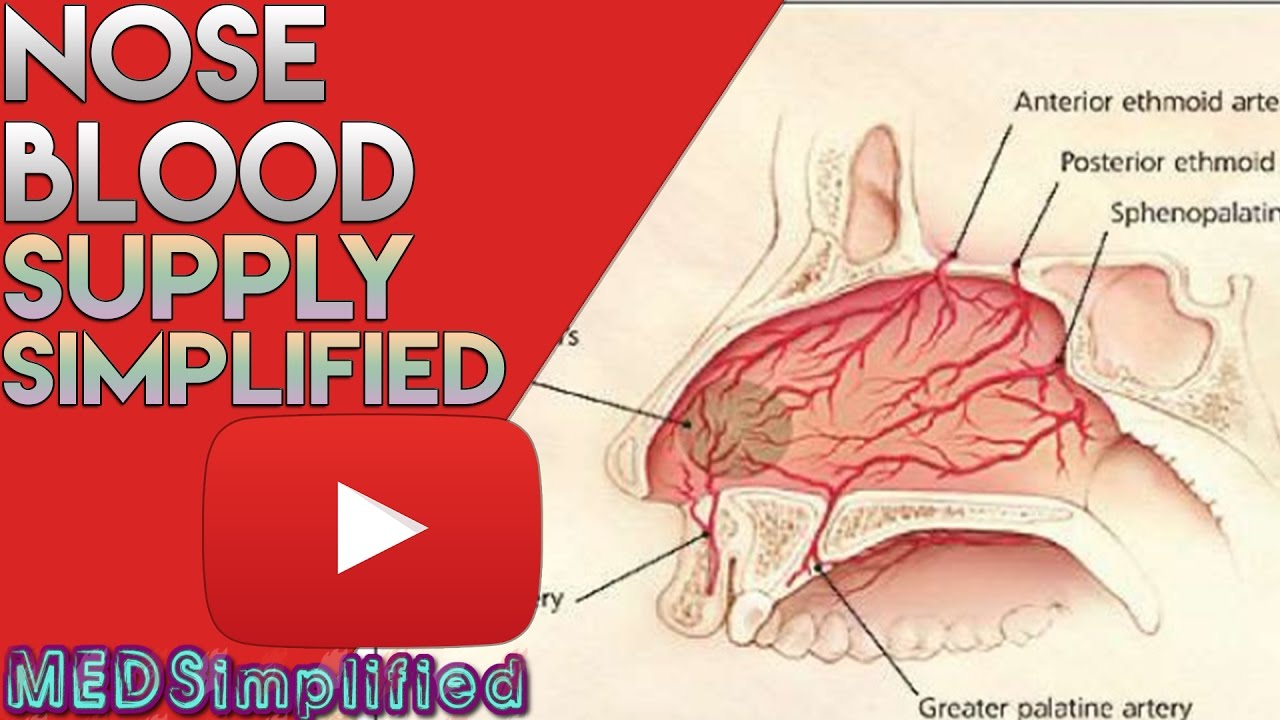 Nose Anatomy Nasal Blood Supply
 Nasal Cavity And Paranasal Sinuses Flashcards Quizlet
 The External Nose Muscles Innervation Teachmeanatomy
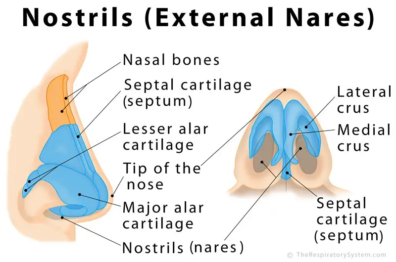 Nostrils Definition Functions Anatomy Pictures
 Nose Revision Surgery And Surgeons Nasal Valve Collapse

 Facial Landmarks An Overview Of Dental Anatomy
 The External Nose Muscles Innervation Teachmeanatomy
 Racgp Traumatic Nasal Injuries In General Practice
 Nasal Anatomy Dr Evan Ransom
 Acfs Archives Of Craniofacial Surgery
Nose Structures External Nose Cartilage Philtrum Naris

 Figure 1 From Anatomy Of The External Nasal Passages And
 Acfs Archives Of Craniofacial Surgery

 Anatomy And Physiology Of The Nose And Throat
 Human Nose Wikipedia
 Human Nose Wikipedia
 Seer Training Nose Nasal Cavities Paranasal Sinuses
 Surgical Anatomy And Physiology Of The Nose Springerlink
Belum ada Komentar untuk "External Nasal Anatomy"
Posting Komentar